Introduction
Pulmonary arterial hypertension is a progressive and life-threatening pulmonary vascular disease characterised by elevated mean pulmonary artery pressure and increased pulmonary vascular resistance (PVR). Genetic predisposition, anatomical anomalies, vascular pathologies, and associated comorbidities contribute to the aetiology of pulmonary arterial hypertension. Reference Ivy, Rosenzweig and Abman1,Reference Kovacs, Bartolome and Denton2 Hepatic arteriovenous malformations are rare vascular anomalies that can lead to high-output shunting and low-resistance circulation. The excessive blood flow from the arterial to the venous system results in pulmonary circulatory overload, increased pulmonary artery pressure, and high-output cardiac failure. Reference Botha, Rasmussen and Carlan3–Reference Subramanyan, Narayan and Costa6
In this case report, we present the stepwise management of an infant with Down syndrome, severe pulmonary arterial hypertension due to a large patent ductus arteriosus, and a giant hepatic arteriovenous malformation, successfully eliminating the need for liver transplantation.
Case report
A three-month-old male infant was referred to our clinic for liver transplantation after being diagnosed with severe pulmonary arterial hypertension and a giant hepatic arteriovenous malformation at an external centre. The patient had a known diagnosis of Down syndrome and hypothyroidism. His weight and height were below the third percentile for his age. On physical examination, he was tachycardic (146 bpm), tachypnoeic (62 breaths per minute), and had an oxygen saturation of 89% in room air. He was receiving furosemide (2 mg/kg/day), enalapril (0.1 mg/kg), and sildenafil (1.5 mg/kg three times daily).
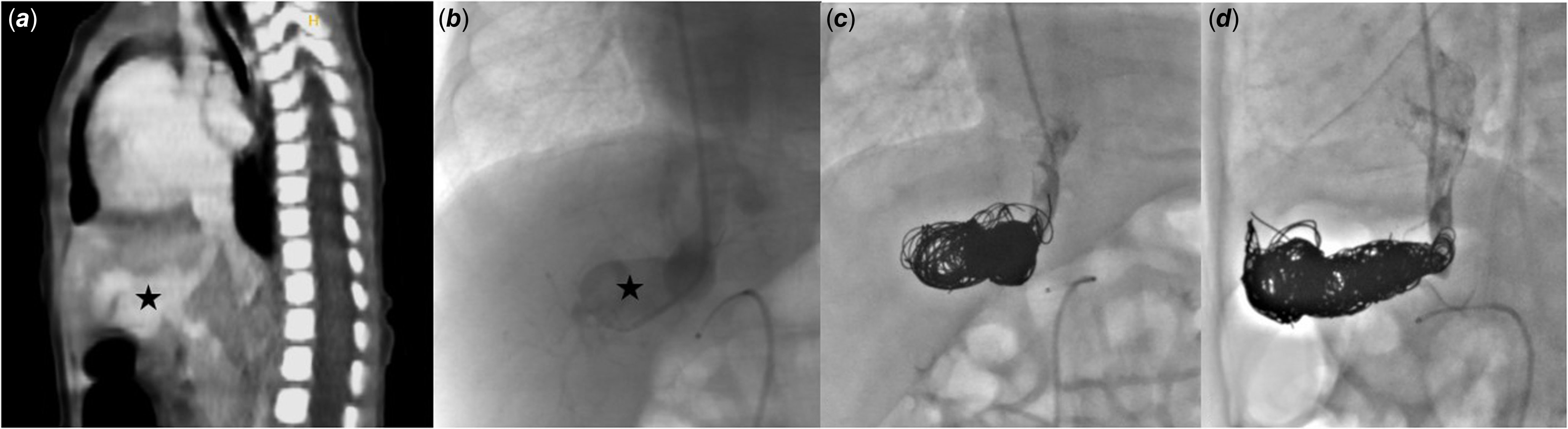
Echocardiographic evaluation revealed significant right atrial and right ventricular enlargement, leftward deviation of the interventricular septum, and left ventricular compression. A 5-mm atrial septal defect and a 3-mm patent ductus arteriosus, both exhibiting bidirectional shunting, were observed. Severe tricuspid regurgitation was present, and the right ventricular systolic pressure was calculated as 80 mmHg (Figure 1). Thoracic and liver CT demonstrated a giant parenchymal hepatic arteriovenous malformation measuring approximately 41 × 18 mm in the medial segment of the left liver lobe, involving the left hepatic vein, left portal vein, coeliac trunk, and the distal main hepatic artery. Notably, the coeliac trunk exhibited diffuse dilation along its entire course, and a marked increase in the calibre of the inferior vena cava was observed at its cardiac entry level (Figure 2(a)). Additionally, pulmonary artery enlargement and radiological findings consistent with pulmonary congestion were detected.
( a ) Transthoracic echocardiography image demonstrating right atrial and right ventricular dilatation. ( b ) Leftward bowing of the interventricular septum, and significant tricuspid regurgitation. ( c ) Images showing bidirectional shunting at the atrial level.

During cardiac catheterisation under general anaesthesia, following written informed consent obtained from the patient’s legal guardian, haemodynamic assessment revealed that pulmonary artery pressure was 60/23 (39) mmHg, systemic arterial pressure was 71/31 (49) mmHg, PVR was 6.30 WU, pulmonary vascular resistance/systemic vascular resistance (PVR/SVR) ratio was 0.57, pulmonary blood flow (Qp) was 4.92 L/min, and Qp/Qs ratio was 1.30. Angiography indicated a giant hepatic malformation with an aneurysmal structure, supplied by multiple tortuous branches originating from the main hepatic artery and intrahepatic portal system. The angiogram demonstrated drainage from the hepatic aneurysm into the hepatic veins, subsequently emptying into the inferior vena cava at the right atrial junction. Access to the left hepatic vein was obtained via the right jugular vein, and the aneurysmal sac was reached from the hepatic vein through a draining vein using a microcatheter. The aneurysm sac was embolised using a total of 17 Azur® CX peripheral coils (HydroCoil, Terumo Medical Corporation, Somerset, NJ, USA), ranging in size from 8 to 20 mm (Figure 2). A post-procedure angiogram confirmed complete occlusion of the aneurysm sac with no residual shunting. The procedure was completed without complications. The patient was discharged on dual anti-congestive therapy and sildenafil, with a planned monthly follow-up.
( a, b ) Computed tomography and angiographic images demonstrating a giant hepatic arteriovenous malformation (star). ( c, d ) Follow-up angiogram showing complete occlusion of the giant hepatic arteriovenous malformation using 17 Azur® CX peripheral coils ranging from 8 to 20 mm in size.

At the three-month follow-up, the patient showed significant clinical improvement, with reduced tachypnoea and a weight gain of 20 g/day. Follow-up echocardiography demonstrated a decrease in right ventricular end-diastolic diameter (from 24 mm to 22 mm) and an increase in left ventricular size (from 17 mm to 20 mm). However, significant tricuspid regurgitation persisted, and right ventricular systolic pressure was calculated as 85 mmHg. Despite the improvement in symptoms, persistent signs of pulmonary hypertension prompted a second catheterisation for further assessment of hepatic malformation occlusion, invasive measurement of pulmonary artery pressure, and evaluation of patent ductus arteriosus closure feasibility due to its suspected role in pulmonary arterial hypertension. During the second catheterisation, haemodynamic measurements showed a pulmonary artery pressure of 54/25 (38) mmHg, a systemic arterial pressure of 60/31 (42) mmHg, a PVR of 4.27 WU, a PVR/SVR of 0.36, a Qp of 6.79 L/min, and a Qp/Qs of 2.42. Selective contrast injection into the coeliac and hepatic arteries confirmed complete occlusion of the previously embolised aneurysm, with no evidence of new fistulization.
Injection into the descending aorta revealed a tubular patent ductus arteriosus measuring 3.5 mm in diameter and 7 mm in length. During the balloon occlusion test, pulmonary artery pressure decreased to 41/13 (25) mmHg, simultaneous systemic arterial pressure was measured at 66/37 (50) mmHg, PVR decreased to 3.26 WU, and the PVR/SVR ratio dropped to 0.22. Patent ductus arteriosus closure was deemed safe and was successfully performed using an 8-mm Lifetech Cera™ vascular plug. The patient was discharged the following day on existing medical therapy.
At the two-month follow-up, the patient showed marked symptomatic improvement and a reduction in right heart chamber size. The shunt through the 5-mm atrial septal defect had shifted to a left-to-right direction, and right ventricular systolic pressure had decreased to 40 mmHg (Figure 3). Our follow-up focuses on optimising clinical outcomes and ensuring long-term stability. Pulmonary arterial hypertension remains a priority, with regular non-invasive monitoring of pulmonary pressures and right ventricular function. The atrial septal defect is closely monitored for its haemodynamic impact, while serial imaging is used to assess hepatic arteriovenous malformation stability and residual shunting. A multidisciplinary team ensures comprehensive care.
( a, b ) Transthoracic echocardiography images showing reduced right atrial and ventricular dilation and increased left ventricular size, with moderate tricuspid regurgitation. ( c, d) Images showing left-to-right shunting at the atrial level.

Discussion
This is the first documented paediatric case of Down syndrome with severe pulmonary arterial hypertension, a giant hepatic arteriovenous malformation, and structural heart anomalies, successfully managed through a multidisciplinary and stepwise treatment approach. Symptoms were successfully managed, leading to haemodynamic stabilisation and ultimately preventing the need for liver transplantation.
Given the complexity of the patient’s condition, a careful and individualised approach was necessary to achieve optimal outcomes. Initially, the hepatic malformation was embolised to control the significant haemodynamic burden, aiming to reduce volume overload and prevent pulmonary congestion. This intervention facilitated clinical improvement; however, due to the persistence of pulmonary arterial hypertension findings, attention was then directed towards addressing the patient’s other cardiovascular anomalies.
In patients with severe pulmonary arterial hypertension, cardiac defects often act as pop-off mechanisms, reducing right ventricular overload and alleviating cardiac workload. Reference Ivy, Rosenzweig and Abman1 Therefore, a thorough assessment is essential to determine whether these defects contribute to pulmonary arterial hypertension pathogenesis and require closure or if they function as protective mechanisms that should be preserved. An inappropriate intervention could irreversibly worsen the prognosis. Reference Ivy, Rosenzweig and Abman1
In paediatric patients, atrial septal defect is not considered a primary cause of pulmonary arterial hypertension. Reference Kovacs, Bartolome and Denton2 Thus, in our case, the focus was not on atrial septal defect closure but rather on evaluating the haemodynamic impact of the patent ductus arteriosus and ensuring the safety of its closure. Moreover, tubular patent ductus arteriosus pose a greater risk for PH progression due to their wide anatomical structure and low spontaneous closure rates, leading to prolonged left-to-right shunting and increased pulmonary vascular burden. Therefore, careful evaluation of the patent ductus arteriosus haemodynamic impact is crucial, considering the patient’s age, pulmonary haemodynamics, and patent ductus arteriosus morphology. Reference Chinawa, Chukwu, Chinawa and Duru7 To ensure a safe and effective intervention, we performed a balloon occlusion test, confirming that patent ductus arteriosus closure could be safely undertaken.
The management of hepatic arteriovenous malformations involves several therapeutic strategies, including hepatic artery ligation or banding, transcatheter embolisation, and liver transplantation, as documented in the literature. Reference Botha, Rasmussen and Carlan3–Reference Subramanyan, Narayan and Costa6 In cases where aneurysmal structures are supplied by multiple vascular collaterals, sequential closure of individual shunts may be impractical and insufficient, as persistent collateral flow can maintain haemodynamic instability. Therefore, total occlusion of the aneurysmal sac may be a more effective and safer approach, eliminating the risk of residual shunting and reducing the likelihood of recurrent symptoms. Coil embolisation has emerged as a preferred method in such scenarios due to its high filling capacity, ease of deployment, and ability to achieve permanent occlusion. By effectively blocking abnormal vascular connections, coil embolisation prevents ongoing high-output cardiac failure and pulmonary overcirculation, contributing to overall hemodynamic stabilisation. Reference Jameel, Amin and Arif5,Reference Subramanyan, Narayan and Costa6 The choice of embolisation technique and materials should be tailored to the anatomical characteristics of the malformation, ensuring complete occlusion while minimising procedural risks.
In infants with severe pulmonary arterial hypertension, a thorough evaluation of both cardiac and extracardiac defects is crucial for optimal management. A comprehensive, multidisciplinary approach, incorporating interventional radiology, paediatric cardiology, pulmonology, and other relevant specialties, plays a key role in ensuring effective diagnosis and treatment. This case highlights the importance of recognising that the clinical presentation may be multifactorial rather than attributed to a single defect, emphasising the need for an integrated and individualised treatment approach.