INTRODUCTION
Chagas disease, or American trypanosomiasis, caused by the protozoan Trypanosoma cruzi is the largest parasitic disease burden in the American continent affecting approximately 8 million people from the southern USA to northern Chile. Even though the enforcement of public health programmes towards vector elimination in some Latin American countries has decreased the incidence of new infections, the disease is still endemic in large areas (WHO, 2013).
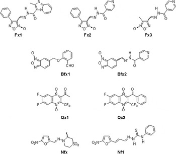
The current treatment of Chagas disease depends on two nitroheterocycles, nifurtimox (Nfx, Fig. 1) and benznidazole. Although effective for acute infections both drugs are not efficient in chronic infections and cause undesirable side effects; therefore, there is an urgent need for the development of safe and effective drugs (Cerecetto and González, Reference Cerecetto and González2010). Extensive efforts have been directed to the development of new chemotherapeutic agents but they have been deficient in the final stages of drug development studies, i.e. tolerance/safety, selectivity, drug-resistance, scaling-up, pharmacokinetic and pharmacodynamic properties (González and Cerecetto, Reference González and Cerecetto2011).
Chemical structures of the compounds herein studied: N-oxide-containing heterocycles (Fx1, Fx2, Fx3, Bfx1, Bfx2, Qx1, Qx2), Nifurtimox (Nfx) and nitrofurane (Nf1).

In an ongoing effort to discover new anti-T. cruzi agents, our group has been exploring the moiety N-oxide as a pharmacophore for this kind of drug (Cerecetto and González, Reference Cerecetto and González2008; Boiani et al. Reference Boiani, Piacenza, Hernández, Boiani, Cerecetto, González and Denicola2010). Within this framework, we have investigated the trypanosomicidal activity of different N-oxide-containing heterocycles, and from 200 synthesized compounds, two furoxans, i.e. Fx1, and Fx2, two benzofuroxans, i.e. Bfx1, and Bfx2, and two quinoxaline 1,4-dioxides, i.e. Qx1, and Qx2 (Fig. 1), were selected based on their excellent in vitro activities (Merlino et al. Reference Merlino, Benítez, Chavez, Da Cunha, Hernández, Tinoco, Campillo, Páez, Cerecetto and González2010; Benítez et al. Reference Benítez, Cabrera, Hernández, Boiani, Lavaggi, Di Maio, Yaluff, Serna, Torres, Ferreira, Vera de Bilbao, Torres, Pérez-Silanes, Solano, Moreno, Aldana, López de Ceráin, Cerecetto, González and Monge2011; Hernández et al. Reference Hernández, Rojas, Gilman, Sauvain, Lima, Barreiro, González and Cerecetto2013). Those agents were active on T. cruzi epimastigotes, trypomastigotes and intracellular amastigotes from different parasite strains, i.e. Tulahuen 2, Y, Colombiana and the CL Brener clone. Additionally, Fx1, Fx2, Qx1 and Qx2 did not exhibit mutagenicity in the Ames test and displayed adequate in vivo behaviour in an acute model of Chagas disease (Benítez et al. Reference Benítez, Cabrera, Hernández, Boiani, Lavaggi, Di Maio, Yaluff, Serna, Torres, Ferreira, Vera de Bilbao, Torres, Pérez-Silanes, Solano, Moreno, Aldana, López de Ceráin, Cerecetto, González and Monge2011; Hernández et al. Reference Hernández, Rojas, Gilman, Sauvain, Lima, Barreiro, González and Cerecetto2013).
Whereas some efforts have been made to elucidate the mode of action of this kind of compounds, knowledge on this subject is still incomplete. Thus, it has been demonstrated that some benzofuroxans, analogues of Bfx1 and Bfx2, are strong inhibitors of parasite dehydrogenase activity affecting mitochondrial membrane potential (Boiani et al. Reference Boiani, Piacenza, Hernández, Boiani, Cerecetto, González and Denicola2010). Accordingly we have proved that Qx1 and Qx2, in contrary to Nfx, decrease mitochondrial dehydrogenase activity thus diminishing the excreted catabolites acetate and succinate in the parasite (Benítez et al. Reference Benítez, Cabrera, Hernández, Boiani, Lavaggi, Di Maio, Yaluff, Serna, Torres, Ferreira, Vera de Bilbao, Torres, Pérez-Silanes, Solano, Moreno, Aldana, López de Ceráin, Cerecetto, González and Monge2011). New knowledge about the mode of action of these kinds of compounds (Fig. 1) may lead to the discovery of new drug targets that help overcome problems related to drug toxicity and drug resistance. Among the different strategies that have been used to elucidate the mechanism of action of anti-T. cruzi drugs (De Castro and Meirelles, Reference De Castro and Meirelles1990; Menna-Barreto et al. Reference Menna-Barreto, Beghini, Ferreira, Pinto, De Castro and Perales2010) we have successfully applied 1H NMR for the evaluation of T. cruzi-excreted catabolites (Boiani et al. Reference Boiani, Aguirre, González, Cerecetto, Chidichimo, Cazzulo, Bertinaria and Guglielmo2008; Caterina et al. Reference Caterina, Perillo, Boiani, Pezaroglo, Cerecetto, González and Salerno2008; Benítez et al. Reference Benítez, Cabrera, Hernández, Boiani, Lavaggi, Di Maio, Yaluff, Serna, Torres, Ferreira, Vera de Bilbao, Torres, Pérez-Silanes, Solano, Moreno, Aldana, López de Ceráin, Cerecetto, González and Monge2011).
Studies on the parasite death phenotype when treated with these compounds are have not been done. This aspect is relevant considering that, for other pathologies and drugs, such knowledge is leading to the design of novel drugs targeting critical points in the death process (Ricci and Zong, Reference Ricci and Zong2006; MacKenzie and Clark, Reference MacKenzie and Clark2008; Tan and White, Reference Tan and White2008). Different cell death phenotypes caused by chemotherapeutic agents have been described in T. cruzi (Menna-Barreta et al. Reference Menna-Barreto, Salomão, Dantas, Santa-Rita, Soares, Barbosa and de Castro2009a ). Interestingly, in protozoa a variety of drug stimuli converge to the same pathway of death, suggesting an intense cross-over between the three types of programmed cell death (PCD), i.e. apoptosis (type I PCD), autophagy (type II PCD) and programmed necrosis (type III PCD). Different techniques have been employed in the study of T. cruzi cell death, i.e. destructive techniques such as flow cytometry, fluorescence microscopy, Western blot, agarose-gel electrophoresis and ultrastructural analysis, or less destructive ones such as protease inhibition analysis (Alvarez et al. Reference Alvarez, Kosec, Sant'Anna, Turk, Cazzulo and Turk2008a , Reference Alvarez, Kosec, Sant'Anna, Turk, Cazzulo and Turk b ; Jiménez et al. Reference Jiménez, Paredes, Sosa and Galanti2008; Irigoín et al. Reference Irigoín, Inada, Fernandes, Piacenza, Gadelha, Vercesi and Radi2009; Menna-Barreto et al. Reference Menna-Barreto, Salomão, Dantas, Santa-Rita, Soares, Barbosa and de Castro2009a ). Recently, we have described the use of 1H NMR spectroscopy (Benítez et al. Reference Benítez, Pezaroglo, Martínez, Casanova, Cabrera, Galanti, González and Cerecetto2012) as a non-invasive method that allows the visualization of the phenomenon of mobile lipid accumulation following the induction of either apoptosis or cytostasis.
The aim of the present study was to analyse some aspects of the mode of action and cell death phenotype induced by the N-oxides shown in Fig. 1. The results indicated a clear difference in the mechanism of action of the newly studied N-oxides compared with the previous ones (Boiani et al. Reference Boiani, Piacenza, Hernández, Boiani, Cerecetto, González and Denicola2010) and particular cell death pathways, for some of them, unrelated to the kind of N-oxide.
MATERIALS AND METHODS
Chemicals
N-oxides Fx1, Fx2, Fx3, Bfx1, Bfx2, Qx1 and Qx2 as well as the nitrofurane, Nf1 (Fig. 1), were obtained as previously described (Merlino et al. Reference Merlino, Benítez, Chavez, Da Cunha, Hernández, Tinoco, Campillo, Páez, Cerecetto and González2010; Aravena et al. 2011; Benítez et al. Reference Benítez, Cabrera, Hernández, Boiani, Lavaggi, Di Maio, Yaluff, Serna, Torres, Ferreira, Vera de Bilbao, Torres, Pérez-Silanes, Solano, Moreno, Aldana, López de Ceráin, Cerecetto, González and Monge2011; Hernández et al. Reference Hernández, Rojas, Gilman, Sauvain, Lima, Barreiro, González and Cerecetto2013). Nfx was purchased from Bayer. Other chemicals were purchased from Sigma-Aldrich unless otherwise indicated.
Cell cultures
Trypanosoma cruzi epimastigote forms (Y strain) were cultured at 28 °C for 5–7 days (exponential phase of growth) under aerobiosis in axenic BHI-tryptose milieu (33 g L−1 brain-heart infusion, 3 g L−1 tryptose, 0·02 g L−1 hemin, 0·3 g L−1 D-(+)-glucose, supplemented with 10% (v/v) calf serum, 200 000 units L−1 penicillin and 0·2 g L−1 streptomycin). For TUNEL and PI-staining studies, T. cruzi epimastigote forms (Y strain and CL Brener clone) were cultured at 28 °C for 5–7 days (exponential phase of growth) under aerobiosis in axenic Diamond milieu (12·5 g L−1 yeast extract, 12·5 g L−1 tryptose, 12·5 g L−1 tryptone, 106 mm NaCl, 29 mm H2KPO4, 23 mm HK2PO4, 7·2 pH, 7·5 mm hemin supplemented with 10% (v/v) calf serum, 75 units mL−1 penicillin and 75 mg L−1 streptomycin).
1H NMR study of the excreted catabolites
For 1H NMR spectroscopic studies, parasites in exponential phase of growth are resuspended in fresh milieu. One mL containing 10 million T. cruzi (Y strain) treated for 2 days with each studied compound at the IC50 doses (Fx1 14·8±2·2 μ m, Fx2 7·95±0·06 mm, Bfx1 4·8±0·6 mm, Bfx2 13·3±0·1 mm, Qx1 1·6±0·4 mm, Qx2 1·8±0·1 mm, Nfx 6·5±0·2 mm, Nf1 1·3±0·5 mm and Fx3 >300·0 mm,), were centrifuged at 3000 g for 10 min. Before measuring, 0·01 mL of DMF, as internal standard, and 0·09 mL of D2O were added to 0·5 mL of the supernatant. Spectra were registered with water suppression in 5 mm NMR (Aldrich, USA) sample tubes. The chemical displacements used to identify the respective metabolites were previously confirmed by adding each analysed metabolite to the studied supernatant as well as by a control solution with 4 mg mL−1 of each metabolite in buffer phosphate, pH = 7·4. Each run was done, at least, in triplicate and Student's t-test was used to analyse the significance of the changes. The chemical shifts (δ, ppm) and multiplicity of the analysed catabolites are: Lac, 1.316, d; Ala, 1.466, d; Ace, 1.904, s; Pyr, 2.357, s; Suc, 2.392, s; Gly, 3.547, s. For quinoxaline di-N-oxides, Qx1 and Qx2, a dose–response study was performed using IC50, 3×IC50 and 5×IC50. Two controls were used, a control with fresh milieu and a control with parasites, with the corresponding concentration of parasites and DMSO used in the samples.
Mitochondrial dehydrogenase activities
Mitochondrial dehydrogenase activities were measured in 24-well plates. Twenty million per mL T. cruzi epimastigotes (Y strain) were washed twice at 3000 g for 10 min and resuspended in PBS-glucose (5·5 μ m). Then 600 μL were loaded in each well and 20 mm of each of the studied compounds were added. The assay was performed in quadriplicate and untreated parasites were maintained as controls corresponding to the given time of treatment. The cultures were incubated at 28 °C. At different incubation times the epimastigotes were counted and the colorimetric MTT dye-reduction assay was performed. For this purpose, 25 μL of a solution containing 4 mg mL−1 of MTT in PBS-glucose (5·5 mm) were added to each well and plates were incubated for an additional 2 h at 28 °C. The reaction was stopped by the addition of 100 μL SDS-isopropanol (10% SDS, 50% isopropanol, H2O) and incubated for an additional 2 h at 28 °C. The absorbance was measured at 570 nm. Under these conditions, compounds did not interfere with the reaction mixture. Percentage of mitochondrial dehydrogenase activities (Pmdh) were determined using untreated parasites’ activities as 100%.
Glucose uptake studies (Boiani et al. Reference Boiani, Boiani, Alicia, Hernández, Chidichimo, Cazzulo, Cerecetto and González2009)
Trypanosoma cruzi epimastigotes (Y strain, 100×106 parasites mL−1) were washed twice with PBS-glucose and resuspended in PBS-glucose (5·5 mm). 800 μL of this suspension were transferred to a 24-well cell-culture plate. Then, Qx2 was added dissolved in DMSO (8 μL), at IC50 and 3×IC50 concentrations and incubated for 4 h. After centrifugation at 3000 g for 10 min the parasite-free supernatant was treated with 500 μL Benedict's reagents at reflux during 5 min. Glucose concentration was determined, measuring at 744 nm using a calibration curve. Negative controls were made with DMSO or Qx2 at IC50 and 3×IC50.
Cell death phenotype studies
1H NMR-VML spectroscopy analysis
Cell sample preparation
Treated or un-treated (control) cells (150×106) were harvested and centrifuged for 10 min at 3000 g . The pellet was washed three times in PBS, re-suspended in PBS (500 μL), transferred to a 5 mm NMR tube (ALDRICH, USA) and D2O (90 μL) was added. The mixture was homogenized prior to acquire the spectrum.
NMR spectra acquisition
1H NMR experiments were recorded at 20 °C in a Bruker Avance DPX-400 spectrometer, operating at 400·132 mHz, with a 5 mm broadband inverse geometry probe. The acquisition parameters were 90° pulse (zgpr, avance-version v 1.7.10.2, 1D sequence with f1 presaturation), 128 scans, and spectral width of 14·983 ppm. The acquisition time was 1·3664 s. Signal intensities were calculated by performing appropriate baseline corrections and then integrating the area under each of the resonances using MestRe-C NMR software (http://mestrelab.com/). Spectra were analysed using the Topspin 1.3 software package. The integrated regions were 1·20–1·35 ppm for CH2 and 0·80–0·90 ppm for CH3. The visualized regions were 3·10–3·30 ppm for Cho and 2·80 and 5·40 ppm for polyunsaturated fatty acids (PUFA).
Statistical analysis
Values are expressed as means±s.e.m. of at least three independent experiments. Statistical comparisons were performed with unpaired Student's t-tests by using OriginPro 8 software. P<0·05 was considered statistically significant.
TUNEL assay and staining with PI
After each treatment parasites (600 μL, 10×106 mL−1) were collected by centrifugation at 3000 g , washed twice in PBS, resuspended in the same buffer and placed on a slide. After drying at room temperature the cells were fixed with methanol (70%) and washed in PBS. For TUNEL assays cells were made permeable with 0·2% Triton X-100. Afterwards, cells were incubated with a reaction mix containing dUTP-FITC (Fluorescein isothiocyanate). Nuclei were counterstained with DAPI (4′,6-diamidin-2-phenylindol) (1 mg mL−1). Treatment of parasites with H2O2 (500 μ m, 30 min of incubation at 28 °C) was included as a positive control (Benítez et al. 2012) and parasites without treatment were included as a negative one.
For PI-staining, after the permeabilization step 30 μL of PI solution (1 μg mL−1) was added, mixed and immediately observed at 400× using Nikon Eclipse E400 microscopy fluorescence-microscopy. Pictures were captured with a Nikon Coolpix 4500 digital camera. Results were quantified counting 200 cells in duplicate from three independent experiments.
Ultrastructural analysis
Parasites treated with Fx1, Bfx1 or Nf1 were processed for TEM analysis. After washing three times in PBS, the parasites (an optimum amount of 400×106) were fixed in 2·5% glutaraldehyde (40 min/room temperature) and post-fixed in a solution containing 1% OsO4, 0·8% potassium ferricyanide and 2·5 mm CaCl2 (30 min/room temperature). Afterwards the cells were dehydrated in an ascending alcohol series following by acetone and embedded in epoxy-resin (Araldita Durcupan, FLUKA). Thin sections, 0·5 μ m, were stained with methyleneboraxic blue (1%) and examined in a Nikon Eclipse E200 microscope. Ultrathin sections were stained with uranyl acetate and lead citrate during 10 min and examined in a Jeol JEM 1010 transmission microscope operated at 80 kV. Controls for autophagy (starved parasites in PBS for 24 h) (Benítez et al. Reference Benítez, Pezaroglo, Martínez, Casanova, Cabrera, Galanti, González and Cerecetto2012) and normal parasites (parasites incubated with the compounds dissolvent, DMSO) were included.
Protease inhibition analysis
Epimastigotes (10 million mL−1, strains Y or Tulahuen 2) were incubated for 2 h with the protease inhibitor 3-methyladenine (3-mA, 20 mm, Sigma-Aldrich) at 28 °C. Afterwards, the parasites were washed with PBS (3× at 3000 g for 10 min), resuspended in culture milieu, and incubated with Fx1, Bfx1 or Nf1 at 28 °C. Cell quantification was performed using a Neubauer chamber during 5 days. Three independent experiments were performed. Untreated parasites were used as control.
RESULTS
Effect of N-oxides on T. cruzi epimastigotes-excreted catabolites by 1H NMR
Parasite-excreted catabolites in presence of N-oxides could give information on the biological pathways affected by these compounds. Consequently, we assayed this parameter by 1H NMR spectroscopy. This technique has proved to be a useful tool in the elucidation of the mechanism of drug action (Sánchez-Moreno et al. Reference Sánchez-Moreno, Fernández-Becerra, Castilla and Osuna1995; Mesa-Valle et al. Reference Mesa-Valle, Castilla-Calvente, Sánchez-Moreno, Moraleda-Lindez, Barbe and Osuna1996; Fernandez-Ramos et al. Reference Fernandez-Ramos, Luque, Fernández-Becerra, Osuna, Jankevicius, Jankevicius, Rosales and Sánchez-Moreno1999; Boiani et al. Reference Boiani, Aguirre, González, Cerecetto, Chidichimo, Cazzulo, Bertinaria and Guglielmo2008; Caterina et al. Reference Caterina, Perillo, Boiani, Pezaroglo, Cerecetto, González and Salerno2008; Benítez et al. Reference Benítez, Cabrera, Hernández, Boiani, Lavaggi, Di Maio, Yaluff, Serna, Torres, Ferreira, Vera de Bilbao, Torres, Pérez-Silanes, Solano, Moreno, Aldana, López de Ceráin, Cerecetto, González and Monge2011; Sánchez-Moreno et al. Reference Sánchez-Moreno, Gómez-Contreras, Navarro, Marín, Ramírez-Macías, Olmo, Sanz, Campayo, Cano and Yunta2012).
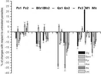
For that purpose we compared the 1H NMR spectra of supernatants from the parasites treated with N-oxides Fx1, Fx2, Bfx1, Bfx2, Qx1 and Qx2 with those from untreated T. cruzi epimastigotes (Y strain) and from Nfx, the inactive N-oxide Fx3 and the active nitrofurane Nf1 (Fig. 1). All compounds were tested at their IC50, except for compound Fx3 that was assayed at 300 μ m because its IC50 was not determined due to solubility problems. We have mainly focused on the carboxylic acid salts lactate (Lac), acetate (Ace), pyruvate (Pyr) and succinate (Succ) and on alanine (Ala) and glycine (Gly), which are the most relevant modified catabolites (Fig. 2).
Percentage of the end products excreted by T. cruzi epimastigote Y strain to the milieu expressed with respect to untreated parasites.

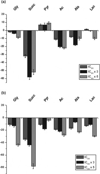
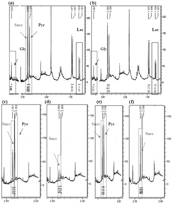
With the exception of nitrofurane Nf1 all the N-oxides decreased the excreted Gly and Ace. The most effective compound in reducing the amount of released Gly was the furoxan Fx3 while the quinoxaline di-N-oxide Qx2 induced a marked decrease of Ace. All compounds, except Bfx2, decreased the release of Succ, the best inhibitors being the quinoxalines Qx1 and Qx2 (Benítez et al. Reference Benítez, Cabrera, Hernández, Boiani, Lavaggi, Di Maio, Yaluff, Serna, Torres, Ferreira, Vera de Bilbao, Torres, Pérez-Silanes, Solano, Moreno, Aldana, López de Ceráin, Cerecetto, González and Monge2011). Regarding the release of Pyr no pattern was observed; this catabolite increased after incubation of parasites with Fx1, Fx2, Bfx1 and Qx1 while it diminished when treated with Fx3, Bfx2 and Qx2. Benzofuroxans and quinoxaline di-N-oxides decreased the amount of excreted Ala, with Bfx1 and Qx2 being the most active. Two furoxans, Fx2 and Fx3, increased the amount of released Lac while no significantly increase in the concentration of release Lac was observed after incubation of parasites with the rest of the heterocycles containing N-oxides. When dose–response studies were performed with quinoxaline di-N-oxides a decrease of Lac was observed for both derivatives, Qx1 and Qx2, at a dose five-fold the corresponding IC50 (Fig. 3), with Qx2 being the best inhibitor of Lac-release. A clear dose–response was observed for the inhibition of Gly, Succ and Ace excretion. Figure 4 shows representative examples of the spectra generated for selected compounds.
Catabolites excreted by parasites treated with compounds Qx1 (a) and Qx2 (b). Dose–response studies.

Relevant regions of the 1H NMR spectra for the assayed excreted-catabolites. (a), (c) and (e) Untreated-parasites; (b), (d) and (f) Parasites treated with Fx2, Qx1 and Qx2 at its IC50, respectively. For experimental details see Materials and methods section. All the spectra were recorded in D2O at 295·16 K. Relevant changes in catabolite concentrations are highlighted.

Effect of N-oxides on mitochondrial dehydrogenases
Since most of the studied N-oxides were capable of decreasing both the release of Succ and the release of Ace, we studied the effect of N-oxides on mitochondrial dehydrogenases. Succ biochemical pathway in the mitochondrion involves a succinate dehydrogenase (complex II-respiratory chain) while Ace requires Succ and the action of an acetate-succinate CoA transferase (Bringaud et al. Reference Bringaud, Rivière and Coustou2006; Opperdoes and Coombs, Reference Opperdoes and Coombs2007).
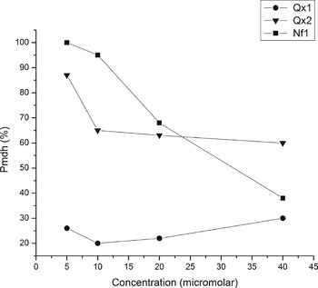
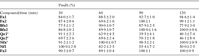
Mitochondrial dehydrogenase activities (Pmdh) for live parasites treated with 20 μ m of different N-oxides with respect to untreated control was assessed using the MTT assay performed at a short incubation period, no more than 120 min; this procedure was previously described for Leishmania (Maarouf et al. Reference Maarouf, De Kouchkovsky, Brown, Petit and Robert-Gero1997). The following compounds were tested: active N-oxides Fx1, Fx2, Bfx1, Bfx2; quinoxalines Qx1, Qx2; Nfx (Benítez et al. Reference Benítez, Cabrera, Hernández, Boiani, Lavaggi, Di Maio, Yaluff, Serna, Torres, Ferreira, Vera de Bilbao, Torres, Pérez-Silanes, Solano, Moreno, Aldana, López de Ceráin, Cerecetto, González and Monge2011) as the reference drug; and inactive furoxan Fx3 and nitrofurane Nf1 (Table 1). Only the nitrofurane Nf1, and unlike the nitrofurane Nfx, significantly decreased the mitochondrial dehydrogenase activity in a time-dependent manner while the benzofuroxan Bfx1 showed a moderate effect. A dose–response study was performed with Qx1, Qx2 and Nf1 using a 90 min incubation time because it was the time at which a more significant change in Pmdh was observed (Fig. 5). Curiously, compounds showing similar IC50 against T. cruzi (Qx1 1·6±0·4 mm, Qx2 1·8±0·1 mm and Nf1 1·3±0·5 mm) showed different behaviours regarding mitochondrial dehydrogenases, with Qx1 being an absolute inhibitor at all the assayed doses.
Mitochondrial dehydrogenases inhibition by Qx1 (●), Qx2 (▲) and Nf1 (■). The enzymatic activities were determined at 90 min of incubation. For experimental details see Materials and methods.

Mitochondrial dehydrogenase activities, as a percentage of untreated parasites, in live parasites treated with the studied N-oxides and reference compounds

Effect of Qx2 on glucose uptake
Since quinoxaline Qx2 was able to decrease the release of Succ, Ace, Ala and Lac we studied the ability of this compound to modify glucose uptake by T. cruzi epimastigotes. The consumption of glucose by epimastigotes of T. cruzi is characterized by the excretion, under aerobic conditions, of reduced products such as Succ, Ace and Ala (Cazzulo, Reference Cazzulo1992). On the other hand, most trypanosomatids produce Lac from glucose although often as a minor end product, i.e. bloodstream trypomastigotes, and additionally Lac excretion is considerably reduced in mutants showing a reduced glucose consumption rate (Coustou et al. Reference Coustou, Besteiro, Rivière, Biran, Biteau, Franconi, Boshart, Baltz and Bringaud2005).
The glucose uptake (glu-upt) for live parasites treated with the quinoxaline Qx2, at the corresponding IC50 and at 3×IC50 doses, was assessed using the colorimetric Benedict assay performed at 4 h of incubation (Boiani et al. Reference Boiani, Boiani, Alicia, Hernández, Chidichimo, Cazzulo, Cerecetto and González2009). Clearly, Qx2 diminished the glucose uptake to 33% at the IC50 dose (1·9 mm glu-upt compared with 3·0 mm glu-upt when DMSO was used). Contrarily, at a dose three times the IC50 an increase in glucose was observed, probably as a result of the loss of cellular integrity promoted by this high N-oxide concentration (−1·3 mm glu-upt compared with 3 mm glu-upt when DMSO was used). These results are in agreement with the 1H NMR study of excreted catabolites.
N-oxides and cell death phenotype
1H NMR-VML spectroscopy analysis of T. cruzi epimastigotes treated with N-oxides
Increased methylene resonances in 1H NMR spectra resulting from the accumulation of VMLs correlate with the onset of apoptosis in several drug-treated cell models (Blankenberg et al. Reference Blankenberg, Storrs, Naumovski, Goralski and Spielman1996) reflected in an increase in the CH2/CH3-ratio (Mikhailenko et al. Reference Mikhailenko, Philchenkov and Zavelevich2005) and in some cases an increment in the signals from PUFA (Hakumäki et al. Reference Hakumäki, Poptani, Sandmair, Ylä-Herttuala and Kauppinen1999). The modification of signals from choline (Cho), phosphatidylcholine (PTC) and phosphocholine (PC) has been associated with apoptosis and cell loss (Milkevitch et al. Reference Milkevitch, Shim, Pilatus, Pickup, Wehrle, Samid, Poptani, Glickson and Delikatny2005). Additionally, apoptotic processes in T. cruzi epimastigotes can be visualized through modifications in the VML profiles. Thus, the increment on the CH2/CH3-ratio and changes in the ‘choline region signals’ were indicative of apoptosis while necrosis was associated, in some cases, with changes on the ‘choline region signals’; no modifications were observed in autophagic cell-death processes (Benítez et al. Reference Benítez, Pezaroglo, Martínez, Casanova, Cabrera, Galanti, González and Cerecetto2012).
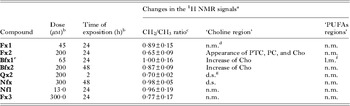
Table 2 summarizes the effect of the studied compounds on cell death processes as measured by 1H NMR-VMLs. Experimental conditions were determined for each compound by light microscopy observation, trypan blue staining, mobility and morphology. For all the N-oxides, except Qx1, the CH2/CH3 ratios were statistically lower than 1·0, indicating absence of cell death by apoptosis (Benítez et al. Reference Benítez, Pezaroglo, Martínez, Casanova, Cabrera, Galanti, González and Cerecetto2012). Despite this, the results for Fx2 from the PUFAs and choline-containing lipids protons data were reminiscent of those we observed previously for apoptotic conditions (Benítez et al. Reference Benítez, Pezaroglo, Martínez, Casanova, Cabrera, Galanti, González and Cerecetto2012). On the other hand, the same protonic regions indicate that Qx2 induces a necrotic process, similar to the one previously observed for Nfx (Benítez et al. Reference Benítez, Pezaroglo, Martínez, Casanova, Cabrera, Galanti, González and Cerecetto2012). Both benzofuroxans, Bfx1 and Bfx2, produced an increment of protons from choline-moiety. Finally, Fx1, Fx3 and Nf1 did not modify the protons signals from ‘choline’ and ‘PUFAs’ regions suggesting an autophagic process (Benítez et al. Reference Benítez, Pezaroglo, Martínez, Casanova, Cabrera, Galanti, González and Cerecetto2012).
Changes in the visible mobile lipids of T. cruzi treated with the different studied compounds analysed by 1H NMR

a Respect to untreated parasites.
b The doses and time of exposition was determined for each compound using light microscopy.
c Expressed respect to untreated parasites.
d ‘n.m.’: not detected modifications.
e Using Tulahuen 2 strain.
f ‘l.m.’: little modifications.
g ‘d.s.’: disappearance or decrease of signals.
In order to better clarify the type of cell death induced by N-oxides we applied TUNEL and PI-staining techniques on epimastigotes treated with the studied compounds.
TUNEL and PI-staining assays
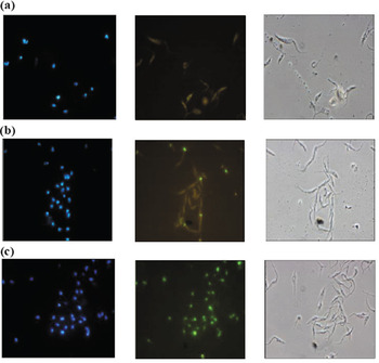
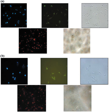
Treatment of epimastigotes with the different N-oxides did not induce positive TUNEL results (Fig. 6, Table S1) confirming that any of those compounds induce parasite apoptosis under the assayed conditions. These results were in agreement with those obtained measuring the CH2/CH3 ratios by 1H NMR-VML. On the other hand, PI-staining experiments showed that N-oxides Fx2, Qx1 and Qx2, similarly to Nfx (Benítez et al. Reference Benítez, Pezaroglo, Martínez, Casanova, Cabrera, Galanti, González and Cerecetto2012), induced parasite necrosis under the same conditions applied in the 1H NMR experiments (Table 2) (see examples in Fig. 7).
TUNEL assays (left, DAPI-staining; centre, FITC-staining; right, phase contrast; 1000×). (a) Treatment with Bfx1 (72 μ m, 24 h); (b) Treatment with Nf1 (19·5 μ m, 48 h); (c) Positive control (H2O2: 500 μ m, 30 min of incubation at 28 °C).

Examples of results from the TUNEL and PI-staining assays (Up: left, DAPI-staining; centre, FITC-staining; right, phase contrast; 1000×. Down: left, PI-staining; right, phase contrast; 400×). (a) Treatment with Qx1 (8 μ m, 24 h); (b) Treatment with Qx2 (18 μ m, 3 h).

In addition, these experiments confirmed that neither apoptosis nor necrosis were operative, as the main cell death phenotype, for N-oxides Fx1, Bfx1, Bfx2 and the nitrofurane Nf1.
Ultrastructural characterization of cell death phenotypes in parasites treated with N-oxide compounds
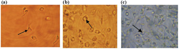
Light microscopy observation of T. cruzi epimastigotes treated with Fx1, Bfx1 and Nf1 compounds showed a common pattern of structural changes (Fig. 8). Particularly, a big vacuole was observed in the parasitic cytoplasm. Consequently, we selected these three compounds to perform ultrastructural analysis by TEM.
Light microscopy (640×). (a) Treatment with Fx1 (50 μ m, 24 h); (b) Treatment with Bfx1 (65 μ m, 24 h); (c) Treatment with Nf1 (5·1 μ m, 24 h). Notes: epimastigotes of T. cruzi Tulahuen 2 strain; the arrows show the cytoplasmic vacuole.

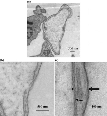
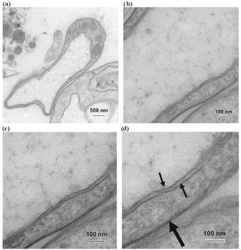
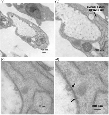
Transmission electron microscopy analysis of T. cruzi epimastigotes treated with Fx1 showed important ultrastructural changes, the most relevant being a big cytoplasmic vesicle, with low matrix electron density and concentric double membranes (Fig. 9). This morphology resembles the autophagic phenotype observed by the action of some naphthoimidazoles (Menna-Barreto et al. Reference Menna-Barreto, Corrêa, Cascabulho, Fernandes, Pinto, Soares and de Castro2009b ) and the ‘BigEye’ phenotype observed in bloodstream forms of T. brucei when endocytosis is disrupted by the knockdown of clathrin heavy chain (Allen et al. Reference Allen, Goulding and Field2003; Frearson et al. Reference Frearson, Brand, McElroy, Cleghorn, Smid, Stojanovski, Price, Guther, Torrie, Robinson, Hallyburton, Mpamhanga, Brannigan, Wilkinson, Hodgkinson, Hui, Qiu, Raimi, van Aalten, Brenk, Gilbert, Read, Fairlamb, Ferguson, Smith and Wyatt2010). Similarly, the nitrofurane Nf1 induced concentric membrane structures surrounded, in some cases, by endoplasmic reticulum (Fig. 10). Contrarily, the benzofuroxane Bfx1 showed a clear autophagic pattern, such as concentric membrane structures and autophagosomes surrounded by endoplasmic reticulum (Fig. 11).
Transmission electron microscopy of parasites treated with Fx1. (a) 12 000×; (b) Detail of (a) (40 000×); (c) Detail of (b) (200 000×). Note: big arrow: plasmatic membrane; small arrows: double membrane of vesicle.

Transmission electron microscopy of parasites treated with Nf1. (a) 25 000×; (b) Detail of (a) (100 000×); (c) Detail of (b) (150 000×); (d) Detail of (c) (200 000×). Note: big arrow: plasmatic membrane; small arrows: double membrane of vesicle.

Transmission electron microscopy of parasites treated with Bfx1. (a) 15 000×; (b) Detail of (a) (30 000×); (c) Detail of (b) (100 000×); (d) Detail of (b) (150 000×). Note: arrows: double membrane of vesicle.

Evaluation of autophagy induced by the N-oxides Fx1, Bfx1 and the nitrofurane Nf1
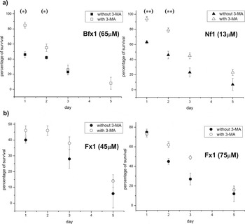
It is well-known that autophagic cell death can be inhibited by suppressing autophagosome formation with autophagic inhibitors, such as 3-methyladenine (3-mA), a non-specific inhibitor of a class III phosphatidylinositol 3-kinases required for autophagy (Klionsky et al. Reference Klionsky, Abdalla, Abeliovich, Abraham, Acevedo-Arozena, Adeli and Agholme2012). The pre-incubation of epimastigotes with 3-mA reversed the trypanosomicidal effect of the N-oxide Bfx1 and the nitrofurane Nf1 at least during the first 2 days of the studies (Fig. 12a). Probably, the effect of 3-mA was not evident from the third day as a result of the parasite inhibitor consumption. 3-mA per se did not interfere with the number of parasites (data not shown). On the other hand, there was no alteration in the parasite survival percentages when 3-mA-pre-incubated parasites were treated with Fx1 at two different doses (Fig. 12b).
Pre-incubation of Bfx1-, Nf1-, and Fx1-treated epimastigotes with protease inhibitor 3-MA. Data represent the mean±s.d. of at least three independent experiments. (*) P<0·003; (**) P<0·0003.

DISCUSSION
Many efforts have been made to identify the mechanism of action of anti-T. cruzi agents. Our interest focused on studying the mode of action of the most relevant N-oxide-containing heterocycles from our in-house chemical library. It was previously described that some benzo[1,2-c][1,2,5]oxadiazole N-oxides (benzofuroxans) and quinoxaline N1,N 4-dioxides were strong inhibitors of parasite dehydrogenase activity. Aiming to discriminate the mechanism of action by some of these families of compounds, levels of parasitic low-molecular weight thiols, reactive oxidant species, mitochondrial membrane potential and excreted catabolites were studied previously (Boiani et al. Reference Boiani, Piacenza, Hernández, Boiani, Cerecetto, González and Denicola2010; Benítez et al. Reference Benítez, Cabrera, Hernández, Boiani, Lavaggi, Di Maio, Yaluff, Serna, Torres, Ferreira, Vera de Bilbao, Torres, Pérez-Silanes, Solano, Moreno, Aldana, López de Ceráin, Cerecetto, González and Monge2011).
Most of the studied N-oxides decreased the release of both succinate and acetate suggesting that those compounds are acting on the mitochondria. Therefore we focused our study on the effect of N-oxides on mitochondrial dehydrogenases. None of the newly studied N-oxides were able to affect these dehydrogenases, though the quinoxalines Qx1 and Qx2, as well as Nf1, the nitrofurane used as reference, inhibited that enzyme. Considering that these compounds present similar IC50 against T. cruzi epimastigotes but different behaviour on mitochondrial dehydrogenases, and taking into account that the metabolism of carboxylic acids is related to glucose metabolism (Cazzulo, Reference Cazzulo1992; Coustou et al. Reference Coustou, Besteiro, Rivière, Biran, Biteau, Franconi, Boshart, Baltz and Bringaud2005), we analysed glucose consumption by the parasite in the presence of this N-oxide. This study may be indicating that Qx2 also affects the integrity or some pathway into the glycosome (Bringaud et al. Reference Bringaud, Rivière and Coustou2006; Opperdoes and Coombs, Reference Opperdoes and Coombs2007).
Several efforts have been made to identify the type of cell death in parasitic protozoa (Rodrigues and De Souza, Reference Rodrigues and De Souza2008) but none with regard to N-oxides.
Both mobile lipids analysed by 1H NMR (Benítez et al. Reference Benítez, Pezaroglo, Martínez, Casanova, Cabrera, Galanti, González and Cerecetto2012) and TUNEL assay showed that the studied N-oxides do not induce T. cruzi death by apoptosis under the assayed conditions. However, according to propidium iodide staining both quinoxaline dioxides, Qx1 and Qx2 as well as the 1,2,5-oxadiazole N-oxide Fx2 and the clinically used nitrofurane, Nifurtimox, were able to induce necrosis of parasites in the assayed conditions.
Light microscopy observations indicated that the Fx2-structurally related N-oxides Fx1 and Bfx1, together with the nitrofurane Nf1, induced very particular structural changes. This is interesting considering that structural similarities in these families of compounds are not related to the type of cellular death. Therefore, we performed ultrastructural analysis by TEM and the use of protease inhibitor to confirm or discard an autophagy process. Autophagy is a self-degradation process presented in eukaryotes, implicated in the removal and/or remodelling of damaged cellular structures. In yeasts and mammals, autophagosome formation involves the assembling of a pre-autophagosomal structure, close to endoplasmic reticulum cisternae (Yorimitsu and Klionsky, Reference Yorimitsu and Klionsky2007).
Ultrastructural analysis and pre-incubation with the protease-inhibitor 3-methyladenine suggested the induction of an autophagic phenotype in T. cruzi epimastigotes treated with Bfx1 and Nf1. Morphological characteristics such as the appearance of concentric membrane structures and autophagosome-like bodies were commonly observed in treated parasites, with the latter surrounded by endoplasmic reticulum. Additionally, the pre-incubation of epimastigotes with the autophagic inhibitor abolished the effect of Bfx1 and Nf1, at least for 48 h. However, the ultrastructural and protease-inhibitor data showed a very different behaviour for Fx1. In this case, the presence of a concentric membrane vesicle, such as an autophagosome, could indicate an autophagic phenotype; however, endoplasmic reticulum in close proximity to the vacuole was not observed. Additionally, there was no effect of the inhibitor 3-methyladenine in parasite survival. Consequently, a ‘BigEye’ phenotype may be proposed as a consequence of Fx1 action, as observed in T. brucei bloodstream forms when endocytosis is disrupted by the knockdown of clathrin heavy chain (Allen et al. Reference Allen, Goulding and Field2003; Frearson et al. Reference Frearson, Brand, McElroy, Cleghorn, Smid, Stojanovski, Price, Guther, Torrie, Robinson, Hallyburton, Mpamhanga, Brannigan, Wilkinson, Hodgkinson, Hui, Qiu, Raimi, van Aalten, Brenk, Gilbert, Read, Fairlamb, Ferguson, Smith and Wyatt2010). However, to consider that this process is operative in Fx1 treated T. cruzi epimastigotes needs further investigation.
Our results in T. cruzi treated with different classes of N-oxides has led to an understanding of mechanism of action and protozoa cell death that could help develop new therapeutic strategies.
Supplementary material
To view supplementary material for this article, please visit http://dx.doi.org/S003118201300200X.
FINANCIAL SUPPORT
This work was supported by CSIC-Grupos 611 (Uruguay), PEDECIBA (Uruguay), FONDECYT 1090124 and 1130113 (to NG), FONDECYT 1110053 (to GC), CONICYT-PBCT Anillo ACT 112 ‘Research in the Design of Pharmacological and Immunological Strategies for the Control of Parasitic and Neoplasic Aggressions’ (Chile), and RIDIMEDCHAG-CYTED. D.B. is a fellow of ANII.