Introduction
The most common subtypes of dementia in later life are Alzheimer's disease (AD) and dementia with Lewy bodies (DLB). Distinguishing between AD and DLB during life can be difficult as symptoms between the two conditions often overlap especially in the advanced stages of the disease. The clinical diagnostic criteria for DLB also lack sensitivity so that many cases are missed or misdiagnosed (Litvan et al., Reference Litvan, Bhatia, Burn, Goetz, Lang and Mckeith2003). Furthermore, neuroimaging changes in dementia can also overlap with those seen in normal aging (Minati et al., Reference Minati, Grisoli and Bruzzone2007).
Structural neuroimaging, including magnetic resonance imaging (MRI), is an important part of the clinical work up for all patients with dementia. More recently an advanced MRI method, diffusion tensor imaging (DTI), has emerged as a technique to assess the microstructure of the brain. It is thought that DTI may be a more sensitive neuroimaging procedure compared to conventional structural MRI, as it may detect subtle preclinical changes (Ringman et al., Reference Ringman2007). DTI is an advanced MRI technique that allows assessment of the tissue microstructure by measuring the incoherent motion of water molecules (Le Bihan et al., Reference Le Bihan, Breton and Lallemand1986). The diffusion of molecules in tissues varies depending on the interactions with obstacles such as tissue membranes and fibers (Le Bihan, Reference Le Bihan2007). White matter tracts in the brain have a fibrous internal structure, meaning that the diffusion of water along these tracts is directionally dependent; a property known as anisotropy (Mori and Zhang, Reference Mori and Zhang2006). Water molecules will diffuse further along a tract if aligned in the same direction, as theoretically there are “fewer obstacles to prevent movement” (Mori and Zhang, Reference Mori and Zhang2006). There are two important measures that can be derived from DTI; Mean diffusivity (MD) and Fractional anisotropy (FA). MD represents the overall displacement of water molecules whereas FA quantifies the directionality of local tract structure. Typically DTI abnormalities are represented by increased MD and/or reduced FA. For example, an FA value of 0 would indicate that the diffusion of water molecules is the same in all directions, whereas a value of 1 would indicate diffusivity of molecules in a single direction. When there is damage to the walls of the axons, FA will decrease as the water molecules adopt a more random direction of diffusion. It is thought that when combined with the macro-structural changes on conventional MRI scanning, DTI may be used to assist with earlier diagnosis of different dementia subtypes.
There have been a number of diffusion imaging studies in AD (Vasconcelos et al., Reference Vasconcelos, Brucki, Jackowiski and Bueno2009), the majority of which have compared AD and similar aged controls or with subjects with mild cognitive impairment. The most consistent findings are that posterior brain regions seem to be more affected than anterior regions during the early stages of AD, then, as the disease progresses, the limbic and frontal structures also become involved followed lastly by the primary sensorimotor regions, which have been shown to correlate with the clinical manifestations of the disease (Medina and Gaviria, Reference Medina and Gaviria2008). There have been fewer studies in DLB and with varying results (Watson and O'Brien, Reference Watson and O'Brien2012). Some studies report widespread diffusivity changes in comparison to healthy controls (Bozzali et al., Reference Bozzali2005; Lee et al., Reference Lee2010), whereas others report little to no change (Firbank et al., Reference Firbank2007; Kantarci et al., Reference Kantarci2010).
Most DTI studies in DLB have utilized analysis methods that focus on cortical structures and key neuronal WM tracts. The most consistent findings have been changes in the Inferior Longitudinal Fasciculus (ILF) and precuneal WM tracts, which form part of the ventral and dorsal visual streams respectively (Ota et al., Reference Ota2009; Kantarci et al., Reference Kantarci2010; Kiuchi et al., Reference Kiuchi2011; Watson et al., Reference Watson2012a). These findings are particularly interesting given that visuoperceptual impairments and visual hallucinations are common in DLB (McKeith et al., Reference McKeith2005). A study in 2010 by Kantarci et al., revealed increased diffusivity in the amygdala in DLB compared to controls (Kantarci et al., Reference Kantarci2010), which also correlated with the UPDRS-III scores (Kantarci et al., Reference Kantarci2010). Given that α-synuclein accumulation in the amygdala has also been observed in DLB, this may implicate the amygdala as an important structure in the pathogenesis of DLB.
To our knowledge this is the first study utilizing DTI to assess brainstem structures, such as the midbrain and pons in DLB. There has also been a relative lack of studies investigating amygdala and thalamic diffusivity change in subcortical structures, which have been implicated in the pathogenesis of DLB that may help distinguish DLB from other dementias and normal aging (Watson et al., Reference Watson2012a). Therefore using region of interest (ROI) methods, we investigated diffusion parameters in subjects with DLB, AD, and similar aged controls in thalamus, precuneus, pons, midbrain, and amygdala. We also studied the relationship between the diffusion measures and various clinical and neuropsychological variables in AD and DLB.
Methods
Subjects
Seventy one people with dementia (36 AD, 35 DLB) over the age of sixty were recruited from a community dwelling population of patients referred to geographically representative Old Age Psychiatry, Geriatric Medicine or Neurology Services. Informed consent was obtained from all subjects and where appropriate, their nearest relative provided written informed consent. Thirty-five healthy controls of similar age and gender were also included in the study. They were recruited from relatives and friends of those with dementia, or volunteered via advertisements in local community newsletters. Those with evidence of dementia, depression, or a history of neurological, psychological, or psychiatric problems were excluded. All subjects were aged over 60 and did not have any contraindications for MRI. The research was approved by the local ethics committee.
Diagnosis and assessment
Diagnosis and assessment of study participants has been reported (Watson et al., Reference Watson, O'Brien, Barber and Blamire2012b). Diagnoses of AD and DLB were made in accordance with NINCDS-ADRDA (McKhann et al., Reference McKhann, Drachman, Folstein, Katzman, Price and Stadlan1984) and DLB consensus criteria (McKeith et al., Reference McKeith1996, Reference McKeith2005) respectively, by consensus agreement among three experienced raters (RB, JO'B, and RW) blinded to all information from the MRI scans.
Assessment of global cognitive measures included the CAMCOG, which incorporates the Mini-Mental State Examination (MMSE; Folstein et al., Reference Folstein, Folstein and McHugh1975). Depressive symptoms were measured using the 15-item Geriatric Depression Scale (Sheikh and Yesavage, Reference Sheikh and Yesavage1986). Motor parkinsonism was assessed with the Unified Parkinson's Disease Rating Scale Part III (UPDRS-III) (Fahn, Reference Fahn and Elton1987).
For participants with dementia, neuropsychiatric features were assessed using the Neuropsychiatric Inventory (NPI) (Cummings et al., Reference Cummings1994), cognitive fluctuations were assessed using the Clinician's Assessment of Fluctuation (Walker et al., Reference Walker2000), and functional abilities were assessed using the Bristol Activities of Daily Living (Bucks et al., Reference Bucks, Ashworth, Wilcock and Siegfried1996).
MRI acquisition
Subjects underwent magnetic resonance (MR) scanning on a 3T MRI system (Intera Achieva scanner, Philips Medical Systems, Eindhoven, Netherlands) with an eight channel receiver head coil, within two months of the study assessment. DTI images were acquired using a Pulsed Gradient Spin Echo (PGSE) sequence and multislice single shot EPI readout, with TE = 71 ms and TR = 2,524 ms. The image volumes were angulated such that the axial slice orientation was standardized to align with the AC-PC line with 2 mm in-plane resolution, 6 mm slice thickness, and matrix size of 128 (anterior-posterior) × 128 (right-left) × 24 (superior-inferior). The scan was accelerated with a sensitivity encoding (SENSE) factor of 2 in the anterior-posterior direction and reconstructed with the Cepstral Loudness Enhanced Algorithm (CLEAR) algorithm. Diffusion weighting was achieved by applying diffusion sensitizing gradient pulses with measurements made in 16 directions with a b value of 1,000 mm s−2.
DTI preprocessing and analysis
The functional MRI of the Brain (FMRIB) software library (FSL) program was used to process and analyze raw DTI data (Smith et al., Reference Smith2004) accessed at www.fmrib.ox.ac.uk/fsl.
To correct for the distorting effect of eddy currents, we adapted the approach of Shen et al. (Reference Shen2004) and used an affine registration, in FSL's FLIRT (FMRIB's Linear Image Registration Tool) to register pairs of diffusion weighted images together. The eddy corrected diffusion weighted images were then transformed using a rigid body registration to the b = 0 mm s−2 image. The MD and FA images were calculated using FSL tensor analysis of the aligned diffusion weighted images at each brain voxel.
ROI analysis
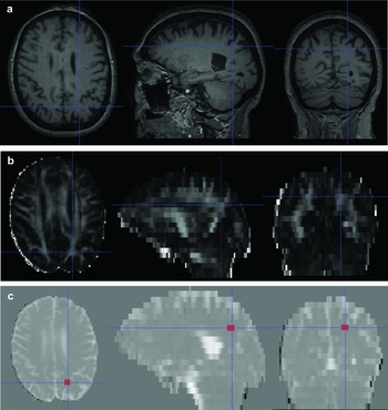
For each subject, diffusion images (FA, MD) were initially coregistered to their corresponding T1 weighted MRI scans. Using the ITK-snap software (Yushkevich et al., Reference Yushkevich2006), a series of fixed square ROIs were manually placed on selected brain regions including thalamus (L, R), precuneus (L, R), pons, midbrain, and amygdala (L, R) onto the subjects’ T1 MRI scan as shown in Figure 1 A–E respectively. The regions varied in size: for thalamus, pons, and midbrain, 4 × 4 pixels; and for precuneus and amygdala, 2 × 2 pixels. These ROIs were drawn according to a set protocol assessing axial, sagittal, and coronal views along with information from a human brain atlas (Mai et al., Reference Mai, Assheuer and Paxinos1997). Optimal placement of regions was undertaken in order to try and avoid cerebrospinal fluid and WM hyperintensities. We identified a white matter tract for the precuneus, located using a consistent method for each scan (Figure 2). The T1 MRI-defined ROIs were then projected onto their FA and MD images where their co-ordinates (cross-hairs) were recorded and subsequently mean FA and MD counts per voxel within these regions were calculated. For thalamus, precuneus, and amygdala, left and right values were averaged.
Axial sections showing the location of ROIs in (a) thalamus; (b) precuneus; (c) pons; (d) midbrain; and (e) amygdala. Image on the left is the structural T1 weighted MRI scan; Image on the right is their FA scan.

Precuneal ROI placement. The structural T1 MRI (a) was co-registered with the corresponding FA image (b). The MD image (c) was then overlaid onto an FA image and the co-ordinates for the location of the precuneus were recorded. The ROI was then drawn, taking care to ensure where possible that the region was placed to the voxel closest to the center of the cross-hairs.

Statistical analysis
Statistical analysis was performed using Statistical Package for Social Sciences (SPSS) version 19 (www.ibm.com/software/analytics/spss/products/statistics). Data were assessed for normality by visual inspection of variable histograms and use of the Shapiro-Wilk test. Where appropriate, demographical and imaging variables were examined using parametric (Analysis of one way variance (ANOVA), two sample t-test) and non-parametric (χ 2e, Mann Whitney U) methods. Bonferroni post hoc tests were performed as required. To assess the relationship between FA and MD values with clinical measures, a one-tailed Pearson's partial correlation coefficient was used, controlling for age and MMSE score. After adjusting for multiple statistical testing, a corrected p-value of <0.05 was considered significant. To quantify intra-rater agreement, a two-way mixed intra-class correlation coefficient (ICC) was computed, with a 95% confidence interval.
Rater agreement
Intra-observer agreement was assessed for each ROI measurement in FA and MD by selection of three subjects at random by an independent observer and repeat measurements undertaken five times on five consecutive days.
Exclusion of subjects
From the initial cohort of 106 subjects, four were excluded (two AD, one DLB, one Control) due to poor quality MR data, mostly caused by motion artifacts.
Results
Subject demographics
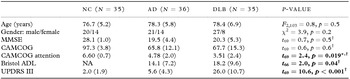
Demographic data for AD, DLB, and control subjects are summarized in Table 1. No significant differences in age (F = 0.8, p = 0.5) or sex (χ2 = 3.9, p = 0.2) were observed between groups. Mean MMSE and CAMCOG scores were similar among dementia groups, while as expected UPDRS-III and CAMCOG attention scores significantly differed between DLB and AD (p < 0.02).
Subject demographics

*Post hoc tests: NC > DLB, AD (p < 0.001).
†Student's t-test (AD vs. DLB).
Note: Values expressed as mean (1SD). Bold text denotes significant group differences. DLB = dementia with Lewy bodies; AD = Alzheimer's disease; NC = Normal Control; MMSE = Mini-Mental State Examination; CAMCOG = Cambridge Cognitive Examination; Bristol ADL = Bristol Activities of Daily Living; UPDRS III = Unified Parkinson's Disease Rating Scale, Part III.
DTI measures
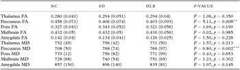
FA and MD for AD, DLB, and control subjects are presented in Table 2. Significant differences were observed in the white and gray matter tracts in precuneus for both FA (p ≤ 0.03) and MD (p ≤ 0.007) between dementia groups and controls. There were no differences observed between AD and DLB for either FA (p = 1.0) or MD (p = 1.0). Similarly there were no significant differences in diffusion parameters between DLB and AD in the thalamus, amygdala, midbrain, or pons.
FA and MD values

**Post hoc Bonferroni test; Control > AD (p = 0.026), Control > DLB (p = 0.016).
***Post hoc Bonferroni test; AD > Control (p = 0.005), DLB > Control (p = 0.007).
Note: Otherwise post hoc Bonferroni test non-significant. Values expressed as mean (1 SD).
Clinical correlates
The relationship between diffusivity measures (FA, MD) and MMSE and UPDRS-III was investigated where appropriate in controls, AD, and DLB. Controlling for age, a positive correlation was observed between MMSE and FA in the pons in DLB (p = 0.009), and in precuneus in AD (p = 0.03). Since there was a relationship between MMSE and DTI measures, MMSE along with age were then subsequently controlled for when investigating the partial correlation with selected clinical measures (UPDRS-III and CAMCOG attention subscale scores). As a result, a positive relationship (controlling for age and MMSE) between MD and UPDRS-III score was identified in the amygdala in DLB (r’ = 0.486, p = 0.006); however, we did not find a significant association between UPDRS and other regional DTI measures.
Rater agreement
Overall there was excellent intra-rater reliability with an average ICC of 0.867 for FA and 0.793 for MD. Intra-class correlation coefficient values for intra-rater agreement for each structure are summarized in Table 3.
ICC values for intra-rater agreement

Discussion
This study utilized DTI as a means of assessing in vivo neurodegenerative changes in five key structures in a well characterized cohort of DLB, AD, and control subjects. The key findings of this study were: (i) mean FA in the precuneus was reduced in DLB and AD subjects, compared to controls; (ii) precuneal MD was elevated in AD and DLB subjects compared to controls; (iii) there were no significant differences in FA and MD in the thalamus, pons, midbrain, and amygdala between dementia subjects and controls or between dementia sub-groups; and (iv) controlling for age and MMSE, there was a statistically significant positive relationship between amygdala MD and UPDRS-III score in DLB.
Precuneal FA was significantly reduced in DLB and AD compared to controls. The precuneus, which is part of the superior parietal lobe, is involved in visuospatial processing, episodic memory retrieval, and self-processing operations (Cavanna and Trimble, Reference Cavanna and Trimble2006). Therefore, the finding of reduced FA in the precuneus in subjects with DLB is of interest given the characteristic visuoperceptual problems of the condition (McKeith et al., Reference McKeith2005).
Similar changes have been reported previously (Firbank et al., Reference Firbank2007). In that study, which involved 16 subjects with DLB, 15 with AD and 16 controls, a significant decrease in FA in the precuneus in DLB was found (p = 0.009). It is possible that the reduced FA in the precuneus reflects microstructural damaged to the WM tracts. Our findings also add to data already obtained from SPECT studies, which demonstrate significant hypoperfusion in the precuneus in DLB subjects (Colloby et al., Reference Colloby2002; Kemp et al., Reference Kemp2005), showing the precuneus is potentially a key region affected in DLB. Subjects with AD also demonstrated reduced FA in the precuneus. This may reflect the overlap in the pathophysiology of AD and DLB. It may also reflect the nature of the disease process in AD, where typically there is more global atrophy than DLB, causing neuronal destruction and secondary WM degeneration (Whitwell et al., Reference Whitwell2007). Therefore, although the precuneus appears to be affected in both conditions, it may be that in DLB it is more selectively damaged and in AD affected as part of the process of global atrophy (Watson et al., Reference Watson2012a). However, given the apparent degree of overlap in these measures in AD and DLB, the technique is unlikely to be effective in terms of diagnostic utility in classifying AD and DLB, and therefore of limited use for individual case detection.
A positive correlation between precuneal FA and MMSE was observed in subjects with AD (p = 0.032). This is similar to findings in a study by Frisoni et al., who used voxel-based morphometry to detect gray matter loss in AD. They found a correlation between gray matter loss in the precuneus and MMSE score (Frisoni et al., Reference Frisoni2002). The degeneration of precuneal gray matter and the resulting disruption in connectivity, reflected by decreased FA, could contribute to the decline in overall cognitive function. However, this correlation was not observed in DLB, highlighting the differences in the pathological processes between AD and DLB. Motor parkinsonism was assessed using UPDRS-III score and correlated with elevated MD in the amygdala in DLB (r′ = 0.49, p = 0.006). The relationship between UPDRS-III score and MD in the amygdala was in agreement with an earlier DTI study by Kantarci et al. (Reference Kantarci2010) involving 30 subjects with DLB, demonstrating a relationship between amygdala diffusivity and UPDRS-III score (p = 0.005). The authors hypothesized that there was unlikely to be a direct structural-functional link between changes in the amygdala and parkinsonism (Kantarci et al., Reference Kantarci2010). Rather, it was proposed that the high burden of Lewy bodies in the amygdala correlated with the high burden in the substantia nigra, causing features of parkinsonism (Kantarci et al., Reference Kantarci2010). It is possible that the severity of parkinsonism, as measured by UPDRS-III, reflects Lewy body burden in the amygdala. These findings are interesting as various studies have shown that α-synuclein, a major component of Lewy bodies, aggregates in the amygdala (Popescu et al., Reference Popescu, Lippa, Lee and Trojanowski2004). Therefore, despite the correlation being modest, our finding of a relationship between UPDRS-III score and increased diffusivity in the amygdala supports the notion that the amygdala may be an important structure underpinning some of the key clinical features of DLB and could be assessed further using an alternative imaging technique such as multimodal MRI.
Strengths of this study include the size of the DLB cohort, which is at present the largest cohort to complete a DTI study to date. The dementia groups were well matched for age and educational level. Although care was taken to ensure the control group did not display any features of cognitive impairment or dementia, some may well have preclinical disease, as has been shown studies of healthy older adults (Goldman et al., Reference Goldman2001; Rodrigue et al., Reference Rodrigue2012). Clinical measures were carefully preselected to assess potential correlations in order to decrease the risk of type I errors. There were several study limitations. First, the lack of pathological diagnosis, as with all antemortem neuroimaging studies. However, inclusion was based on well-validated clinical diagnostic criteria. Second, like the majority of previous studies assessing DTI in DLB, manual ROI placement along with their associated errors were also used in this study. We attempted to reduce these errors by using the same method to locate structures in which the region was being placed. After the region had been placed in the selected area the sagittal, axial, and coronal views were then checked to ensure correct alignment. Third, the crossing fiber algorithm was not implemented as part of the preprocessing stream. As such, the images were not modeled for the number of crossing fibers. However, since we followed a strict protocol of ROI placement, we predict that such effects would be similar across all subjects and not significantly affect results. Thus, the findings should be interpreted with this in view. Forth, although excellent intra-rater agreement was observed, inter-rater agreement was not measured; therefore a full examination of the techniques reproducibility could not be achieved. Lastly, we may have included cerebrospinal fluid (CSF) or white matter hyperintensities (WMH) in the sampled regions, which would have affected the diffusion parameters leading to inconsistent results. In order to minimize this, we utilized relatively small ROI's, which meant they were less likely to encroach on CSF or WMH's that may surround the structure. Future studies could involve volumetric techniques given that cases with WMH were not excluded.
To our knowledge, this was one of the largest DLB cohorts that were studied using DTI, and the first to assess midbrain, pons, and thalamus. Elevated MD and reduced FA were found in the WM tracts of the precuneus in DLB and AD compared to similar aged controls, suggesting that the precuneus is an important region of change in dementia, and potentially a marker of disease severity in AD. In DLB, amygdala diffusivity was associated with UPDRS-III score, which when combined with the known pathological changes further highlights the importance of the amygdala in the pathogenesis of DLB. Diffusion tensor MRI may help shed new light on the different neurobiological changes underpinning the key clinical features of DLB.
Conflict of interest
None.
Description of authors’ roles
J. O'Donovan completed the region of interest analysis, interpreted the results, and wrote the paper. R. Watson designed the study, co-supervised the project, interpreted results, assessed study subjects and provided diagnostic ratings and reviewed the paper. S. Colloby assisted with study design, co-supervised the project, completed the statistical analysis, and reviewed the paper. A. Blamire reviewed the paper and secured funding for the project. J. O'Brien co-supervised the project, reviewed the paper, and secured funding of the project.
Acknowledgments
We thank Dr Robert Barber for providing assistance with subject diagnostic rating as well as Josh Wood, Assistant Psychologist, and the members of North East Dementias and Neurodegenerative Diseases Research network (NE-DeNDRoN) for their assistance with recruitment and assessment of study participants. We are also grateful to all of the study volunteers for their involvement. The study was supported by the Sir Jules Thorn Charitable Trust (grant number 05/JTA), The Jean Shanks Foundation, and the UK NIHR Biomedical Research Centre and Biomedical Research Unit in Lewy body dementia awarded to the Newcastle upon Tyne Hospitals NHS Foundation Trust and the NIHR Biomedical Research Centre and Biomedical Research Unit in Dementia awarded to Cambridge University Hospitals NHS Trust and the University of Cambridge.