Introduction
Bone is the second most transplanted tissue after blood, and the incidence of bone defects are alarmingly increasing due to trauma or diseases [Reference Jones1, Reference Amini, Laurencin and Nukavarapu2]. Traditionally, bone defect repair includes clinical treatments involving autologous and allogeneic transplantation. Autografts are the “gold standard” in bone defect repair as they do not cause any immunogenic reactions and are histocompatible [Reference Amini, Laurencin and Nukavarapu2, Reference Baroli3, Reference Dimitriou, Jones, McGonagle and Giannoudis4, Reference Yaszemski, Payne, Hayes, Langer and Mikos5, Reference Dusseldorp and Mobbs6]. However, the transplant procedures are costly, they involve multiple surgeries, and the patient can suffer from various complications associated with an operation, such as bleeding, inflammation, and injuries [Reference Ebraheim, Elgafy and Xu7]. Allografts, although less expensive than autografts, have limited applications because of possible immunoreactions and donor shortage [Reference Greenwald, Phil, Boden, Goldberg and Khan8].
Tissue engineering is an alternative approach where a scaffold is directly implanted in vivo to promote tissue formation and ingrowth in situ. The scaffold is a three-dimensional engineered material which acts as a temporary template for guiding bone repair and is usually combined with biomolecules [Reference Jones1, Reference O’Keefe and Mao9, Reference Rezwan, Chen, Blaker and Boccaccini10, Reference Boccaccini, Fleck, Schubert, Roether, Philippart, Boccaccini, Fleck, Schubert and Roether11]. A wide variety of materials are considered as scaffold materials for bone tissue engineering, ranging from natural and synthetic polymers to bioceramics, such as hydroxyapatite (HA), bioactive glass, and polymer–ceramic composites [Reference Rezwan, Chen, Blaker and Boccaccini10, Reference Boccaccini, Fleck, Schubert, Roether, Philippart, Boccaccini, Fleck, Schubert and Roether11, Reference Chen, Thompson and Boccaccini12, Reference Baino and Vitale-Brovarone13, Reference Shuai, Huang, Feng, Gao, Shuai, Xiao, Deng, Peng and Wu14, Reference Moncal, Heo, Godzik, Sosnoski, Mrowczynski, Rizk, Ozbolat, Tucker, Gerhard, Dey, Lewis, Yang and Ozbolat15, Reference Elsayed, Zocca, Schmidt, Günster, Colombo and Bernardo16]. Biodegradable polymers are of two types: synthetic and naturally derived polymers. Synthetic polymers have the risk of toxicity, have less predictable biocompatibility, and can induce inflammatory responses. Naturally derived polymers, on the other hand, are biocompatible, abundant, biodegradable, and do not induce any toxicity because of their natural origin [Reference Kumbar, Laurencin and Deng17].
Alginate is a naturally occurring anionic polysaccharide obtained from seaweeds and is a linear polymeric acid comprising 1,4-linked α-mannuronic acid (M) and 1,4 linked β-guluronic acid (G) monomers [Reference Srinivasan, Jayasree, Chennazhi, Nair and Jayakumar18]. They are widely considered as an ideal scaffold-forming material in tissue engineering because of their nontoxicity, biocompatibility, abundance, and low-cost involvement [Reference Rehm and Moradali19]. They can quickly be dissolved in aqueous solutions without the aid of any solvents at ambient temperature conditions [Reference Zhao, Zhang, Fu, Xie and Chen20]. Also, they can be easily modified with peptides, proteins, and other inorganic biomaterials. Scaffolds fabricated from alginate are weak in strength, hence limiting their applications in bone tissue engineering [Reference Lin and Yeh21]. A possible approach to improve these properties and overcome these limitations is the formation of a composite with inorganic materials, such as calcium phosphate, HA, and bioactive glass. The main driving force behind the formation of such a composite is the fact that bone itself is a natural composite comprising 70% HA and 30% collagen [Reference Boccaccini, Fleck, Schubert, Roether, Philippart, Boccaccini, Fleck, Schubert and Roether11]. Bioactive glasses are preferred over HA and calcium phosphate as a filler in tissue engineering scaffolds because bioactive glass can bond to both soft and hard tissues without forming an intervening fibrous tissue due to the formation of a carbonated HA layer on the bioactive glass surface in body fluids [Reference Srinivasan, Jayasree, Chennazhi, Nair and Jayakumar18].
Bioactive glasses and glass–ceramics (GC) are attractive biomaterials in bone repair owing to their biocompatibility, osteoconductivity, and osteoinductivity. They are successfully used in clinical treatments as a bone repair material, a bone filler, and as an adjuvant in bone grafts [Reference Jones1, Reference Gerhardt and Boccaccini22]. They can bond with the bone, termed as bioactivity, and are employed in various orthopedic applications for facilitating bone ingrowth [Reference Hench, Splinter, Allen and Greenlee23, Reference Abdollahi, Chih, Ma and Cerruti24]. Hence, the addition of bioactive glass inclusions would confer enhanced bioactivity to the alginate matrix having inherently poor bioactivity. Dissolution products released from the bioactive glass can also upregulate gene expression and promote osteogenesis [Reference Jones1]. Thus, cell adhesion and cell–material interactions of alginate can be improved by the addition of bioactive glass. Also, the addition of bioactive glass as reinforcement in the alginate matrix can increase the stiffness and compressive strength of the scaffold [Reference Rezwan, Chen, Blaker and Boccaccini10]. Therefore, making a composite by incorporating bioactive glass–ceramics would improve the mechanical stability of the weak alginate polymer for mimicking various bone functions. Bioactive glass synthesis is carried out by two methods: the conventional melt quenching technique and the sol–gel method. Bioactive glasses prepared by the sol–gel method have better homogeneity, higher surface area, and higher purity compared to melt quenched glasses [Reference Sepulveda, Jones and Hench25, Reference Sakka26].
There are limited numbers of previous reports describing the fabrication of alginate–bioactive glass composites for biological applications [Reference Srinivasan, Jayasree, Chennazhi, Nair and Jayakumar18, Reference Zhao, Zhang, Fu, Xie and Chen20, Reference Mouriño, Newby, Pishbin, Cattalini, Lucangioli and Boccaccini27]. Gallium cross-linked alginate bioglass composite was prepared for potential use in bone tissue engineering [Reference Mouriño, Newby, Pishbin, Cattalini, Lucangioli and Boccaccini27]. Alginate–bioactive glass composite scaffold was prepared for tissue engineering applications, where the glass was in the system SiO2–CaO–P2O5 [Reference Srinivasan, Jayasree, Chennazhi, Nair and Jayakumar18, Reference Zhao, Zhang, Fu, Xie and Chen20]. However, in this work, the bioactive GCs in the system SiO2–Na2O–CaO–P2O5 were used to fabricate the composite scaffold containing alginate and GCs. The glass–ceramics contain the Na2Ca2Si3O9 (combeite) crystalline phase in a glassy matrix and that has been reported to show higher flexural strength than compact bone, while showing enhanced bioactivity and lower elastic modulus in comparison to Cerabone® (Japan) [Reference Peitl, Zanotto, Serbena and Hench28]. The composite scaffold fabrication has been carried out by a freeze-drying technique. The effects of the addition of bioactive glass–ceramics on properties such as mechanical behavior, porosity, in vitro bioactivity, and biocompatibility were evaluated.
Results and discussion
Characterization of scaffolds
Microstructure analysis
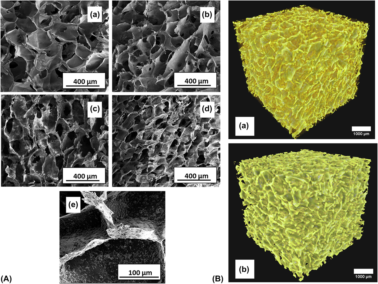
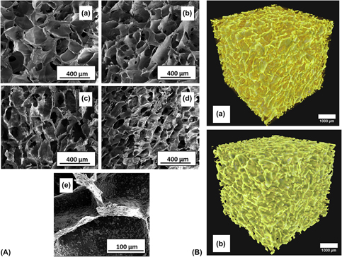
In bone tissue engineering, well-interconnected macropores are required for nutrient and oxygen supply and tissue ingrowth [Reference Savina, Cnudde, D’Hollander, Van Hoorebeke, Mattiasson, Galaev and Du Prez29, Reference Peter, Sudheesh Kumar, Binulal, Nair, Tamura and Jayakumar30]. They facilitate the migration and proliferation of mesenchymal cells and osteoblasts [Reference Hannink and Arts31]. Figure 1(a) shows the field emission scanning electron microscopy (FESEM) image of the pure alginate scaffolds and alginate–GC composite scaffold with different weight percentages of GC powder. It can be seen from the microstructure that both the pristine and composite scaffolds displayed the uniform distribution of macropores. The pore size was quantified from the FESEM images using ImageJ software, and pore size was found to be in the range of 110–380 µm. The average pore size decreased with the increase in the GC content. The decrease in pore size with the addition of GC is possibly because of the modification of ice crystal size during the freezing process due to the interaction of GC with water molecules [Reference Shiraishi, Anada, Honda, Masuda, Sasaki and Suzuki32]. Also, it was observed that the GC particles were well dispersed along the pore walls of the composite scaffolds. This is a very critical aspect because this exposure helps them to be in direct contact with the body fluids, thus enhancing the bioactivity of the scaffold. Hence, the pore structure of the present scaffold is well suited for tissue engineering applications.
(A) FESEM image of the alginate–GC composite scaffolds. Images show the typical morphology of scaffold; (a) pure alginate and morphology of GC–alginate composite scaffolds; (b) GC1 (c) GC2, and (d) GC3. (e) Distribution of the GC particles along the walls of the GC3 scaffold; (B) 3D reconstructed images of different samples: (a) ALG, (b) GC3.

Computed tomography is an imaging technique that is utilized to visualize and analyze the microstructural features in materials, and finds extensive application in the analysis of porous materials [Reference Maire33]. X-ray microtomography was performed on pristine alginate (ALG) and composite (GC3) samples for qualitative analysis of pore characteristics in the samples. Figure 1(b) shows the 3D reconstructed images of pristine alginate (ALG) and composite (GC3) samples. The samples showed a highly porous architecture with pores uniformly distributed throughout the scaffold structure. Also, there was no significant variation in the pore distribution and pore interconnectivity with the incorporation of GC particles. This is a promising outcome considering the macroporosity requirements of a bone tissue engineering scaffold.
Phase and structural analysis
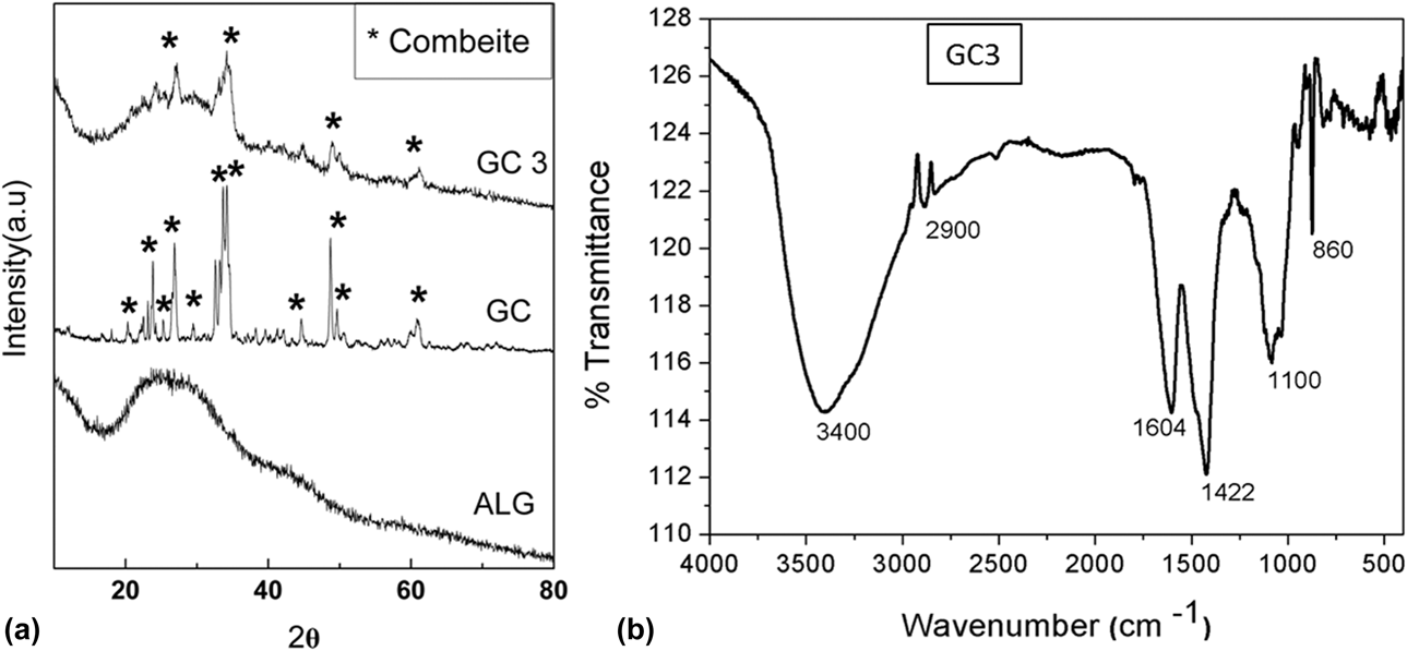
Figure 2(a) shows the X-ray powder diffraction (XRD) pattern of the pure GC powder, pristine alginate, and composite scaffolds. The pure alginate scaffold shows a typical amorphous pattern with a broad band between 20° and 50° 2θ. The XRD pattern for the GC shows the peaks for the combeite (Na2Ca2Si3O9) phase [Reference Thomas and Bera34]. The XRD pattern of the alginate–GC composite scaffolds shows the amorphous background corresponding to the alginate component and some small peaks corresponding to the combeite phase of the GC. Hence, the results reveal the composite behavior of the alginate–GC scaffolds.
(a) XRD patterns of the pure alginate scaffold (ALG), pure GC powder (GC), and alginate–GC composite scaffold (GC3); (b) FTIR spectra of the alginate–GC composite scaffold.

Figure 2(b) shows the FTIR spectra of the alginate–GC composite scaffold (GC3). The spectrum shows a broad peak at 3400 cm−1 due to the absorption band of the OH group [Reference Srinivasan, Jayasree, Chennazhi, Nair and Jayakumar18, Reference Kazy, Sar, Singh, Sen and D’Souza35]. A minor peak observed at 2900 cm−1 is ascribed to –CH stretching of alkanes [Reference Mishra, Basu and Kumar36]. The peaks at 1604 and 1422 cm−1 correspond to the carboxyl and carbonyl groups of sodium alginate, respectively [Reference Kazy, Sar, Singh, Sen and D’Souza35, Reference Mishra, Basu and Kumar36]. The peak at 1100 cm−1 corresponds to Si–O stretching vibration, whereas the peak at 860 cm−1 is attributed to Si–O–Si bending vibration present in the GC [Reference Kazy, Sar, Singh, Sen and D’Souza35]. The peak at 615 cm−1 corresponds to the stretching vibration of phosphate groups [Reference Srinivasan, Jayasree, Chennazhi, Nair and Jayakumar18]. Hence, the FTIR spectra showed functional groups that are present in both the alginate and GC, thus confirming the composite nature of the scaffold.
Porosity
A high degree of porosity is required for enhanced osteogenesis [Reference Hannink and Arts31]. It is observed that porosity of the scaffolds has an inverse relationship with GC concentration. The pristine alginate scaffold showed a porosity of 87.6 ± 0.51%, while the composite scaffolds GC1, GC2, and GC3 exhibited percent porosities of 85 ± 0.571, 84 ± 0.6, and 82.5 ± 0.69, respectively. All the scaffolds showed a porosity higher than 80%, which is sufficient to support vascularization, osteogenesis, and osteointegration.
Swelling behavior
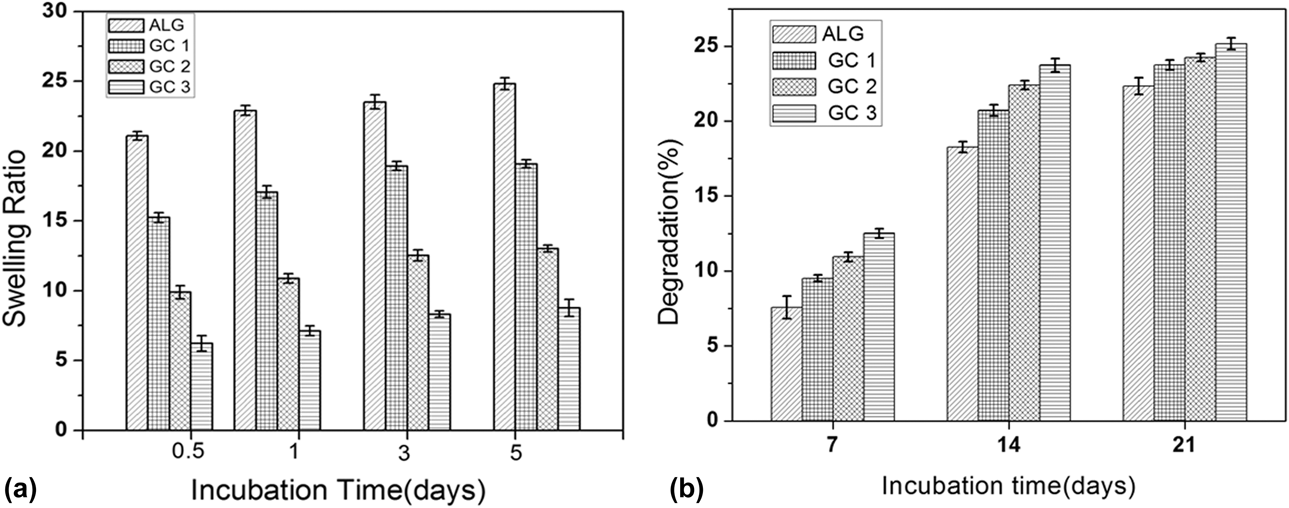
Figure 3(a) shows the change in swelling ratio of pure alginate and alginate–GC composite scaffolds with incubation time. There was a decrease in swelling with an increase in the concentration of the GC powder. The 5-day swelling ratio for the pure alginate scaffold was 24, and it decreased to 9 for the GC3 composite scaffold. This shows that the presence of GC has enabled the controlled swelling in the scaffold. This can be attributed to the strong interaction between the polymer and ceramic particles in the composite and partially to the decreased porosity. Although swelling helps in the nutrient supply to the interior of the scaffold, higher swelling deteriorates the mechanical properties of the scaffold, so a controlled swelling is desirable [Reference Peter, Sudheesh Kumar, Binulal, Nair, Tamura and Jayakumar30]. The controlled swelling ratio observed in the GC–alginate composite scaffold is favorable as it helps to maintain a mechanical–biological property balance in the scaffold.
(a) Swelling ratio of pure alginate and alginate–GC composite scaffolds as a function of incubation time in PBS; (b) degradation (in weight percentage) of pure alginate and alginate–GC composite scaffolds as a function of incubation time in PBS.

Degradation behavior
For an ideal scaffold, the rate at which the implant degrades should be at a similar rate to that of new tissue formation [Reference Rezwan, Chen, Blaker and Boccaccini10]. Polymer degradation involves 3 steps: hydrolysis, enzymatic cleavage, and dissolution. In the case of alginate, the degradation is dependent on the ionic medium in which the scaffold is kept [Reference Kuo and Ma37]. Figure 3(b) shows the percentage degradation of pure alginate and alginate–GC composite scaffolds with incubation time in phosphate-buffered saline (PBS) solution. To control the degradation behavior of alginate, it is cross-linked with Ca2+ ion. When sodium alginate is kept in a calcium ion solution, the sodium ion is replaced by calcium ion in the polymer and each calcium ion is cross-linked with two polymer strand moieties. The dissolution of ions into the solution is reduced due to this cross-linking. Also, more calcium ions are encapsulated in the scaffold because of the concentration gradient, which aided in the bridging of the gel network. It was observed that the major degradation occurred within the first 14 days. The increased degradation rate showed by the composite scaffold compared to the pure alginate one is due to the quick dissolution of GC particles in the PBS solution. It was observed that after 14 days, the degradation rates of the composite and pure alginate scaffolds were almost similar, although composite scaffolds showed slightly higher rates. This could be due to the addition of GCs, which may have weakened the sodium–calcium ion interaction in the system. Overall, the alginate–GC scaffolds showed a controlled degradation behavior.
Bioactivity
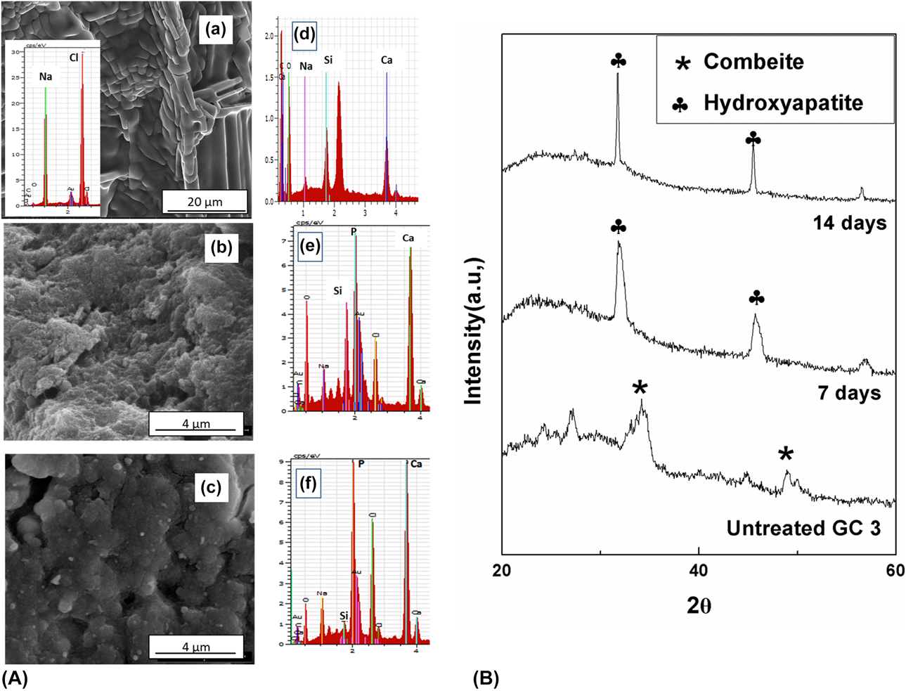
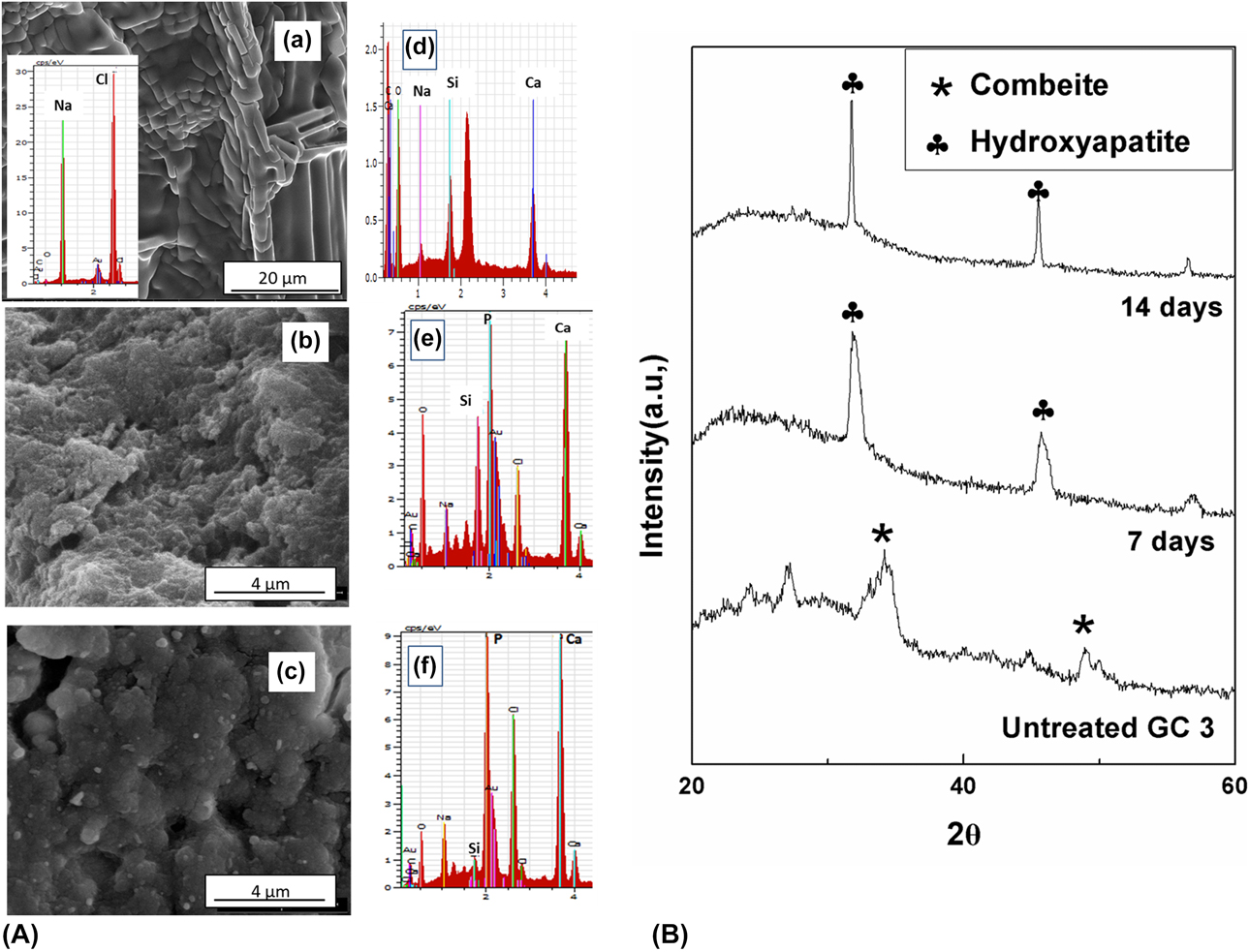
Bioactivity is the capability of a material in vitro to bond with the host bone tissue [Reference Jones1]. The essential condition to evaluate bioactivity is the formation of an apatite layer on its surface at ideal body conditions [Reference Kokubo and Takadama38]. Bioactivity of the pure alginate and the GC-containing composite scaffolds was evaluated in a simulated body fluid (SBF) for a period of 7 and 14 days. Figure 4(A) shows the FESEM image and EDAX of the scaffolds. In case of pure alginate scaffold [Fig. 4(A)(a)], there was the deposition of sodium chloride salts mainly on the surface of the pristine scaffolds. The EDAX spectrum showed that there was no deposition of apatite in the pure alginate scaffold. The FESEM images of the composite scaffolds showed HA-rich layer deposition on the surface. EDAX analysis was used to confirm the formation of apatite on the surface of the composite scaffolds. The EDAX spectra of the incubated scaffold showed an increase in the intensities of calcium and phosphorous compared to that of the untreated scaffold [Fig. 4(A)(d)]. The very small silicon peak after 14 days is due to the growth of a thick apatite layer on the surface. The apatite formation on the composite scaffolds was also evaluated by XRD analysis. Figure 4(B) shows the XRD pattern of the alginate–GC scaffold before and after soaking in the SBF solution for 7 and 14 days. The XRD pattern of the composite shows peaks at 31.8° and 46.2° 2θ ascribed to the (211) and (222) planes of HA. There was a sharp increase in the intensity of peaks with an increase in the incubation time, indicating the increased deposition of the apatite layer with time. Hence, the presence of GC facilitated the formation of an apatite layer on the composite scaffold, and all these results confirm the satisfactory bioactivity of the composite scaffolds.
(A) FESEM images and EDAX of scaffolds for bioactivity studies: (a) image and EDAX of the ALG scaffold after soaking in SBF solution for 14 days, FESEM images of the GC3 scaffold after soaking in SBF solution for (b) 7 days and (c) 14 days. EDAX of (d) the untreated scaffold and the GC3 scaffold after soaking in SBF solution for (e) 7 days and (f) 14 days. (B) XRD pattern of the GC3 scaffold before soaking in SBF solution and after soaking in SBF for 7 and 14 days.
Mechanical behavior
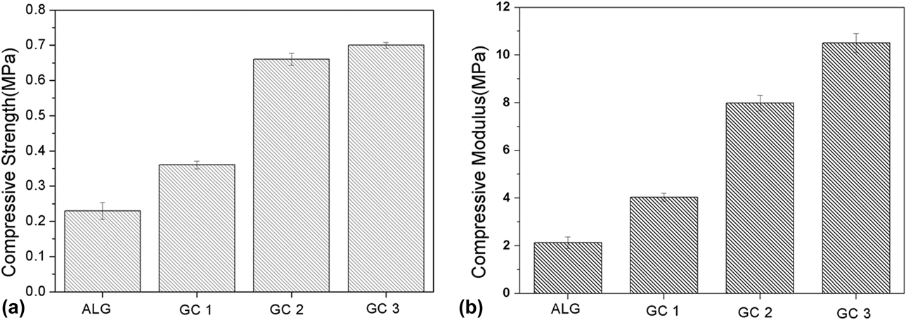
The mechanical property of the scaffold is essential in bone tissue engineering as good mechanical properties are required by scaffolds to maintain their shape during transplantation and to resist loads in the body after implantation [Reference Lin and Yeh21]. Hence, it is vital to reinforce pure alginate scaffolds, which are soft and weak. GC was used as the reinforcing agent in this study to improve the mechanical behavior of the alginate scaffolds. Figures 5(a) and 5(b) show the compressive strength and compressive modulus of the different scaffolds, respectively. As is evident from the plot, the compressive strength of the scaffolds increased with increasing GC content, and there was a drastic increase in strength in the case of GC2 and GC3 scaffolds. Maximum compressive strength and modulus of 0.7 and 10 MPa, respectively, were achieved for the GC3 composite scaffold. Hence, the addition of GC powder has a remarkable reinforcing effect on the weak alginate scaffold. The presence of the GC phase in the composite resists the crack propagation under compressive load, consequently improving the mechanical behavior of the composite scaffolds. The achieved compressive strength is sufficient for safe laboratory handling and effective manipulation of the scaffold by the surgeon during implantation.
(a) Compressive strength of different scaffolds; (b) compressive modulus of different scaffolds.

Cell viability assay
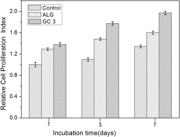
The ability of scaffolds to maintain their cytocompatibility and their potential to facilitate cell proliferation are critical factors in tissue engineering [Reference Chan and Leong39]. MTT assay was used to study the cytocompatibility and osteoblast cell proliferation of the composite scaffolds cultured in MG-63 cell line. Cells cultured on the tissue culture plate were used as the control. Figure 6 shows the relative cell proliferation index for ALG and GC3 scaffolds normalized with respect to the control. As is evident from the plot, both the pristine and composite scaffolds showed a higher proliferation rate compared to the control. Among the scaffolds, the GC3 scaffold showed a higher cell proliferation index, suggesting the superior ability of the composite scaffold to promote cell proliferation. Also, it was evident from the plot that there was a significant level of increase in cell proliferation with incubation time, and a relative cell viability index value of 1.97 was obtained by day 5 for the GC3 scaffold. Hence, MTT assay results confirmed that the alginate–GC composite did not induce any cytotoxicity in the cells, while also promoting cell proliferation of MG-63 cells.
Relative cell proliferation index of ALG and GC3 scaffolds at different time intervals with respect to the control.

Cell adhesion and morphology
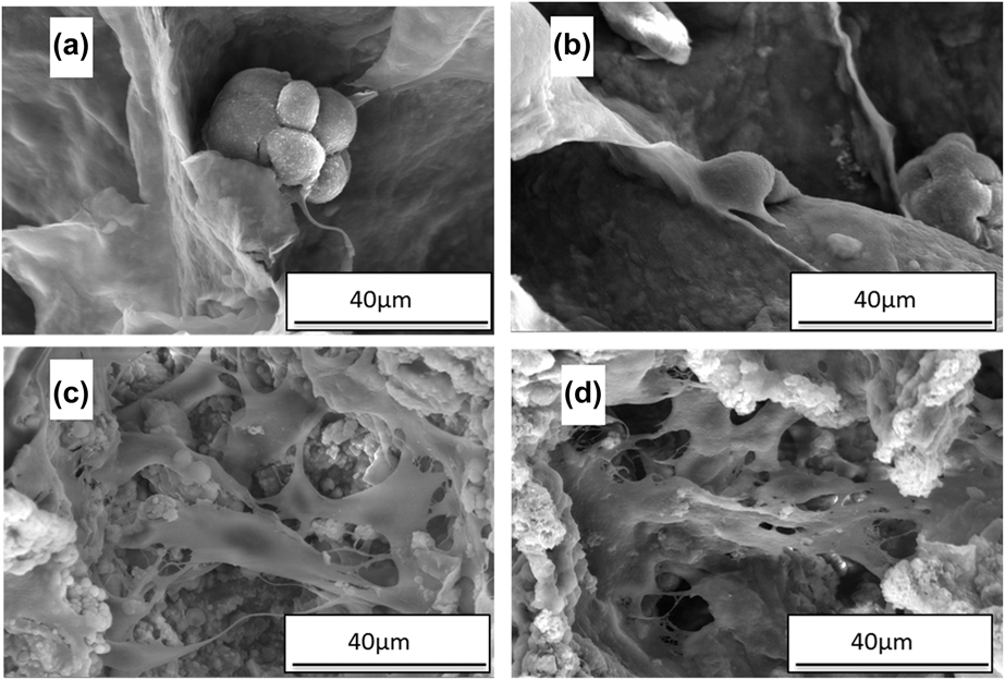
The adhesion of osteoblasts plays a vital role in the expression of various cell functions, such as cell proliferation and differentiation [Reference Duan, Yang, Mei, Tang, Li, Shi, Mao, Zhang and Cai40]. The adhesion, proliferation, and morphology of the cultured cells on the composite scaffold were analyzed using FESEM. Figure 7 shows the FESEM micrographs of MG-63 cells seeded on pristine alginate scaffolds and alginate–GC composite (GC3) scaffolds. The micrographs revealed that the cells were attached to the surface of the scaffolds. The pristine alginate scaffolds presented a spherical morphology and were distributed scarcely on the scaffold surface. On the contrary, in the case of the composite scaffolds, a large number of osteoblasts were well attached, well spread, and proliferated on the scaffold surface. The cells on the composite scaffolds adapted an extended morphology and were anchored on the surface with their numerous filamentous microextensions. Also, a higher number of osteoblasts were found to spread and proliferate on the composite scaffold surface after an incubation period of 14 days in comparison to 7 days. The presence of GC particles provided a suitable environment for the adhesion and proliferation of osteoblasts. This enhanced cell adhesion and proliferation behavior can be attributed to the enhanced bioactivity, high surface area, and roughness imparted by the GC particles in the composite. Hence, these observations are consistent with the MTT results and revealed the ability of the scaffold to facilitate cell adhesion and proliferation.
FESEM images of MG-63 morphology and attachment on: pure alginate scaffolds after seeding for (a) 7 days and (b) 14 days; composite scaffold (GC3) for (c) 7 days and (d) 14 days.

Alkaline phosphatase activity
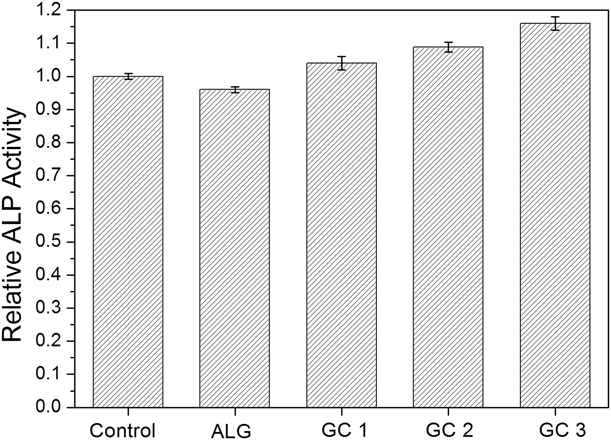
Alkaline phosphatase (ALP) activity is widely used as a standard early phenotypic marker for osteoblasts [Reference Hsu, Liang, Hung and Kuo41]. The bone-forming ability of the MG-63 cells cultured on the composite scaffolds was studied by measuring the p-nitrophenol produced, and the results were normalized with the control. Figure 8 shows the relative ALP activity of the scaffolds after 14 days of cell incubation. It can be observed that all the composite scaffolds exhibited considerable ALP activity with respect to the control. Also, the ALP activity increases with the increase in GC in the composite scaffold. Among the samples, the GC3 showed the highest ALP activity, measuring a 1.16-time increase with respect to the control. The other two composite scaffolds showed 1.09 (GC2) and 1.06 (GC1), respectively, while the pure alginate scaffold showed only 0.96 times with respect to the control. Considering that the control surface used in this study was tissue culture plastic, which is an idealized cell culture surface and not a bioinert surface, the ALP activity obtained in this study is sufficient to support the osteogenic potential of the composite scaffolds. The increased ALP activity of the composite scaffold is because the ionic dissolution products released from the GC particles can upregulate osteocalcin expression, hence improving the ALP activity. Also, the increased sensitivity of the osteoblast toward high silicon concentration of the GC enhanced ALP production [Reference Andrade, Valério and Goes42, Reference Xynos, Edgar, Buttery, Hench and Polak43].
Relative ALP activity of different scaffolds.

In vitro mineralization
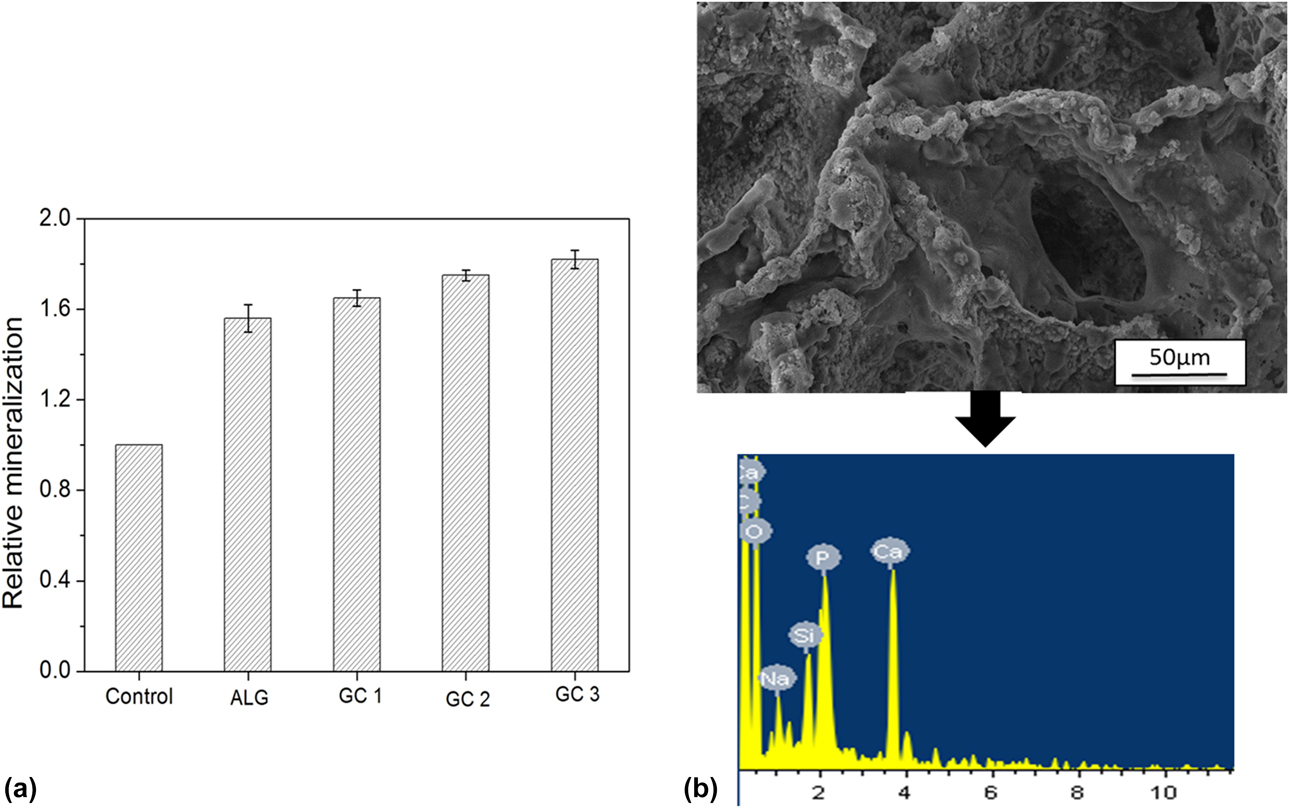
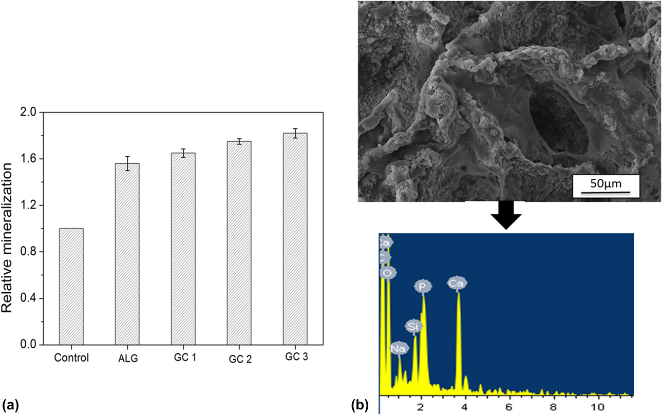
Alizarin red S (ARS) staining is usually used to evaluate the formation of calcium-rich deposits in a cell-cultured experiment, which can be used to quantify the mineralization behavior of osteoblast cells [Reference Hsu, Liang, Hung and Kuo41, Reference Gregory, Gunn, Peister and Prockop44]. ARS can bind specifically to calcium salts, and hence, the scaffolds cultured with MG-63 cells were studied using ARS calorimetric assay. Figure 9(a) shows the relative mineralization (calcium deposition) normalized with respect to the control. ARS staining results followed a similar trend, as shown by the ALP results. It can be seen that the GC-based composite scaffolds showed higher mineralization than the pristine alginate scaffolds. The GC3 scaffold showed a 1.82-fold mineralization compared to the control, whereas the pure alginate showed the least with a 1.56-fold mineralization. The formation of calcium deposition on the composite scaffold (GC3) was also confirmed by FESEM and EDAX analysis [Fig. 9(b)]. A large number of calcium depositions were observed on the surface of the scaffold. EDAX study showed peaks for calcium and phosphorus, which confirmed the cell-mediated mineralization in the scaffold. Thus, the intracellular mineralization was consistent with the extracellular mineralization study carried out under physiological conditions (SBF). The enhanced mineralization showed by the composite scaffolds can be explained as follows: the dissolution products from the GC particles can create an extracellular environment, which facilitates osteoblast expression and extracellular matrix deposition [Reference Tsigkou, Jones, Polak and Stevens45, Reference Varanasi, Owyoung, Saiz, Marshall, Marshall and Loomer46]. The presence of GC in the scaffold plays a critical role in osteogenesis as the osteoblast has higher sensitivity toward silicon ions released from GCs [Reference Xynos, Edgar, Buttery, Hench and Polak43, Reference Gregory, Gunn, Peister and Prockop44, Reference Varanasi, Owyoung, Saiz, Marshall, Marshall and Loomer46].
(a) Relative mineralization behavior of different scaffolds; (b) SEM and EDAX of the composite scaffold seeding with MG-63 cells for 14 days showing calcium deposition.
Conclusions
In summary, the alginate–GC composite scaffold was successfully fabricated by a freeze-drying technique. A macroporous well-interconnected pore structure (with pore size 110–380 µm) was obtained in the scaffold. A high degree of porosity (higher than 80%) was obtained for all the scaffolds even after the addition of GC particles. The obtained porosity and pore structure are sufficient to support vascularization, bone ingrowth, and nutrient supply. The composite scaffold showed excellent in vitro bioactivity where the GC particles acted as binding sites for HA attachment, and this can induce bone formation in the scaffold even in the absence of osteogenic factors. Mechanical behavior analysis showed that the compressive strength and modulus increased with an increase in the GC content, which reinforces the alginate matrix. A compressive strength of 0.7 MPa and modulus of 10 MPa were obtained for the GC3 scaffold. The composite scaffold showed excellent biocompatibility with good cell attachment and proliferation. Osteoblast cells showed improved ALP activity and mineralized matrix formation in the composite scaffold. The results revealed the ability of the GC components to induce osteogenesis and osteoblast differentiation. Thus, the alginate–GC scaffolds obtained in this study can serve as a promising biomaterial for bone tissue engineering.
Materials and methods
Fabrication of scaffold
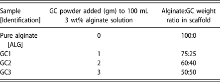
The GC with the composition (in mol%) SiO2–49.3, Na2O–23.4, CaO–25.6, and P2O5–1.7 was prepared by a sol–gel process as reported earlier [Reference Thomas and Bera34]. 3 wt% alginate solution was prepared by dissolving sodium alginate in Millipore water with continuous stirring for 3 h. After a clear homogeneous solution was obtained, the sol–gel-derived GC powder was added to the alginate solution according to the different compositions shown in Table I. A 30-mM calcium chloride solution was added to the slurry for in situ cross-linking. The mixed slurry was further dispersed ultrasonically for 15 min and then dispensed into vials. The vials were placed in a deep freezer and frozen for 12 h. The frozen samples were freeze-dried at −80 °C under vacuum conditions for 48 h. The obtained scaffolds were again cross-linked by dipping in 100 mM calcium chloride solution for 24 h and again freeze-dried for 24 h to obtain the final scaffolds. An additional cross-linking step was carried out to provide the required stability of the scaffolds. A similar double stage cross-linking has also been adopted by others [Reference Lin and Yeh21].
Compositions of the samples used for the study.

Characterization of scaffolds
The microstructure of the pure and composite scaffolds was analyzed using a field emission scanning electron microscope (FEI Nova Nano, Oregon). The pore size was quantified from the FESEM images using ImageJ software (NIH, Maryland). The pore characteristics of the representative samples were studied using synchrotron-based X-ray micro-computed tomography (SRµCT) (Imaging beamline, Indus-2, RRCAT, Indore). The experimental set-up for SRµCT consists of high-precision sample manipulation and rotation stages, and a CCD camera with an X-ray fluorescent screen with fiber-optics coupling. The samples were mounted on the sample holder and scanned using a synchrotron X-ray monochromatic beam of energy 10 keV. The angle step was fixed at 0.2 and the acquisition time per image was 600 ms. 2D radiographic projections obtained from SRµCT were flat-field-corrected, and tomographic cross-section slice images were reconstructed using filtered back-projection techniques. The 2D slices were stacked together and volume-rendered to obtain the final 3D images.
X-ray diffraction analysis of the pure and composite scaffolds was performed in a Rigaku machine (Japan) using Cu Kα radiation. The XRD pattern was obtained with a step size of 0.05. The characteristic functional groups present in the composite scaffolds were examined using FTIR spectroscopy (PerkinElmer, Massachusetts). The pellets for FTIR measurement were prepared by mixing 2 mg of the sample with 200 mg of potassium bromide. The transmission spectra for the scaffolds were measured in the range 4000–400 cm−1.
Porosity measurement
The porosity of the scaffolds was calculated using the following equation:
 $${P_1} = \left[ {1 - {{{\rho _{\rm{a}}}} \over {{\rho _{\rm{g}}}}}} \right] \times 100\quad ,$$
where ρa is the density of the scaffold and ρg is the theoretical density of component powders.
Swelling ability
The swelling ability of the scaffolds was evaluated in PBS up to five days. After recording the dry weight (W d) of all the scaffolds, they were immersed in PBS solution (pH 7.4) at 37 °C for different time intervals. After the predetermined time, the scaffolds were taken out, the excess solution present on the scaffold surface was blotted, and the wet weight was recorded as W w. The swelling ratio (S w) was determined using the following equation:
 $${\rm{Swelling}}\;{\rm{ratio}}\;\left( {{S_{\rm{w}}}} \right) = {{{W_{\rm{w}}}-{W_{\rm{d}}}} \over {{W_{\rm{d}}}}}\quad .$$
Three samples were studied for every given time interval.
Biodegradation behavior
Biodegradation of the scaffolds was evaluated in terms of weight loss after incubation in PBS solution at 37 °C for 7, 14, and 21 days. After every predetermined time interval, the scaffolds were removed, washed with deionized water, and freeze-dried. The dried scaffold was weighed, and the degradation percentage was calculated using the following formula:
 $${\rm{Degradation}}\;\left( {\rm{\% }} \right) = {{{W_0} - {W_t}} \over {{W_0}}} \times 100\quad ,$$
where W 0 is the weight of the original sample measured before immersion in PBS and W t is the weight of the sample measured after immersion in PBS at a given time. Three samples were studied for every given time interval.
In vitro bioactivity
Bioactivity of the scaffolds can be assessed in vitro by monitoring the formation of HA on their surface in a SBF. The SBF was prepared by the procedure described by Kokubu and Takadama [Reference Kokubo and Takadama38]. The scaffolds were immersed in the SBF and kept in an incubator at 37 °C. The SBF was replaced twice every week to counter the decrease in cation concentration in the solution during the study [Reference Chen, Thompson and Boccaccini12, Reference Pirayesh and Nychka47]. After the given incubation time, the samples were removed from the SBF solution, rinsed in deionized water to remove the adsorbed minerals, and freeze-dried for 24 h. The dried samples were characterized using XRD, FESEM, and EDAX to assess the possible formation of the HA phase following different immersion periods in the SBF.
Mechanical behavior
The compressive strength of the scaffolds was determined by using a universal testing machine (Tinius Olsen, United Kingdom). Compressive modulus was determined from the linear region of the stress–strain curve. The specimens used for the study were cylindrical samples of height 15 mm and diameter 10 mm. Five parallel samples were tested for every scaffold, and the final results were calculated averagely.
Cell viability assay
For the cell viability assay, the scaffolds were cut into uniform dimensions of 1 cm diameter and 2 mm thickness. The biocompatibility studies were performed using the MG-63 cell line, which was maintained in DMEM with 10% FBS at 37 °C, 5% CO2, and 95% humidity. Cells cultured on a tissue culture plate were used as the control. The cells were seeded on the scaffolds in a 24-well plate at a cell concentration of 1 × 10⁴ cells/mL. The culture media of 0.6 mL was maintained for each sample. The plate was then kept in the incubator for adherence. After the given time interval, 20 µL of MTT solution at a concentration of 5 mg/mL was added to each well and incubated for 4 h. MTT was removed followed by the addition of dimethyl sulfoxide, which solubilizes the formazan crystals. This was followed by the measurement of absorbance at 595 nm, from which the cell proliferation index was determined.
Cell adhesion and morphology
The scaffolds with dimensions of 1 cm diameter and 2 mm thickness were used for the study. The scaffolds were initially preconditioned for 24 h with the cell culture medium. The cells were seeded on the scaffolds in a 12-well plate at a cell concentration of 3 × 10⁵ cells/0.6 mL and incubated for 4 h to allow adherence of cells on the scaffold. Following this, 1 mL fresh medium was added, incubation was carried out at 37 °C in 5% CO for 7 and 14 days, and the medium was changed every two days. Following incubation, the scaffolds were repeatedly rinsed with PBS (devoid of Ca and Mg). The cells were then fixed with 2.5% glutaraldehyde solution for three hours, washed with PBS, and dehydrated in a series of ethanol gradations (30, 50, 70, 90, 95, and 100%). Finally, the samples were dried, gold sputter–coated, and observed using ESEM (FEI Nova Nano).
ALP activity
ALP activity of the osteoblast cells cultured on the scaffolds was studied using the p-nitrophenol phosphate-based colorimetric assay. Cells cultured on the tissue culture plate were used as the control. After 14 days of cell incubation, the scaffolds were dissolved in 55 mM sodium citrate buffer followed by centrifugation. The collected cells were permeabilized with 0.1% PBST (0.1% Triton X-100 [Sigma Aldrich, Missouri] in PBS) for 20 min. This is followed by the incubation of the cell lysate with p-nitrophenol phosphate for 2 h at 37 °C, and the absorbance was read at 405 nm using a double-beam UV-visible spectrophotometer (Systronics, India).
In vitro mineralization
Calcium deposition due to in vitro bone mineralization by the encapsulated cells was studied by an ARS staining–based calorimetric assay. Cells cultured on the tissue culture plate were used as the control. After 14 days of incubation, the cells were fixed with 4% paraformaldehyde for 20 min. After fixation, the scaffolds were washed with DPBS and stained with 0.2% Alizarin red for two hours at 37 °C. Mineralization was quantified by performing a cetylpyridinium chloride-based colorimetric assay. The stained scaffolds were dissolved with 55 mM sodium citrate buffer, and the cells stained with Alizarin red were spun down. Bound Alizarin red was eluted using 10% cetylpyridinium chloride solution and incubated for 30 min. The absorbance of the eluted dye was read at 562 nm using a double-beam UV-visible spectrophotometer (Systronics). Calcium mineralization on the scaffolds was also observed and confirmed by FESEM and EDAX analysis.
Acknowledgments
This research was financially supported by the National Institute of Technology, Rourkela, India. The authors would like to thank Prof. Indranil Banerjee and Mr. VS Sharan Ratnam, Department of Biotechnology and Medical Engineering, NIT, Rourkela, for their assistance.