Introduction
Meloidogyne incognita, the root-knot nematode, is a member of the group of sedentary plant-parasitic nematodes (PPNs) that cause serious damage (to the tune of hundreds of billions of dollars) to crop production worldwide (Elling, Reference Elling2013). As an obligate endoparasite, M. incognita resides permanently inside the roots of its host throughout its lifecycle, and to derive nutrients, this organism has developed a specialized organ called the stylet. The pharynx of M. incognita is continuous with the stylet lumen and aids with the ingestion of food material into the intestine (Eisenback and Hunt, Reference Eisenback, Hunt, Perry, Moens and Starr2009). Pharyngeal development occurs during embryogenesis, and several genes responsible for its development, most of which belong to the Notch-like receptor family, have been identified in Caenorhabditis elegans. Notch pathways are known to regulate the aspects of growth and patterning in metazoans (Rudel and Kimble, Reference Rudel and Kimble2001). Pharyngeal cells are produced by two distinct molecular pathways, ABa and EMS. The ABa pathway is dependent on the Notch receptor orthologue glp-1 (abnormal germline proliferation) (Mango, Reference Mango2007), which is a homologue of the Drosophila Notch gene (Roehl et al. Reference Roehl1996) and induces germline proliferation during pharyngeal embryonic development. Glp-1 is structurally and functionally similar to another Notch-related receptor, lin-12, and these two proteins have similar conserved motifs that contribute to the functional roles of these proteins as membrane-bound receptors involved in Notch signalling (Yochem and Greenwald, Reference Yochem and Greenwald1989).
With recent advances in the generation of genomic datasets for nematodes, many studies have focused on identifying and characterizing genes with varied functional roles and determining their conservation throughout evolution. Various secretory and effector proteins have been characterized over the past few years (Banerjee et al. Reference Banerjee2017). Misp12 is a potential root-knot nematode effector expressed in the dorsal oesophageal gland (Xie et al. Reference Xie2016). The SXP/RAL-2 secretory protein was identified in M. incognita and is expressed in the subventral pharyngeal glands (Tytgat et al. Reference Tytgat2004). However, there are limited reports concerning the identification and characterization of genes involved in nematode development. Among the genes involved in organ development in nematodes, collagen genes are the most studied, and the M. incognita collagen genes, Mi-col-1 and lemmi-5, and the Globodera pallida collagen genes, col-1 and col-2, have been identified and characterized (Wang et al. Reference Wang, Deom and Hussey1998; Gray et al. Reference Gray, Curtis and Jones2001). Chitin synthase, which functions in the production of eggshells, has been reported in C. elegans and M eloidogyne artiellia (Veronico et al. Reference Veronico2001; Fanelli et al. Reference Fanelli2005). However, with the exception of the study conducted by Calderón-Urrea et al. (Reference Calderón-Urrea2016), who studied and compared the early development of M. incognita with that of C. elegans, almost no detailed studies have investigated genes associated with the early development of M. incognita.
Several reports detail the RNAi-mediated knockdown of specific secretory genes that affect parasitism in PPNs (Rehman et al. Reference Rehman, Gupta and Goyal2016). However, only some of the known developmental genes have been targeted by host-mediated gene silencing in the management of PPNs. The Rpn7 gene, which is essential for the integrity of the 26S proteasome, has been targeted in attempts to control the root-knot nematode M. incognita, and the silencing of this gene resulted in a reduction in nematode motility (Niu et al. Reference Niu2012). Two genes essential for body wall formation in M eloidogyne graminicola, namely the Mg-pat-10 and Mg-unc-87 genes, which function in muscle contraction and the maintenance of structural myofilaments, have been identified and suppressed in vitro through soaking, resulting in 91% and 87% reductions in infectivity, respectively (Nsengimana et al. Reference Nsengimana2013). nhr-48, a nuclear receptor gene that regulates various developmental, reproduction and pathogenicity processes in nematodes, has been silenced in M. incognita, resulting in delayed development and reduced reproduction (Lu et al. Reference Lu2016). Despite these efforts, few developmental genes have been targeted in nematode control efforts.
The present study reports the identification and characterization of the glp-1 gene of M. incognita. A host-mediated RNAi silencing approach was adapted to determine the potential of Mi-glp-1 as a candidate gene for the control of nematode infection.
Materials and methods
Genome-wide identification of the glp-1 gene in M. incognita
The M. incognita genome database was downloaded from http://www6.inra.fr/Meloidogy ne_incognita (Abad et al. Reference Abad2008), and local nucleotide and protein M. incognita databases were created through a BioEdit local BLAST search for the identification of the glp-1 gene in M. incognita. A tblastn search was conducted against these databases using the C. elegans glp-1 (CAA79620.1) gene as the query and default parameters (expected threshold value 10; maximum number of aligned sequences displayed 100). All the sequences that met the requirements were subjected to gene predictions through GeneMark and FGENSH analyses, and the genes that did not contain the known conserved domains and motifs detailed in the Pfam database (http://pfam.janelia.org/) were removed (Finn et al. Reference Finn2016). MiV1ctg1087 (Minc16055), the single contig obtained with the maximum possible score, was selected as the probable Mi-glp-1 for further experimentation. In addition to Minc16055 in M. incognita, we identified glp-1 orthologues in four other Meloidogyne species (spp.), namely M. hapla (Mh10g200 708_Contig1018), M. floridensis (contig nMf_1_1_scaf00321), M. javanica (MJ01378, MJ05005) and M. chitwoodi (MC01544, MC00257), and in G. pallida (GPLIN_000999900.1), a cyst nematode, using their genome, transcriptome and/or expressed sequence tag (EST) databases. The derived sequences were downloaded from their genome assembly or transcriptomic databases or EST clusters available at either NCBI, NEMBASE4 (www.nematode.org) and/or nematode.net V4.0 (Opperman et al. Reference Opperman2008; Elsworth et al. Reference Elsworth, Wasmuth and Blaxter2011; Cotton et al. Reference Cotton2014; Lunt et al. Reference Lunt2014; Martin et al. Reference Martin2015).
Phylogenetic and gene structure analyses
The identified GLP-1 amino acid sequences in PPNs, including M. incognita, M. hapla, M. floridensis, M. javanica, M. chitwoodi and G. pallida, along with the reported GLP-1 sequences of free-living nematode species, e.g. C. elegans, Caenorhabditis briggsae, Caenorhabditis japonica and Caenorhabditis remanei, and the Notch-like protein of the marine worm Priapulus caudatus (retrieved from NCBI), were aligned in MUSCLE using the default parameters (Edgar, Reference Edgar2004). To analyse the clustering pattern, an unrooted phylogenetic tree was constructed based on the neighbour-joining (NJ) method using MEGA 7.0 software (http://www.megasoftware.net/), with Poisson correction, pairwise deletion and the bootstrap value set to 2000 replicates (Kumar et al. Reference Kumar, Stecher and Tamura2016).
The protein sequences were analysed with the Pfam (http://pfam.sanger.ac.uk/) and SMART (http://smart.emblheidelberg.de/) databases to confirm the presence of conserved motifs. SignalP3.0 and TMHMM server v. 2.0 were employed to predict the presence of signal peptide sequences and transmembrane domains (TMDs), respectively. Two other software programs (Kd and Protscale) based on different algorithms were used to determine the presence of TMDs (Kyte and Doolittle, Reference Kyte and Doolittle1982; Gasteiger et al. Reference Gasteiger and Walker2005).
The motif-based sequence analysis tool MEME 4.11.2 (http://meme.sdsc.edu/meme/meme.html) (Bailey et al. Reference Bailey2015) with the following parameters was used to identify conserved motifs in the glp-1 gene of M. incognita: optimal width, 10–300 amino acids and maximum number of motifs 10. The gene architecture of this gene, which depicts the exon/intron arrangement, gene length and upstream/downstream region, was designed using the online Gene Structure Display Server 2.0 (http://gsds.cbi.pku.edu.cn/) (Hu et al. Reference Hu2015) with the coding sequences and corresponding genomic sequences.
Maintenance of pure cultures of nematodes
Tomato (Solanum lycopersicum) seeds were sterilized by soaking 20 min in sterile distilled water, 5 min in 70% ethanol and 15 min in 5% NaOCl and 0.1% Tween 20 followed by washing four times in sterile distilled water and germinated on a mixture of cocopeat, vermiculite and sand (1:1:1) in an Indian Agricultural Research Institute (IARI) glass house. Two-week-old tomato seedlings were infected with a pure culture of M. incognita maintained in our laboratory (Kumar et al. Reference Kumar2017). Egg masses of M. incognita were hand-picked from the roots of the infested plants and then maintained until hatching at 28 °C in Petri plates containing 10–15 mL of sterile water to collect second-stage juveniles (J2s) of M. incognita. These J2s were then used for subsequent inoculation assays using transgenic Arabidopsis lines.
Quantitative real-time PCR analyses
To evaluate the expression at different developmental stages, cDNA from the samples of M. incognita egg masses, infective J2s and mature females as well as roots of infected plants harvested at different time points after inoculation was subjected to quantitative real-time PCR (qRT-PCR). J2s were hatched from freshly picked egg masses, mature females were excised from infected roots, and other stages, namely third-stage juveniles (J3s) and fourth-stage juveniles (J4s), were analysed from nematodes within the roots of infected plants harvested at 10 and 21 days post-inoculation (dpi), respectively. The second molt, which gives rise to J3s, occurs 10 dpi, and the third molt, which yields J4s, occurs approximately 16 dpi (Moens et al. Reference Moens, Perry, Starr, Perry, Moens and Starr2009; Martinuz et al. Reference Martinuz, Schouten and Sikora2013). Total RNA was isolated using a Pure Link RNA Mini Kit (Ambion, Carlsbad, CA, USA) according to the manufacturer's instructions and then subjected to DNase treatment using the Qiagen DNase enzyme. cDNA was synthesized using the SuperScript® III First-Strand Synthesis System (Invitrogen, USA). SYBR-based chemistry was adopted for the qRT-PCR, which was performed in a StepOne Plus™ real-time PCR System. The primers for M. incognita-specific actin and 18S rRNA genes were used as references (Nguyễn et al. Reference Nguyễn2014; Ye et al. Reference Ye, Zeng and Kerns2015) (Supplementary Table S1). Three biological and three technical replicates were included for each sample. The data were analysed using the 2[-Delta DeltaC(T)] method, and real-time data are reported as the means ± standard error (s.e.) of three biological replicates per sample (Livak and Schmittgen, Reference Livak and Schmittgen2001). The data obtained were statistically analysed by analysis of variance (ANOVA), and the significance of the differences between sample means was then determined through Student's t-tests (P < 0.05).
A qRT-PCR analysis was also performed to determine the silencing efficacy of glp-1 at the transcript level in M. incognita. Transgenic Arabidopsis RNAi lines were infected with M. incognita J2s, and the infected roots were harvested at 45 dpi. The mature females were isolated from these infected transgenic RNAi lines and compared with females from control (wild-type) Arabidopsis plants using the above-described protocol for qRT-PCR analysis.
In vivo RNAi silencing
A 419 bp fragment of the Mi-glp-1 gene was amplified using gene-specific primers (Supplementary Table S1) in the sense and antisense directions for double-stranded RNA (dsRNA) construction. The primers were designed based on the region flanking the conserved Lin-12/Notch repeat (LNR) domain found in three tandem copies of Notch-related proteins encoded by the glp-1 gene. The RNAi binary vector pBC6 was used for the design of dsRNA constructs (Yadav et al. Reference Yadav, Veluthambi and Subramaniam2006), and the positive dsRNA constructs were confirmed by double digestion of the sense strand with the BamHI and XhoI restriction enzymes and of the antisense strand with KpnI and SacI. The integrity of the products was further confirmed by Sanger sequencing. A positive dsRNA construct and an empty vector construct were transferred into Agrobacterium tumefaciens GV3101. Arabidopsis thaliana (Col0) plants were then transformed with the designed RNAi constructs driven by the CaMV35S promoter using the floral dip method (Clough and Bent, Reference Clough and Bent1998). T1 seeds were selected on antibiotic selection medium containing kanamycin (50 µg mL−1). Kanamycin-resistant plants were transferred to a greenhouse maintained at 22 °C with a 16 h light/ 8 h dark photoperiod, and the presence of the nematode gene in independent transgenic RNAi lines was confirmed by PCR.
Meloidogyne incognita infection bioassay for evaluating silencing efficacy
All the analyses of transgenic plants were performed with the T3 generation, and 10–15 biological replicates of each line were used. Seeds of T3 transgenic lines expressing glp-1 dsRNA or empty vector and of control (wild-type) Arabidopsis plants were surface-sterilized with 70% alcohol for 2 min and 0.1% mercuric chloride +0.1% sodium dodecyl sulphate for 7 min and then washed in sterile distilled water. The seeds were then maintained on Murashige and Skoog media at 4 °C for vernalization prior to germination, and after germination, the plates were maintained at ±22 °C under a 16 h light/8 h dark photoperiod. Twelve days after germination, the control seedlings and the transgenic seedlings expressing glp-1 dsRNA or empty vector were uprooted from the medium, transferred to a suitable mixture of vermiculite, cocopeat and sand (1:1:1), with one plant per slot (3 inches) in a 24-slot tray, and maintained in a growth chamber with a 16 h light/8 h dark photoperiod. After 1 week, each plant was inoculated with 1000 freshly hatched J2s to evaluate the silencing efficacy in vivo. Nematode infection bioassays were performed using mature females isolated from the root samples collected 45 dpi. The isolated females were used for qRT-PCR and morphometric studies. The number of galls per plant, the number of females and the egg mass per gram of root fresh weight were determined and used as measures for determining the degree of nematode infection. Specifically, the nematode multiplication factor (MF) of M. incognita was calculated as follows: (number of egg masses × number of eggs per egg mass) ÷ nematode inoculum level. The statistical significance of the differences between the means of replicates of transgenic lines and wild-type control plants was determined by ANOVA and Student's t-tests (P < 0.05 or P < 0.01).
The nematodes within all the samples of infected roots were stained with acid fuchsin (Bybd et al. Reference Bybd, Kirkpatrick and Barker1983) and photographed using a Nikon microscope. The size of the stained females isolated from the infected plant samples was measured using the 10× objective of a Nikon microscope equipped with the NIS-Elements D measurement software (Eisenback and Hunt, Reference Eisenback, Hunt, Perry, Moens and Starr2009; Kaur and Attri, Reference Kaur and Attri2013). The stylet and pharyngeal structures of the J2 progeny of females isolated from transgenic RNAi and control plants were also observed with an Zeiss AxioImager.M2m microscope coupled to an AxioCam with a 40× objective, and the data were subjected to statistical analyses as described above.
Detection of glp-1 by Northern hybridization
The small RNA fraction was isolated from the RNAi line, empty vector line and control plants using the LiCl method (Verwoerd et al. Reference Verwoerd, Dekker and Hoekema1989). Twenty micrograms of the small RNA fractions were resolved on 15% polyacrylamide/1X MOPS urea gels and blotted onto a Hybond-N+ membrane (Amersham). The Block-It™ RNAi designer (https://rnaidesigner.thermofisher.com/rnaiexpress), siRNA wizard (http://www.invivogen.com/sirnawizard/) and siDesign centre (http://dharmacon.gelifesciences.com/design-center/) tools were used to identify the most suitable regions for siRNA target design, and primers for probe preparation were designed based on the region that was deemed most suitable for siRNA targeting (Supplementary Table S1). To generate specific DNA probes (183 bp in size), 32P-α-dCTP labelling was performed with a Megaprime DNA Labeling kit (GE Healthcare, Amersham) using gene-specific antisense fragments. Hybridization was performed using PerfectHyb™ Plus according to the manufacturer's instructions (Sigma). The signals were detected by autoradiography.
Results
glp-1 orthologues among nematode species
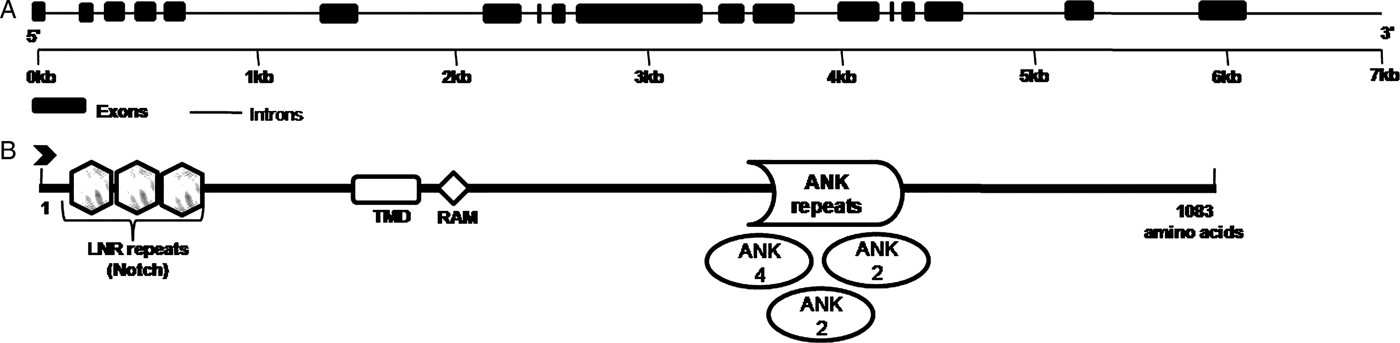
The tblastn analysis performed against the M. incognita genome using the C. elegans glp-1 protein as the query (retrieved from WormBase) and the subsequent Pfam analysis yielded various hits. Among these hits, MiV1ctg1087, the only contig with a high bit score, showed 100% similarity with Minc16055, which was identified through an OrthoMCL analysis of M. incognita as a Ce-glp-1 orthologue by Abad et al. (Reference Abad2008). Thus, functional annotation studies of MiV1ctg1087 involving a Pfam analysis and computer simulation predictions were performed to determine its gene and protein structures. Mi-GLP-1 (Minc16055 or MiV1ctg1087) is a member of the LIN-12/Notch family of receptors and is composed of a series of motifs that are conserved among all Notch receptors. The Ce-glp-1 and Mi-glp-1 genes were nearly identical in size, 7.458 and 7.449 kb, respectively. However, a computational analysis showed differences in the numbers of introns and exons in these orthologous genes: Ce-glp-1 has only nine exons and eight introns (Rudel and Kimble, Reference Rudel and Kimble2001), whereas Mi-glp-1 has 18 exons and 17 introns. This finding indicates that the exon/intron structures of these orthologous genes are not well conserved among nematodes. At the mRNA level, Mi-glp-1 is 3.252 kb in length, whereas Ce-glp-1 comprises 4.326 kb (Fig. 1). The Mi-glp-1 gene encodes a 1083-amino-acid-long truncated protein, as determined through the Pfam analysis, whereas Ce-glp-1 is composed of 1295 amino acids. The gene encoding GLP-4, which is considered a member of the Notch GLP protein family, was recently identified in C. elegans (Rastogi et al. Reference Rastogi2015), and to date, this new member has only been identified in C. elegans and C. briggsae. The genome-wide search performed in this study identified only a single glp-1 contig, which is similar to the findings obtained for C. japonica and C. remanei, each of which has a single glp.
Diagrammatic representation of the predicted structures of the glp-1 gene and its encoded protein. (A) Exon/intron structure of M eloidogyne incognita glp-1. The boxes represent exons, and the black lines represent introns. (B) Architecture of the M. incognita GLP-1 protein. The functional domains are indicated as follows:  LNR,
 TMD,
 RAM domain and
 ANK.

Because glp-1 is a vital gene involved in pharyngeal development during embryogenesis, we also performed a comprehensive species-wide tblastn search for this gene in the genomes and ESTs of various other Meloidogyne species available in NCBI and/or Nematode.net. A glp-1 homologue was identified in various Meloidogyne spp., namely, M. hapla, M. floridensis, M. javanica and M. chitwoodi and in G. pallida, another PPN (details in Supplementary Table S2). We also considered free-living nematodes, including C. elegans, C. briggsae, C. japonica and C. remanei and a marine worm (P. caudatus) Notch receptor gene in our phylogenetic study (Supplementary Fig. S1). The findings revealed that among the homologues identified in Meloidogyne spp., the glp-1 homologue in M. hapla contains all the domains that are present in the Ce-glp-1 gene, whereas Mi-glp-1 does not have an epidermal growth factor (EGF) domain. A motif search including all the species analysed in this study (Supplementary Fig. S2) revealed a conserved ankyrin repeat (ANK).
Protein sequence analysis and phylogenetic analysis of Notch receptor orthologues in selected nematode species
An amino acid sequence analysis of the M. incognita GLP-1 protein revealed a single TMD, an extracellular domain containing the LNRs, the ANK, which is an intracellular domain, and a Pro-Glu-Ser-Thr (PEST) domain. The molecular and biological functions of the LNR and ANK in C. elegans have been described (Austin and Kimble, Reference Austin and Kimble1989; Roehl et al. Reference Roehl1996). Thus, the presence of these motifs in Mi-GLP-1 indicates its probable role in embryogenesis. Specific primers for the LNR were designed, and the region was successfully amplified, confirming the presence of the LNR in M. incognita (data not shown). Interestingly, the presence of a conserved cysteine (‘C’) as the first residue of the LNR was identified in the glp-1 proteins of all nematode species evaluated in this study. Mi-GLP-1 contains one ANK with three copies, three LNR domains at the 5′end, a TMD and an RAM domain but does not have an EGF domain (Fig. 2). GLP-1 induces downstream transcriptional regulators and interacts with them to mediate signalling. Our sequence analysis revealed an ‘RTGGGAA’ DNA-binding site and an RAM domain in Mi-glp-1. These sites have been proposed as binding sites for the LAG-1 (Lin-12 and Glp-1) protein, which is required during embryogenesis to regulate pharyngeal development (Christensen et al. Reference Christensen1996).
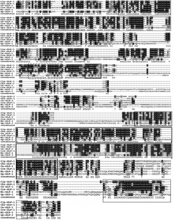
Multiple sequence alignment of GLP-1 in free-living and plant-parasitic nematodes. Black shading indicates conserved amino acids. Rectangular boxes represent different motifs conserved in GLP-1 in these species ( LNR,
 TMD,
 RAM domain,
 ANK and
 PEST domain). Ce, Caenorhabditis elegans; Cbr, Caenorhabditis briggsae; Cjp, Caenorhabditis japonica; Cre, Caenorhabditis remanei; Mi, Meloidogyne incognita; Gp, Globodera pallida.

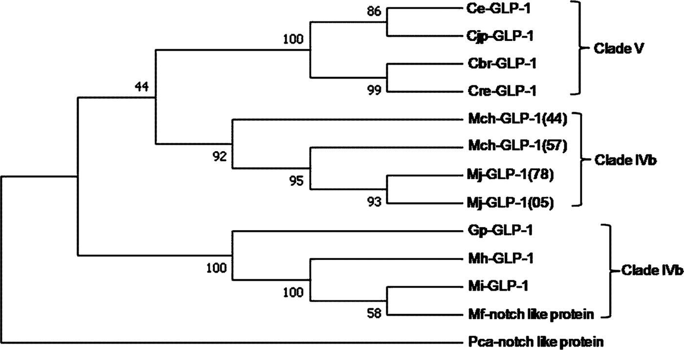
All identified GLP-1 protein sequences belonging to the different nematode species were aligned using MUSCLE (Fig. 2). This alignment revealed the presence of several conserved motifs in these parasitic and free-living nematodes, and the LNR and ANK motifs showed maximal amino acid conservation. A phylogenetic analysis using the amino acid sequences was performed to understand the patterns of relatedness among the glp-1 genes of the nematodes analysed in this study. The phylogenetic analysis of GLP-1 in Meloidogyne and other species, which was conducted using the NJ method, revealed a separate cluster comprised of Mi-GLP-1, Gp-GLP-1, Mh-GLP-1 and Mf-Notch-like protein. As observed in the phylogenetic tree, Mi-GLP-1 appeared to be most closely related to the Mf-Notch-like protein, which lacks an ANK (as demonstrated in the Pfam analysis). However, a higher degree of similarity in the LNR, RAM domain and ANK was found between Mi-GLP-1 and Gp-GLP-1, conclusively demonstrating the close relationship between these two genes (Fig. 3).
Phylogenetic analysis of Notch-like receptors. Neighbour-joining phylogenetic tree of Notch-like receptors from Caenorhabditis elegans (Ce), Caenorhabditis japonica (Cjp), Caenorhabditis briggsae (Cbr), Caenorhabditis remanei (Cre), M eloidogyne chitwoodi (Mch), M eloidogyne javanica (Mj), Globodera pallida (Gp), M eloidogyne incognita (Mi), Meloidogyne floridensis (Mf), M eloidogyne hapla (Mhp) and Priapulus caudatus (Pca, as an outgroup). The percentage of replicate trees in which the associated taxa clustered together in the bootstrap test (2000 replicates) is shown next to the branches. The phylogenetic analysis was conducted using MEGA7.

Transcript abundance of Notch-receptor genes in the developmental stages of M. incognita
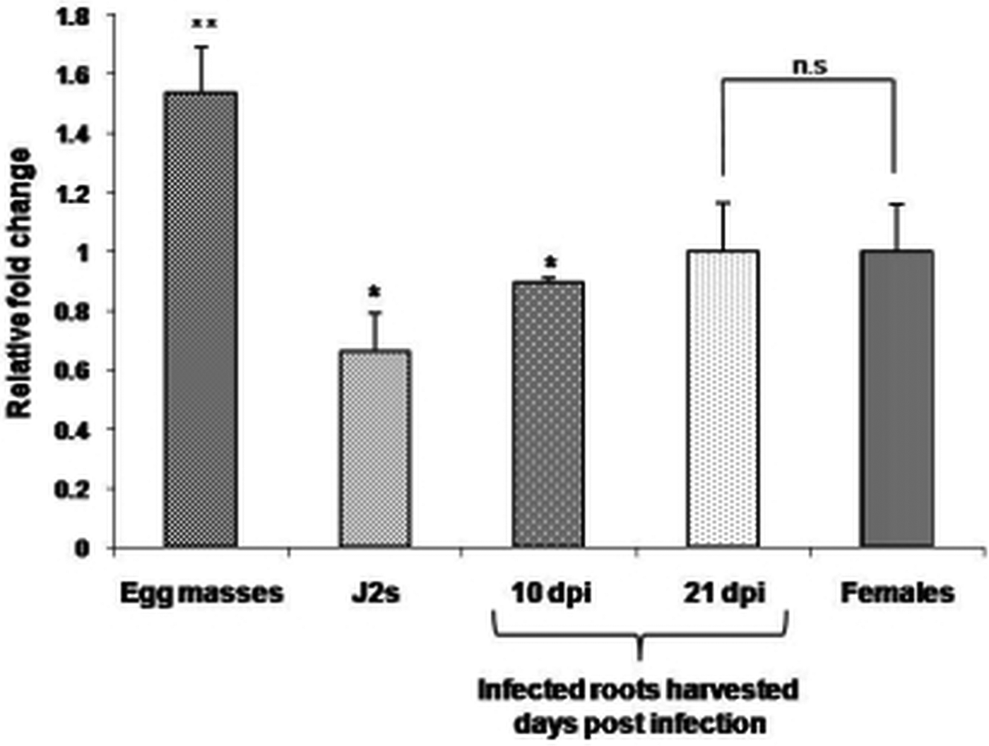
To study the expression pattern of glp-1 in M. incognita development, five different stages were selected: egg masses, J2s, mature females, J3s and J4s (the last two were obtained from the samples of infected plant roots harvested 10 and 21 dpi). Interestingly, glp-1 showed higher expression during early development in M. incognita, i.e. in egg masses (Fig. 4). This finding is consistent with those of previous studies that investigated glp-1 abundance at early developmental stages in C. elegans embryos (Austin and Kimble, Reference Austin and Kimble1989; Crittenden et al. Reference Crittenden1997). A qRT-PCR analysis also revealed that the glp-1 transcript levels were increased in mature females, indicating a possible role for this protein at this developmental stage in M. incognita.
Expression analysis of Mi-glp-1 at different developmental stages in Meloidogyne incognita. The histogram indicates the relative fold change (2–ΔΔCT value) normalized based on the actin gene as an endogenous gene and the female ΔCt value as a calibrator. The asterisks * and ** indicate significant differences at P < 0.05 and P < 0.01, respectively, and ‘n.s.’ indicates no significance.

Determination of the efficacy of glp-1 gene silencing
Nematode infection assay on Arabidopsis RNAi lines
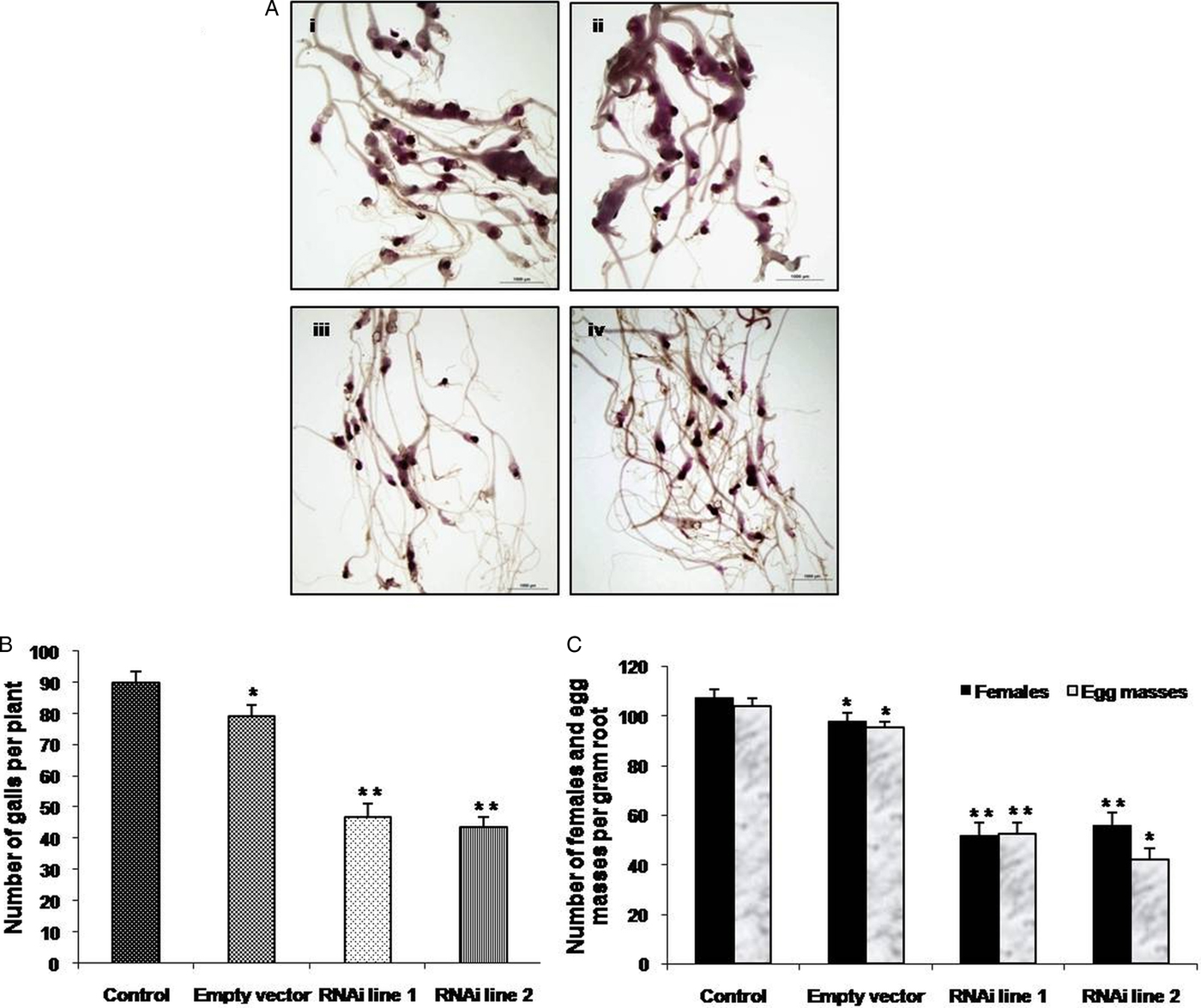
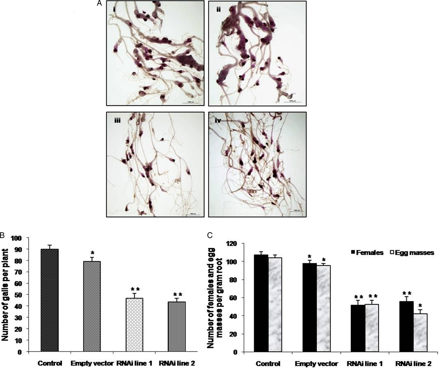
A dsRNA expression construct of the glp-1 gene was designed in the pBC vector (Yadav et al. Reference Yadav, Veluthambi and Subramaniam2006). The constructs were mobilized into Arabidopsis plants for the host-mediated delivery of RNAi of the glp-1 gene. To evaluate the silencing efficacy of Mi-glp-1, T3 seedlings of two independent transgenic lines were infected by parasitic juveniles. Two glp-1 RNAi lines (RNAi lines 1 and 2) were evaluated by staining the nematodes inside their roots and determining the infection level (Fig. 5A). Both glp-1 RNAi lines exhibited reductions in the number of galls (47.8% and 51.3%), females (51.5% and 47.79%) and egg masses (49.3% and 59.4%) compared with the control plants and transgenic plants harbouring an empty vector (Fig. 5A–C). Altogether, both transgenic RNAi lines presented significant reductions in the number of galls, females and egg masses, indicating that the silencing of this gene has a deleterious effect on the growth and development of nematodes. The adult female nematodes were isolated, and their sizes were analysed to determine any phenotypic effects, if any, on M. incognita. The length and width of randomly selected females were measured using a scale on a Nikon microscope (Supplementary Fig. S3). The average length and width of the females dissected from control plants were 396.27 and 248.18 µm, respectively, whereas the average length and width of the females isolated from the RNAi lines were significantly decreased, with values of 231.9 and 140.36 µm, respectively (Supplementary Table S3). Therefore, the nematodes obtained from the RNAi lines showed reductions of 41.4% and 43.4% in their length and width, respectively.
Nematode infection assay. (A) Representative photograph depicting M eloidogyne incognita in infected roots stained with acid fuchsin: (i) wild-type plants, (ii) empty vector line, (iii) RNAi line 1 and (iv) RNAi line 2. (B) and (C) Histogram depicting the number of galls, females and egg masses in the RNAi lines, empty vector transgenic lines and control (wild-type) plants. The asterisks * and ** indicate a significant difference at P < 0.05 and P < 0.01, respectively.
Molecular analyses of RNAi lines through qRT-PCR and Northern blotting
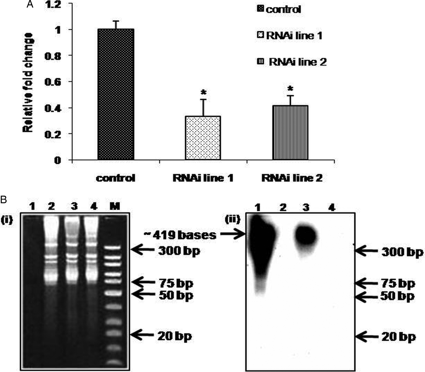
The transcript abundance of the target gene in the females isolated from both RNAi lines showed an approximately 60% reduction compared with that in the females isolated from control plants (Fig. 6A). This decrease in the transcript levels of glp-1 in females obtained from the RNAi lines demonstrated the effect of its silencing through host-delivered dsRNA. A Northern analysis was also performed, and the results confirmed the presence of glp-1 dsRNA in RNAi line 1 (Fig. 6B). One of the key components of host-mediated RNAi was the 419 bp dsRNA of glp-1, which was detected in the transgenic RNAi line.
Molecular analyses of M eloidogyne incognita isolated from transgenic Arabidopsis plants. (A) RT-qPCR-based glp-1 expression analysis of M. incognita females isolated from infected roots of RNAi lines 1 and 2 and females isolated from control plants. (B) Ethidium bromide-stained PAGE gel (before transfer) showing the low-molecular-weight RNA fraction (i) and the corresponding Northern blot for detecting the presence of glp-1 dsRNA (ii). The 183 bp probe corresponding to glp-1 was designed from the LNR domain. In (i) and (ii), lane 1 – gene-specific PCR product as a positive control, lane 2 – empty vector line, lane 3 – RNAi line 1, lane 4 – wild type as a control and lane M – molecular marker.

Effect of silencing on M. incognita fecundity and next-generation J2s
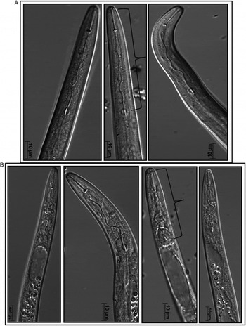
The females that fed on RNAi lines produced fewer numbers of egg masses compared with those that fed on wild-type plants. To further investigate the deleterious effects, if any, of the silencing of the glp-1 gene, the number of eggs present in these egg masses was evaluated, and these eggs were maintained for subsequent hatching. A 26% reduction in the number of eggs was noted in the RNAi lines, and the corresponding MF value was estimated to determine the nematode population. The estimated MF value for the infection of M. incognita in transgenic RNAi lines was calculated to equal 11.57, whereas that for wild-type plants was 31.1 (Supplementary Table S4A). Although J2s with well-developed pharynxes and stylet structures were observed in the samples isolated from the RNAi lines, several J2s showed aberrations around the metacorpus region as well as a shorter distance from the stylet to the metacorpus region of the pharyngeal structures (Fig. 7 and Supplementary Table S4B). Thus, it can be deduced that this effect on J2s is due to the nature in which RNAi is inherited from one generation to the next. Studies in C. elegans have shown inherited effects of silencing even in the absence of the original trigger (Bird et al. Reference Bird2009), and the persistence of silencing effects has also been observed after suppression of the Mi-1 gene of tomato (Gleason et al. Reference Gleason, Liu and Williamson2008).
Phenotypes of stylets and pharynxes of J2s descended from Meloidogyne incognita females isolated from infected plants: (A) wild-type (control) plants and (B) RNAi lines. Curly brackets indicate the length from the mouth to the metacorpus bulb in J2s. Please note shortening of length in few J2s from RNAi lines as compared to control plants.

Discussion
Although several genes related to neuropeptides and secretory pathways involved in the parasitism of PPNs have been reported (Rehman et al. Reference Rehman, Gupta and Goyal2016), detailed information regarding the structural and functional roles of genes with functions in organ development in PPNs, including M. incognita, is lacking. Very few studies have identified genes that play a crucial role in organ development in a PPN. Mi-glp-1, a Notch family member, was identified in this study as an orthologue of Ce-glp-1. In our study, a genome-wide computational approach revealed MiV1ctg1087, a single contig, as Mi-glp-1 in M. incognita. Interestingly, Minc16055 was also annotated as a Notch receptor protein in an identification of microsatellite loci in M. incognita (Castagnone-Sereno et al. Reference Castagnone-Sereno2010). A phylogenetic tree showed 12 orthologues originating from three clusters, all of which were branched, as well as one separate unbranched cluster (P. caudatus) as an outgroup. Notably, the branching of these three clusters originated from a common node. Thus, it could be hypothesized that the Mi-GLP-1 product has putative functions closely related to those of the Ce-GLP-1 protein. Nevertheless, further studies on the function of each conserved domain in Mi-GLP-1 will support its predicted role.
Mi-glp-1 is composed of 7.449 kb, which is similar to the size of Ce-glp-1. However, the corresponding mRNA sizes showed a difference of approximately 1.07 kb, which resulted from differences in the overall gene organization and structure. A similar size difference at the mRNA level was also observed with G. pallida glp-1 (Supplementary Table S2). The observed difference in the exon/intron structure could be due to the scaffold form of the M. incognita genome, which resulted in the formation of a truncated protein. A genome-wide search to identify more members of glp-1 in the M. incognita genome was attempted using Ce-glp-1 as the query sequence, but only a single member was identified. To date, only one other member of the GLP protein family, named GLP-4, has been characterized in C. elegans and C. briggsae (Mango, Reference Mango2007; Rastogi et al. Reference Rastogi2015). At the protein level, Mi-GLP-1 has several conserved motifs that are also present in the corresponding protein in C. elegans. A Pfam analysis showed the presence of a TMD, LNR, an RAM domain and an ANK. The LNR mediates ligand-binding functions, and the ANK and RAM domain are needed for signalling responses (Kelley et al. Reference Kelley1987; Rebay et al. Reference Rebay1991; Heitzler and Simpson, Reference Heitzler and Simpson1993; Artavanis-Tsakonas et al. Reference Artavanis-Tsakonas, Rand and Lake1999). Based on previous experimental evidence, the ANK is thought to be critical for the function of Notch receptors (Roehl and Kimble, Reference Roehl and Kimble1993; Diederich et al. Reference Diederich1994). In addition, three different software programs predicted the presence of a TMD, further indicating that Mi-GLP-1 most likely functions as a receptor protein (Supplementary Fig. S5A and B), although a clear mechanism of interaction has not been determined. A noticeable difference between Ce-GLP-1 and Mi-GLP-1 is the absence of an EGF domain at the 5′ end of the Mi-GLP-1 amino acid sequence, suggesting certain structural and functional differences between the glp-1 gene of C. elegans and that of M. incognita. Hence, the putative GLP-1 protein identified in this study has properties and structures similar to those of a receptor protein with varied domains, each with a specific function. In addition to these conserved domains, DNA-binding sites for ligands such as LAG-1 proteins were also found to be present in Mi-GLP-1. The presence of this type of motif at more than one site on the Mi-GLP-1 Notch receptor suggests that a vital function of GLP-1 is mediated through LAG-1 ligand binding to induce downstream reactions in the signalling pathway. Based on previous studies on Ce-LAG-1 DNA-binding sites, the potential function of this protein in LIN-12/GLP-1 signalling pathways has been proposed (Christensen et al. Reference Christensen1996). Experiments including yeast, two-hybrid assays and in vitro protein co-precipitation have demonstrated direct interactions between LAG-1 and the GLP-1 receptor via these domains in nematodes (Roehl et al. Reference Roehl1996). Further studies are needed to determine the role of these domains in signalling processes. Thus, based on in silico analyses and the available literature, it can be concluded that the gene identified in this study is a Notch receptor protein present in M. incognita.
Our study revealed that Mi-glp-1 is differentially expressed during different developmental stages of M. incognita. The increased transcript level observed in egg masses suggests an important role during embryogenesis. This observation was consistent with previous studies that suggested a possible role for this protein during embryogenesis in C. elegans (Priess et al. Reference Priess, Schnabel and Schnabel1987; Austin and Kimble, Reference Austin and Kimble1989). However, a slight increase in the transcript level was detected in mature females, suggesting another possible role for this protein during later stages of RKN development. Interestingly, similar reports regarding a possible role for glp-1 in the adult stage of C. elegans have not been reported. Future studies examining the localization of this gene product will confirm the role of GLP-1 during female development in M. incognita.
Host-delivered RNAi-mediated root-knot nematode resistance is an established technology, but the identification of better candidate genes remains necessary. In addition, the identification of genes involved in different developmental pathways will enable the targeting of several vital genes. Parasitic and secretory genes have traditionally been the genes of choice in silencing studies. However, there are very few candidate genes from the perspective of nematode development. A few developmental genes, such as Rpn7 (essential for the integrity of the 26S proteasome), nhr-48, FAR-1, splicing factor and integrase genes, have been targeted using RNAi in efforts aiming to control infection with the root-knot nematode M. incognita (Yadav et al. Reference Yadav, Veluthambi and Subramaniam2006; Niu et al. Reference Niu2012; Iberkleid et al. Reference Iberkleid, Sela and Brown Miyara2015; Lu et al. Reference Lu2016). The glp-1 gene, which was identified in this study, appears to be involved in the development, and a dsRNA/siRNA strategy was used for the host-mediated RNAi-based suppression of Mi-glp-1 in M. incognita. Transgenic Arabidopsis lines expressing dsRNA against Mi-glp-1 and infected with M. incognita did not show any phenotypic changes compared with wild-type plants. An approximately 47% reduction in the infection level was recorded in two independent glp-1 RNAi lines, and decreases in the gall formation, female infestation and egg mass production were also observed in these RNAi lines. In addition, an overall decrease in the number of females feeding on RNAi lines was recorded compared with females isolated from control plants. These results unequivocally demonstrated that glp-1 was suppressed in females that fed on the established RNAi lines. The nematode females isolated from control plants had an average length and width similar to the reported body length (ranging from 510 to 690 µm) and width (300 to 430 µm) of a healthy mature M. incognita female (Whitehead, Reference Whitehead1968; García and Sánchez-Puerta, Reference García and Sánchez-Puerta2012). In contrast, the females isolated from the RNAi lines had average lengths and widths of 232 and 141 µm, respectively, indicating an evident decrease in the overall body size in the females that fed on the RNAi lines. However, whether this decrease was a direct result of the low expression of glp-1 is unclear. Direct evidence supporting a role for glp-1 in the growth of M. incognita at later stages in the lifecycle has not yet been reported. However, in this study, a 60% decrease in transcript abundance was noted in the adult nematode females that fed on the RNAi lines. Additionally, to confirm the reduction in expression due to dsRNA/siRNA effects, the dsRNA molecules in the control and transgenic plants (empty vector and RNAi lines) were detected by Northern hybridization. The results confirmed the presence of glp-1-specific dsRNA molecules in the transgenic lines, an important factor for successful host-mediated RNAi (Bass, Reference Bass2000; Agrawal et al. Reference Agrawal2003), but we were unable to identify glp-1-specific siRNAs despite numerous attempts. Several studies have reported a similar problem with other genes, and this difficulty is most likely due to the low sensitivity of the Northern blot analysis (Dinh et al. Reference Dinh, Brown and Elling2014). The ability of M. incognita to ingest large molecules, which has been well established, and the presence of four dicers in Arabidopsis, which likely processed the glp-1-specific dsRNAs (as shown in our Northern analysis) into siRNAs to trigger RNAi responses in M. incognita females feeding on the transgenic RNAi lines might also have played a role. However, future studies comparing the transcript and protein levels in progenies are required to confirm our observations.
To investigate the suppression effect of glp-1 on the fecundity of M. incognita females, egg masses were hand-picked from all females in the infected RNAi lines, control plants and empty vector lines and subsequently incubated to determine their hatching capability, and the number of J2s hatched from each sample was counted. A decrease in the population of J2s obtained from the RNAi lines was noted, and the lower estimated MF value for M. incognita in the transgenic RNAi plants compared with control plants suggested a reduced infectivity of M. incognita on the RNAi lines compared with the wild-type plants. In embryos, glp-1 RNA is maternally donated (i.e. by the mother) in C. elegans (Evans et al. Reference Evans1994); therefore, dsRNA appears to affect even the progeny produced from the affected females. In our J2s, aberrations in the structures of the metacorporeal bulbs and their stylet discs were clearly apparent. A noticeable and significant decrease in the length of the region from the stylet to the metacorpus was also evident. Our study showed an adverse effect of the suppression of the glp-1 gene on the pharyngeal and stylet structures and thereby provides insight into the function of the glp-1 gene in pharyngeal development in M. incognita. Similarly, Geng et al. (Reference Geng2016) reported a degradation of intestinal tissues of M. incognita and identified Sep1, a novel serine protease, as a potent bio-agent from Bacillus firmus for controlling PPN populations. Thus, it can be inferred that the females obtained from transgenic plants exerted effects on the foregut region of their descendants. Our results strongly suggest the transmission of dsRNA-mediated deleterious effects to the next generation of root-knot nematodes. Because a difference in the phenotype of J2s was observed, a decrease in the GLP-1 protein level is possible; however, a more detailed study is required to fully evaluate this hypothesis.
The present work identified and characterized a gene encoding a Notch receptor protein in M. incognita involved in the development of the pharynx during embryogenesis and investigated the evolutionary conservation of its function between free-living nematodes and PPNs. Several common features in the Mi-GLP-1, Ce-GLP-1 and Gp-GLP-1 amino acid sequences were revealed. The in planta production of RNAi has been shown to effectively reduce the infectivity and size of M. incognita females. Deleterious effects were also observed in later generations (J2s) obtained from females isolated from infected transgenic plants. Taken together, our results demonstrate that the host-mediated gene silencing of the nematode glp-1 is an efficient method for inhibiting plant infection. The presence of glp-1 homologues in other PPNs, such as M. hapla, M. floridensis, M. javanica, M. chitwoodi and G. pallida, suggests the wide potential applicability of the silencing of this gene using host-mediated gene silencing approaches for the control of these parasitic nematodes. However, several important questions remain unanswered: (1) Does the glp-1 gene directly determine the pharyngeal development in PPNs or does it interact with other genes to define the development of the anterior region of the pharynx, as shown in free-living nematodes? (2) Does this gene have a specific role in other stages, such as the adult female stage, as indicated from our expression analysis? Providing answers to these questions requires experimental assessment of the phenotypic and molecular function(s) of the glp-1 gene. Although further investigations are essential to fully determine the role of glp-1 in parasitic nematodes, the observations obtained in this study provide a reasonable indication that glp-1 plays a significant role in pharyngeal development in PPNs. The role of GLP-1 as a receptor protein has been well characterized in C. elegans, but its involvement in pathogenic nematodes has not yet been reported. Establishing the involvement of glp-1 in pharyngeal development could further aid the elucidation of various pathways in PPNs, and the identification of genes downstream of the GLP-1 receptor and of its ligands will provide further insights into the various processes and pathways in which glp-1 is involved. These ligands and associated genes can then be targeted to obtain a more in-depth understanding of the cross-talk between different pathways and could eventually be used as targets in RNAi-mediated silencing approaches for PPN management to improve overall crop productivity.
Supplementary material
The supplementary material for this article can be found at https://doi.org/10.1017/S0031182018000641
Acknowledgements
The authors thank the staff of the National Phytotron Facility, IARI, New Delhi, India, for providing space in their greenhouse and growth chambers. The authors would also like to thank Dr Gautam Chawla, Division of Nematology, IARI, India, for providing the Zeiss Axio microscope used in this study and Mr Gaurav for assisting with the use of the Zeiss microscope.
Financial support
The present study was supported by a grant from the ICAR-NASF Project (formerly known as NFBSFARA; code: NFBSFARA/RNA-3022) during the years 2012–2015.
Conflicts of interest
None.
Ethical standards
Not applicable