Introduction
Canola, Brassica napus Linnaeus and Brassica rapa Linnaeus (Brassicaceae), is a major oilseed commodity grown throughout the prairie provinces of Canada. Although the crop is grown primarily in western Canada, the total economic contribution of canola to the Canadian economy is estimated at over $26.7 billion (CAD), and recently, for the first time in Canadian history, canola overtook wheat (Triticum aestivum Linnaeus (Poaceae)) in terms of total harvested area (9.27 versus 8.98 million ha) (LMC International 2016; Statistics Canada 2017). Due to the importance of canola in Canada, insect pests have been well documented (Lamb Reference Lamb1984; Dosdall et al. Reference Dosdall, Herbut and Cowle1994, Reference Dosdall, Soroka and Olfert2011; Mason et al. Reference Mason, Arthur, Olfter and Erlandson1998; Cárcamo et al. Reference Cárcamo, Dosdall, Dolinski, Olfert and Byers2001; Olfert et al. Reference Olfert, Hallett, Weiss, Soroka and Goodfellow2006; Dosdall and Mason Reference Dosdall, Mason and Williams2010).
Recently, a previously undescribed gall midge was discovered inducing flower galls on canola in the provinces of Saskatchewan and Alberta, Canada. The galls were first discovered in 2012 and attributed to infestation by the swede midge, Contarinia nasturtii (Kieffer) (Diptera: Cecidomyiidae), a species that was identified from both Saskatchewan and Manitoba in 2007 (Canadian Food Inspection Agency 2009); however, during our subsequent investigations, it was determined that the inhabitant of the flower galls was not C. nasturtii, but in fact, an undescribed species in the genus Contarinia Rondani. Prior to this discovery, C. nasturtii was the only cecidomyiid known to infest canola in North America, whereas C. nasturtii and the brassica pod midge, Dasineura napi (Loew) (Diptera: Cecidomyiidae), were known to infest canola in Europe (Barnes Reference Barnes1946; Darvas et al. Reference Darvas, Skuhravá, Andersen, L. and B.2000; Hallett and Heal Reference Hallett and Heal2001).
The canola flower midge, described here as Contarinia brassicola Sinclair, new species, belongs to a large cosmopolitan genus with 313 species, most of which live gregariously in flower heads or leaf rolls (Gagné and Jaschhof Reference Gagné and Jaschhof2014). Contarinia belongs to the supertribe Cecidomyiidi and can be separated from other genera of gall midges by the following characters: ovipositor long, retractile, slender, and tapered to a pair of long, narrow cerci; male terminalia with short and simple gonocoxites and evenly tapered gonostyli and aedeagus, cercus and hypoproct subequal in length; tarsal claws without teeth and bent beyond mid-length (Gagné Reference Gagné2018).
Here, we describe the new species from canola and demonstrate how it differs from C. nasturtii. Additionally, we describe the natural history of the insect, known host plants, geographic distribution, and parasitoids. Finally, we conduct a phylogenetic analysis, based on a segment of the mitochondrial cytochrome oxidase subunit I (COI) gene, comparing C. brassicola to other species in the genus.
Materials and methods
Adult gall midges were collected in soil emergence cages or reared from canola flower galls. In 2014–2016, four emergence cages (60 cm × 60 cm, Bugdorm, Megaview Science Company, Taichung, Taiwan) were deployed in early May on four stubble fields that were planted with canola the previous growing season in northeastern Saskatchewan (Table 1). Insects that emerged from the soil were captured in a bottle at the top of the cage filled with a preservative (−50 °C Plumbing Antifreeze, Canadian Tire, Saskatoon, Saskatchewan, Canada). Preservative in the bottles was emptied weekly until cages were removed in August. In the laboratory, insects were sorted and those appearing similar to C. nasturtii were collected and identified. In addition, a Jackson trap baited with a C. nasturtii pheromone lure (Distributions Solida, Saint-Ferréol-les-Neiges, Québec, Canada) was deployed in two of the emergence cages at each site to determine if emerging midges were attracted to the C. nasturtii pheromone. Furthermore, flower galls were collected from two sites near Ridgedale (53.05 ºN, 104.20 ºW) and Nipawin (53.35 ºN, 103.96 ºW), Saskatchewan on 8 July 2016 and 14 July 2017, respectively. Flower galls were brought to the laboratory and placed in petri dishes filled with sterilised, damp, soil-less potting mix (Stringham Reference Stringham1971). Petri dishes with flower galls were maintained in a growth chamber (22 °C, 16:8 hour dark:light, approximately 70% relative humidity) and misted with water daily. In 2016, petri dishes with potting mix were checked daily for adult emergence. In 2017, after 10 days, the potting mix in five petri dishes was sieved to obtain larval cocoons and pupae, the remaining 15 petri dishes were maintained in the growth chamber and checked daily for adult emergence. Adults, larvae, pupae, and pupal exuviae were preserved in 95% ethanol for later description. The new species was compared to specimens of C. nasturtii from Ontario, Canada and the United Kingdom.
Locations and dates of deployment/dismantling of emergence cages located on canola stubble fields (planted in canola the previous year, Spring) and current year canola fields (Summer) to collect adult Contarinia brassicola for species description and to determine emergence phenology.

* Melfort data (2014–2015) from Andreassen et al. Reference Andreassen, Soroka, Grenkow, Olfert and Hallett2018.
Natural history, hosts, and distribution
Samples collected in the emergence cages described above were used to determine the phenology of overwintering C. brassicola in northeastern Saskatchewan (Table 1), as well as to collect adult gall midges for the species description. Furthermore, four emergence cages were placed over canola plants in late July, at four additional fields in the same vicinity as the stubble fields described above, to determine if subsequent generations emerged. Two of the four emergence cages also had a single Jackson trap baited with a C. nasturtii pheromone lure setup in the centre of the cage. These cages and traps were monitored until mid-September or until removal was required for swathing (Table 1).
To determine the distribution of C. brassicola in Saskatchewan and Alberta, a survey for damage symptoms and larvae was conducted in late July of 2014–2016. At each site, 5–10 canola plants were systematically sampled at 10 locations along the field edge and interior (50–100 plants per site), and the number of galled flowers with and without larvae was recorded.
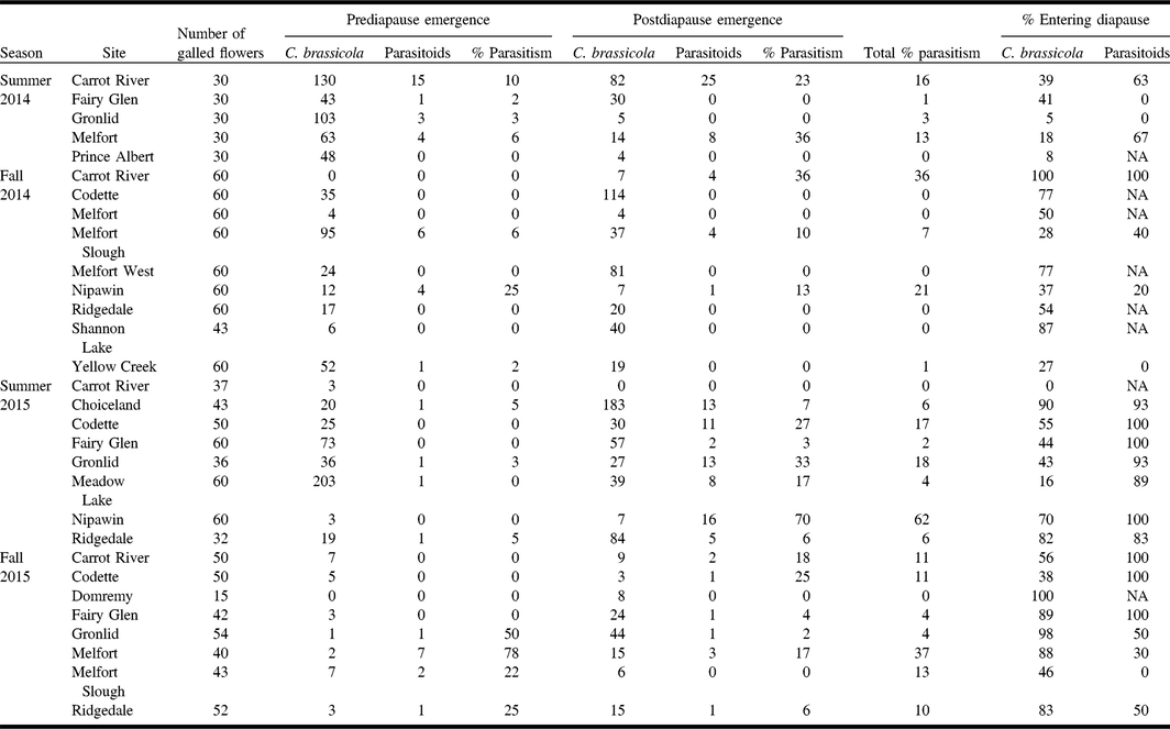
On 19 July 2014, at Carrot River, Saskatchewan (Supplementary Table S1), nine parasitoids were noted antennating and interacting on galled flowers of canola. These were collected and sent to the National Identification Service at the Canadian National Collection of Insects, Arachnids, and Nematodes (CNC) (Ottawa, Ontario, Canada) for species identification. After the initial identification of parasitoids on flower galls, the level of parasitism was determined by collecting flower galls (30–60) in July and September across multiple sites in northeastern Saskatchewan in 2014 and 2015 (Table 2, Supplementary Table S1). Galls from each site were placed into separate plastic bags and transported in refrigerated containers to the laboratory. In the laboratory, a 2-cm layer of moist, sterilised sand, and coconut fibre (1:1) was put in a 1-L plastic container, and the flower galls were placed on top. Half of a Jackson trap sticky liner (Solida, Saint-Ferréol-les-Neiges, Québec, Canada) was placed in each container to capture gall midges and parasitoids. The containers were monitored for emergence for eight weeks, after which no further gall midges or parasitoids emerged. Following the initial emergence period, the sticky liners were removed, substrate moistened with water, containers covered with mesh and placed in a 4 °C walk-in growth chamber to simulate overwintering conditions to determine if some gall midges and parasitoids had entered diapause. After 11–16 weeks in cold storage, the substrate was moistened, and the containers were moved to a growth chamber simulating summer conditions (22 °C, 16:8 hour dark:light, approximately 70% relative humidity), with emergence monitored daily. About 50 to 100 mL of water was added weekly to keep the soil moist. The numbers of gall midges and parasitoids collected pre-diapause and post-diapause, and the percent parasitism by site were determined. Representative parasitoids were sent to the Canadian National Collection of Insects, Arachnids, and Nematodes for morphological identification (and these vouchers were permanently deposited there); gall midge specimens were identified in-house at Agriculture and Agri-Food Canada – Saskatoon.
Diapause and emergence rates for Contarinia brassicola and parasitoids (Gastrancistrus and Inostemma) from infested canola flower galls across multiple sites in northeastern Saskatchewan.

Phylogenetic analyses
Genetic data were collected for 62 gall midge specimens collected from northeastern Saskatchewan (adults and larvae) and east-central Alberta (larvae) for phylogenetic analyses (Supplementary Table S2). In addition, six specimens from a laboratory colony of C. nasturtii established in 2016 (origin: Elora, Ontario, 43.64ºN, 80.40ºW) were included in the analysis. Total genomic DNA was extracted from whole individual specimens with a DNeasy Blood and Tissue Kit with an optional RNase A treatment following the instructions of the manufacturer (Qiagen, Mississauga, Ontario, Canada). A 439-base-pair region of the mitochondrial COI gene was amplified using the primers C1-J-1751 (GGATCACCTGATATAGCATTCCC) and C1-N-2191 (CCCGGTAAAATTAAAATATAAACTTC) (Simon et al. Reference Simon, Frati, Beckenbach, Crespi, Liu and Flook1994). All polymerase chain reaction amplifications were performed in a 25-µL volume containing: approximately 50-ng template DNA, polymerase chain reaction buffer (20-mM Tris–HCl pH 8.4, 50-mM KCl), 2.5-mM MgCl2, 0.5-mM dNTPs, 0.5 mM of each primer, and 1.5-U/µL Taq polymerase. Reaction conditions were 15 minutes at 95 °C, followed by 40 cycles of 95 °C for 40 seconds, 52 °C for 40 seconds, and 72 °C for 40 seconds, and a final elongation at 72 °C for seven minutes performed in a Bio-Rad C1000 thermal cycler (Bio-Rad Laboratories, Mississauga, Ontario, Canada). Two microlitres from each polymerase chain reaction was visualised on a 1.0% agarose gel to confirm amplification. The remaining polymerase chain reaction product was cleaned with a MinElute reaction cleanup kit (Qiagen) and sequenced in both directions using the BigDye v.3.1 sequencing kit (Applied Biosystems Division, Life Technologies, Waltham, Massachusetts, United States of America) on an Applied Biosystems 3720xl capillary sequencer at the Plant Biotechnology Institute, National Research Council of Canada Laboratory (Saskatoon, Saskatchewan). Forward and reverse sequences were reconciled using BioEdit 7.2.5 (Hall Reference Hall1999), and base calls were checked by eye against the chromatograms. The sequences generated were searched against the National Center for Biotechnology Information BLAST non-redundant database to determine the most likely identity of the specimens. Sequences for each unique haplotype are deposited in National Center for Biotechnology Information GenBank (www.ncbi.nlm.nih.gov/genbank) (accession MG922865–MG922876).
To provide phylogenetic context, we also downloaded all COI sequences of described Contarinia species from GenBank, as well as several sequences from Sitodiplosis mosellana (Géhin) (Diptera: Cecidomyiidae) to serve as an outgroup. We manually combined these GenBank sequences with those generated in this study, aligned them with MAFFT version 7.305b (Katoh and Standley Reference Katoh and Standley2013) and trimmed 5′ and 3′ ends of the alignment to remove variable missing data.
We used three search heuristics, Bayesian inference, maximum likelihood, and maximum parsimony, to construct phylogenies from unique COI haplotypes and used default parameters for analyses unless otherwise noted. We used MrBayes version 3.2.6 (Huelsenbeck and Ronquist Reference Huelsenbeck and Ronquist2001; Ronquist and Huelsenbeck Reference Ronquist and Huelsenbeck2003) to conduct tree searches with Bayesian inference. No priors were used, and we implemented the model of evolution predicted by the IQ-TREE version 1.4.2 (Nguyen et al. Reference Nguyen, Schmidt, von Haeseler and Minh2015) model selection procedure (Kalyaanamoorthy et al. Reference Kalyaanamoorthy, Minh, Wong, von Haeseler and Jermiin2017). We ran the analysis for two million generations, sampling every 500 generations, and assessed convergence of the run by observing the average standard deviation of split frequencies and the potential scale reduction factors (which should approach zero and one, respectively), as well as the sampling of posterior distributions of all parameters in Traver version 1.6 (Rambaut et al. Reference Rambaut, Suchard, Xie and Drummond2014) (which should be > 200). We manually removed the first 25% of trees as burn-in and constructed a 50% majority rule consensus tree in PAUP* version 4.0a159 (Swofford Reference Swofford2017). For maximum likelihood tree searches, we used IQ-TREE version 1.4.2 with the model of evolution predicted by the IQ-TREE model selection procedure. We assessed node support with 1000 replicates of both ultra-fast bootstrapping (uf-BS) (Minh et al. Reference Minh, Nguyen and von Haeseler2013) and the Shimodaira/Hasegawa approximate likelihood-ratio test (SH-aLRT) (Guindon et al. Reference Guindon, Dufayard, Lefort, Anismova, Hordijk and Gascuel2010). Finally, we used PAUP* version 4.0a159 to conduct unordered, unweighted, maximum parsimony tree searches (Fitch Reference Fitch1971), with 1000 replicates and holding one tree per replicate, as well as 1000 replicates of bootstrapping (each with 10 replicates using random sequence addition). We visualised trees using FigTree version 1.4.2 (Rambaut and Drummond Reference Rambaut and Drummond2010) and used Trimal version 1.2 (Capella-Gutierrez et al. Reference Capella-Gutierrez, Silla-Martinez and Gabaldon2009) to calculate percent sequence identity between all specimens.
Contarinia brassicola Sinclair, new species
http://zoobank.org/urn:lsid:zoobank.org:act:1F31BB8F-BC0D-4B8B-A60A-6E1556D1B78B
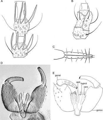
Contarinia brassicola. A, male fourth antennal flagellomere; B, female fourth antennal flagellomere; C, female cerci, dorsal view; D, E, male terminalia, dorsal view. Abbreviations: aed – aedeagus; cerc – cercus; goncx – gonocoxite; gonst – gonostylus; hyprct – hypoproct.

Contarinia brassicola. A, head of pupa, ventral view; B, larval spatula and associated papillae; C, eighth and terminal abdominal segment of mature larva, dorsal view.

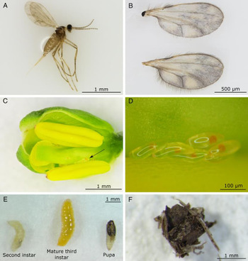
Contarinia brassicola life stages. A, adult female (preserved in 95% ethanol), the pattern of macrotrichia on the wings has been disturbed; B, close-up view of C. brassicola wings showing mottled appearance due to macrotrichia; C, clump of C. brassicola eggs (arrow) in developing canola bud, outer sepal was removed to locate the eggs; D, close-up view of C. brassicola eggs; E, immature stages of C. brassicola including second instar, mature third instar and pupa (removed from cocoon); F, larval cocoon.

Diagnosis. The species is distinguished from C. nasturtii by the female mottled wings created with the presence of dense macrotrichia (wings clear in C. nasturtii), and female first flagellomere is about two times longer than second flagellomere (1.5–1.8 times longer in C. nasturtii); basal male antennal internodes are shorter than length of basal node (nearly subequal to length of basal node in C. nasturtii) and hypoproct (male genitalia) deeply divided by nearly V-shaped notch (U-shaped in C. nasturtii); larval spatula with two rounded teeth (teeth rounded to pointed in C. nasturtii).
Description. Adult. Eye bridge 8–9 facets long. Postvertical peak present. Palpus with four segments: segment 1 short, length subequal to width; segment 2 longer than twice width; segment 3 nearly four times longer than wide; segment 4 more than five times longer than wide. Scape with one ventrolateral seta and several shorter ventromedial setae; 12 flagellomeres in both sexes. Male antennal flagellomeres binodal, with one series of circumfila on each node; basal node slightly shorter than apical node; internode length lesser than length of basal node (Fig. 1A). Female antennal flagellomeres cylindrical, with flagellomere 1 slightly longer than twice the length of flagellomere 2 (Fig. 3A); each flagellomere surrounded by two appressed circumfila connected by two longitudinal bands (Fig. 1B). Clypeus with nine setae.
Thorax. Wing 1.35–1.55-mm long in male (n = 6), 1.56–1.9 mm in female (n = 5). Scutum with setal rows multi-serial; anepisternum with several setulae; anepimeron with row of 5–6 setulae, otherwise pleura bare. Tarsal claw untoothed, empodia extending to bend in claw. Female wing mottled due to the presence of macrotrichia especially along veins (Fig. 3A–B).
Male abdomen. Tergites 1–7 with anterior margin bearing a pair of trichoid sensilla. Tergites 1–6 rectangular with posterior margin with row of setae, setae scattered medially; tergite 7 unsclerotised posteromedially, with several setae at posterior corner; tergite 8 divided into a pair of lateral pigmented ovals, without setae. Sternites 2–7 rectangular with transverse pigmented areas posteriorly and at mid-length; single posterior row of setae and mostly biserial row of setae medially; sternite 8 triangular shaped, with single posterior row of setae and scattered setae medially. Terminalia (Fig. 1D–E): cercus subrectangular, with truncate posterior margin, with three setulae along posterior margin, and one preapical setula on outer margin. Hypoproct deeply divided by V-shaped notch, narrow (compared with Harris Reference Harris1966, fig. 64), tapered apically, bearing single apical setula and one preapical setula. Gonocoxite cylindrical, longer than gonostylus. Gonostylus slender throughout, only slightly broader basally; setulose dorsolaterally on basal quarter; setulae scattered throughout length. Aedeagus glabrous, tapered to narrow apex, extending slightly beyond hypoproct and cercus; apex with narrow spine-like extension.
Female abdomen. Tergites 1–7 rectangular, wider than long, with single posterior row of setae, biserial on tergite 7; scattered setae medially; anterior margin bearing pair of trichoid sensilla. Tergite 8 quadrate without posterior row of setae and anterior margin bearing pair of trichoid sensilla; anterior margin with deep U-shaped notch. Sternites 2–6 rectangular with transverse pigmented areas posteriorly and at mid-length; single posterior row of setae and multiserial row of setae medially; sternite 7 quadrate with single posterior row of setae and scattered setae medially; sternite 8 not evident. Ovipositor elongate, retractable, sparsely covered with sensilla. Cerci tightly appressed, narrow, tapered, and pointed apically (Fig. 1C); sensilla stout, spine-like, evenly distributed, including apical short sensillum; two subapical sensilla stouter and longer.
Pupa. Body elongate-cylindrical. Antennal bases separate, each with short subterminal projection anteriorly (Fig. 2A). Cephalic sclerite with very short, obliquely directed seta on each side. Frons smooth, with three facial papillae near base of palpal sheath. Respiratory organ elongate, very slender, filament-like, slightly arched anteriorly (Fig. 2A). Fine spicules covering posterolateral face of tergite 1; tergites 2–8 with patch of thick, stout spicules anteriorly.
Third instar larva. Yellow (Fig. 3E), generally spindleform, posterior end rounded. Integument lacking projections. Antenna very short, little longer than wide, not extending beyond head capsule; slightly tapered apically. Spatula with two rounded apical teeth (compared with Mamaev and Krivosheina Reference Mamaev and Krivosheina1993, fig. 73.6), separated by wide shallow emargination, with slight posterolateral projection (Fig. 2B). Abdominal segment 8 with prominent pair of spiracles and three pairs of papillae. Terminal segment with eight papillae, six with short sensilla; remaining two papillae enlarged, on conical, blunt-tipped corniform outgrowths (Fig. 2C).
Egg. Clear, glossy, elongate-elliptical, posterior end rounded (Fig. 3C–D).
Material examined (slide mounted). Holotype. male, labelled: “SK [Saskatchewan]: Codette [53.240, −103.950] / 2016 / canola stubble / emer. tps / B. Mori”; “HOLOTYPE / Contarinia / brassicola / Sinclair [red label]”; “CNC / 1079158” (CNC). Paratypes: Saskatchewan: Codette [53.240, −103.950], 2016, canola stubble, emergence traps, B. Mori; CNC database numbers 1079159–1079162 (four males, CNC); Melfort [52.827, −104.599], 2016, canola stubble, emergence traps, B. Mori, CNC database numbers 1079164–1079170 (two males, five females, CNC); Ridgedale [53.462, −104.196], 2016, reared from infested canola flowers, B. Mori, CNC database number 1079163 (two larvae, CNC); SE of Nipawin, 53.352859, −103.979300, larvae coll. 17.vii.2017, B. Mori, CNC database numbers 1079171–1079172 (four pupal exuviae, CNC).
Additional material examined (in alcohol). Canada. Saskatchewan: Codette [53.240, −103.950], 2016, canola stubble, emergence traps, B. Mori (seven males, CNC); Codette [53.240, −103.950], 29.vi.2016, emergence cage, B. Mori (five males, 19 females, CNC); NW of Codette, 53.299668, −104.050366, larvae collected 6.vii.2017, pupae collected 21.vii.2017, B. Mori (nine pupae and cocoons, CNC); Melfort [52.827, −104.599], 2016, emergence cage, B. Mori (five females, CNC); Melfort [52.827, −104.599], 29.vi.2016, emergence cage, B. Mori (seven males, CNC); SE of Nipawin, 53.352859, −103.979300, larvae collected 17.vii.2017, B. Mori (three males, seven females, pupal exuviae, cocoons, CNC); Ridgedale [53.462, −104.196], 2016, reared from infested canola flowers, B. Mori (three females, CNC).
Etymology. The specific name is from the Latin cola (inhabitant) in reference to its host plant, Brassica.
Natural history
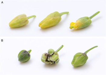
Adults (Fig. 3A) emerge from the soil in the late spring or early summer (Fig. 4A), coinciding with bud formation on Brassica as indicated by collections in spring emergence cages. Females most likely mate soon after emergence (similar to C. nasturtii (Readshaw Reference Readshaw1961) and C. negundifolia Felt (Wilson Reference Wilson1966)) and lay eggs in clusters in developing flower buds (Fig. 3C–D). Eggs hatch and larvae, initially white in colour changing to yellow as they develop (Fig. 3E), feed within the developing flower; the flower bud remains closed and becomes swollen, forming a gall (Figs. 5A, C, 6A). Larvae feed gregariously (1 to > 20), and black, necrotic damage (feeding scar) is often apparent on the style when later instars are present (Fig. 5B). Galled flowers produce no seed and are conspicuous from other normally developing buds and flowers (Fig. 5A, C), especially after they dry, later in the season (Fig. 5C). Occasionally, the style will continue to elongate and protrude from flowers closed around the base although other organisms can also cause this (e.g., aster yellows). The flower galls created by C. brassicola are conspicuously different from the caper-shaped flower galls created by C. nasturtii (Fig. 6A–B). Like C. nasturtii, late-instar C. brassicola are capable of “jumping” and drop from the plant to pupate in the soil. Larvae cover themselves with soil and debris to form cocoons (Fig. 3F) and either enter diapause and overwinter or complete their development and emerge.
Mean number of Contarinia brassicola adults captured per site in emergence cages located on A, canola-stubble fields planted to canola the previous year and placed in spring (2016 on alternate axis) and B, current year canola fields placed in summer across years (2015 on alternate axis). Locations correspond to those in Table 1.

Damage created by Contarinia brassicola to canola. A, flower galls (arrows); B, dissected flower gall (outer flower petals removed) showing the black, necrotic lesions on the style associated with C. brassicola larval feeding (arrows indicate larvae); C, dried, older flower galls located at various places along the elongating raceme, no longer containing larvae.

Flower galls created by Contarinia brassicola (A) and C. nasturtii (B) on canola, B. napus. The middle plant part in B is a C. nasturtii infested bud cluster; this type of damage has not been observed with C. brassicola.

Contarinia brassicola is at least bivoltine in Saskatchewan. An initial overwintering generation of adults emerges in late-June or early-July, and a second generation of adults occurs in August (Fig. 4A–B). A portion of larvae from the first generation enter diapause (Table 2), while the remainder complete their development and emerge as a second generation. The proportion of larvae entering diapause in the second generation appears to be triggered by external factors such as temperature or day length, but diapause does not appear to be obligatory as we were able to rear adults from larvae collected in September (Table 2). We have observed larvae in volunteer canola flowers in July, August, and September, indicating the possibility of a prolonged adult emergence, or three generations per year. The first generation of larvae is most likely to affect the crop (F1 of overwintering adults); however, if flowering is significantly delayed or if flowers are still present on secondary and tertiary racemes in August, the second generation could also impact the crop. Further studies are currently underway to determine the potential impact on yield.
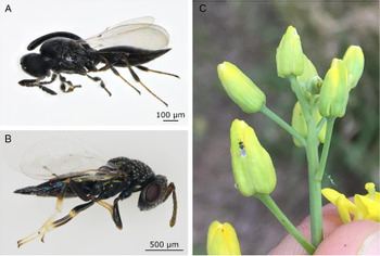
Parasitoid adults collected directly off the flower galls (July 2014) and those reared from flower galls (2014–2015) were identified as Gastrancistrus Westwood (Hymenoptera: Pteromalidae) and Inostemma Haliday (Hymenoptera: Platygasteridae) (Fig. 7). Both species can undergo diapause within their host, and total parasitism varied from 0 to 62% (mean ± standard error: 10 ± 3%) across sites, collection seasons and years (Table 2).
Parasitoids associated with Contarinia brassicola. A, Inostemma; B, Gastrancistrus; C, Gastrancistrus observed in the field on a C. brassicola infested canola (Brassica napus) flower gall.

No C. brassicola were captured on traps baited with C. nasturtii pheromone lures within the emergence cages at either the stubble or canola field sites.
Hosts and distribution
To date, C. brassicola has been found associated only with flowers of canola (B. napus and B. rapa). Preliminary investigations of other potential brassicaceous hosts (Lepidium densiflorum Schrader, Thlaspi arvense Linnaeus, Sinapis arvensis Linnaeus) in the field have yielded no observations of infestation. Further studies are underway to determine the host range of C. brassicola.
Contarinia brassicola is widely distributed across Saskatchewan and Alberta (Fig. 8), and damage and larvae have also been observed by the senior author in the Swan River Valley in Manitoba.
Geographic distribution of Contarinia brassicola as determined through yearly surveys (2014–2016) for damage symptoms and presence of larva. Map produced in SimpleMappr (Shorthouse Reference Shorthouse2010).

Phylogenetic analyses
Comparison of the sequences derived from the individuals in this study with the National Center for Biotechnology Information non-redundant/nucleotide collection database revealed no strong sequence similarities (> 95% sequence identity) to any pre-existing sequences. Two haplotypes (brassicola_hap6 and brassicola_hap8) shared the highest sequence similarity of all specimens generated here with an unidentified Cecidomyiidae specimen (query coverage: 93%, sequence similarity: 94%, e-value: 2e−172, accession: KR660433.1). Although sampling of the genus is limited, the lack of strong sequence similarity with any species in National Center for Biotechnology Information GenBank provides additional support that the specimens collected throughout Saskatchewan and Alberta in this study are a previously unidentified species, described here as C. brassicola.
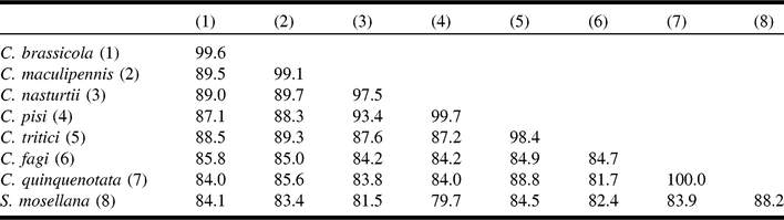
For phylogenetic analyses, we added 31 sequences from GenBank (2 C. fagi Rübsaamen, 11 C. maculipennis Felt, 13 C. nasturtii, 2 C. pisi (Loew), 1 C. quinquenotata (Loew), 2 C. tritici (Kirby), and 4 S. mosellana as an outgroup) to the 62 sequences of C. brassicola and 6 of C. nasturtii generated here, creating a final dataset of 103 specimens and 439 base pairs (33 unique haplotypes). All phylogenetic analyses used the general time-reversible model plus gamma and agreed on a single set of relationships, and all Contarinia species with > 1 specimen were reciprocally monophyletic (Fig. 9). However, support for many of the interspecific relationships within Contarinia was weak and varied between search heuristics; only Bayesian inference analysis strongly supported the placement of C. brassicola as sister to the clade containing C. maculipennis, C. nasturtii, and C. pisi (Fig. 9). Average percent sequence identity between C. brassicola specimens and those of other species were < 90% (Table 3). We did not observe any substantial geographic clustering within C. brassicola, and the most common haplotype (45_brassicola_hap1) was geographically widespread, being found at 5 localities in Saskatchewan (23 specimens) and 10 localities in Alberta (21 specimens) (Supplementary Table S3).
Maximum likelihood consensus tree of unique haplotypes. Three values of branch support are presented: SH-aLRT/uf-BS from maximum likelihood, posterior probability from BI (as a percent), and bootstrap from maximum parsimony. High support values are indicated with black text (SH-aLRT/uf-BS >80/>95, and posterior probability and maximum parsimony bootstrap > 80), and low support with grey text; NA indicates a node that was not resolved in a particular analysis. Unique haplotypes represented by a single sequence are named with their GenBank accession or in the case of sequences generated in this study by a haplotype code (see Supplementary Table S3). In cases where specimens shared a haplotype, haplotype names begin with the number of specimens that exhibited that haplotype, followed by a haplotype code; see Supplementary Table S3 for haplotype codes. Province where haplotypes were found is indicated for Contarinia brassicola (AB: Alberta, SK: Saskatchewan).

Average percent sequence identity within and between species. Parenthetical numbers following species names correspond to column headers.

Discussion
Contarinia brassicola is described from canola grown across the northern agricultural extent of the Canadian prairies in Saskatchewan and Alberta excluding the Peace River Region. At present, C. brassicola appears to be the main Contarinia species found on canola across the prairies, as C. nasturtii has not been found on pheromone traps since we started to conduct surveys in 2006 (J.J.S. and B.A.M., unpublished data), nor were C. brassicola captured on traps baited with C. nasturtii pheromone lures within emergence cages. This is not to say C. nasturtii is no longer present on the prairies, only that we have not observed damage or captured adults in recent years. Contarinia brassicola can be differentiated from C. nasturtii by the shape of the male hypoproct, the length of the female third antennal flagellomere, as well as COI sequence variation.
Contarinia nasturtii was originally described by Kieffer (Reference Kieffer1888) from swollen and closed flowers of Rorippa palustris (Linnaeus) Besser (Brassicaceae), but it is associated with many other damage symptoms, including crinkled and crumpled leaves, swelling, distortion, and twisting of young leaves and shoots, swollen petioles, blind heads, and growth of secondary shoots (“many-necked”) (Barnes Reference Barnes1946; Bardner et al. Reference Bardner, Edwards, Arnold and Rogerson1971). In laboratory testing, C. brassicola does not cause any of these other damage symptoms associated with C. nasturtii. The only observed damage symptom from C. brassicola infestation was galled flowers (Figs. 5, 6A). In addition, we have reared gall midges from thousands of flower galls collected from Saskatchewan, and no C. nasturtii have emerged. In the United Kingdom, the originally described flower damage associated with C. nasturtii has “often been forgotten” as the other damage symptoms are usually the most prevalent (Barnes Reference Barnes1946).
Phylogenetic and morphological analyses support C. brassicola as a distinct species as well as its placement within the genus Contarinia. The phylogenetic analyses found that all sampled Contarinia species were reciprocally monophyletic, including C. brassicola. Interspecific relationships were not strongly supported between search heuristics, but sample sizes of Contarinia were also limited; more comprehensive sampling within the genus will be required to assess relationships between species. Measures of interspecific divergence were high (Table 3) and much higher than some traditionally used thresholds (1%: Ratnasingham and Hebert Reference Ratnasingham and Hebert2007, 2%: Hebert et al. Reference Hebert, deWaard and Landry2010, 3%: Hebert et al. Reference Hebert, Cywinska, Ball and deWaard2003). Although the use of such arbitrary thresholds is debatable (e.g., Meier et al. Reference Meier, Zhang and Ali2008; Yassin et al. Reference Yassin, Markow, Narechania, O’Grady and DeSalle2010), the magnitude of interspecific divergence observed here strongly supports C. brassicola as a distinct species. We found no geographic genetic structure in C. brassicola, indicating potential gene flow across its range. However, use of additional nuclear markers (either nuclear gene sequences, microsatellites, or single nucleotide polymorphisms) will be required to confidently assess species limits and relationships within the genus and may provide better insight into patterns of gene flow.
Both genera of parasitoids found attacking C. brassicola are known to attack other cecidomyiids (e.g., Dorchin et al. Reference Dorchin, Clarkin, Scott, Luongo and Abrahamson2007; Abram et al. Reference Abram, Haye, Mason, Cappuccino and Kuhlman2012). Although the level of parasitism is low (mean 10%) across sites, these species may play a role in regulating C. brassicola populations. The current level of parasitism appears highest in northeastern Saskatchewan, the region where C. brassicola is prevalent and where field damage was first observed (Table 2; Fig. 8). Recently, a single parasitoid in the genus Synopeas Förster has been recorded from C. nasturtii in Québec, Canada (Hallett Reference Hallett and Reddy2017), but no Synopeas species have been found associated with C. brassicola. Gastrancistrus species have not been identified from C. nasturtii but are found associated with other cecidomyiids (Yukawa Reference Yukawa1983; Van Hezewijk and Roland Reference Van Hezewijk and Roland2003). Inostemma opacum Thomson has been found parasitising C. nasturtii in Europe (Abram et al. Reference Abram, Haye, Mason, Cappuccino and Kuhlman2012), but the species of Inostemma reared from C. brassicola is not currently known. Future work will investigate whether or not the parasitoids identified from C. brassicola galls may also parasitise C. nasturtii.
The origin of C. brassicola remains to be determined. However, the genetic, morphological, physiological, and ecological differences between C. nasturtii and C. brassicola suggest that they are not closely related or that speciation occurred long ago. We postulate that C. brassicola is native to North America and may have switched hosts entirely or expanded its host range as the area planted to canola increased over the last 40 years. Host switching or range expansion can occur over a relatively short time span when a species gains access to a novel resource (e.g., canola) (Agosta Reference Agosta2006; Singer and Parmesan Reference Singer and Parmesan2018). In addition, the widespread distribution of C. brassicola, and multiple COI haplotypes, but lack of population structure, indicates that C. brassicola is in a state of panmixia, with high population connectivity. If C. brassicola was a recent invader, genetic diversity at the invasion edge may be reduced due to population bottlenecks and founder events (Austerlitz et al. Reference Austerlitz, Jung-Muller, Godelle and Gouyon1997; Dlugosch and Parker Reference Dlugosch and Parker2008; Fitzpatrick et al. Reference Fitzpatrick, Fordyce, Niemiller and Reynolds2012), which was not observed with C. brassicola. The current evidence suggests C. brassicola is a native, rather than an invasive alien species. In the future, identification of the female-produced sex pheromone(s) followed by worldwide pheromone-baited trapping may provide more insight into the origin of this species.
Supplementary material
To view supplementary material for this article, please visit https://doi.org/10.4039/tce.2018.63.
Acknowledgements
We thank Larry Grenkow, Stephanie Harris, Taylor Kaye, Nancy Melnychuk, Jennifer Holowachuk, Dr. Owen Olfert, Dr. Meghan Vankosky, and a plethora of summer students for their help with field collections. We thank Scott Meers and Shelley Barkley (Alberta Agriculture and Forestry) for surveying and collecting Alberta samples. We would also like to thank Dr. Lubomir Masner and Dr. Gary Gibson for parasitoid identification. We thank Jonathon Williams, Tyler Wist, and Shane Hladun for their photography assistance. We are grateful to all our producer co-operators for allowing field access; in particular, we would like to thank Kent Baxter, Norman Enns, Ian Rushmer, and Jamie Freedman. This work was also aided from the help of Wade Annand, Tim Perkins, Lyle Cowell, and Mark Gordon. Funding for this project was provided by the Canola Council of Canada’s Canola Agronomic Research Program (CARP) funded by SaskCanola, the Alberta Canola Producers Commission, and the Manitoba Canola Growers Association. Further support was provided by the Alberta Crop Industry Development Fund (ACIDF). Michelle Locke (Canadian National Collection of Insects, Arachnids, and Nematodes) kindly assisted in creating the image of the male terminalia. Karen McLachlan Hamilton (Canadian Food Inspection Agency), Graham Thurston (Canadian Food Inspection Agency), Ray Gagné (Smithsonian Institute), and an anonymous reviewer kindly provided comments on earlier versions of the manuscript.