Introduction
In sexual reproduction, successful fertilization requires the union of maternal and paternal gametes, either of which seems to have a significant role to play in successful fertilization and embryonic development (McGrath & Solter, Reference McGrath and Solter1984). Maternal resources are directly available for the developing preimplantation stage embryos that support the development until embryonic genome activation takes place (Li et al., Reference Li, Zheng and Dean2010). RNA profiling of murine zygote has confirmed the presence of around 380 sperm cytosolic RNAs (Park et al., Reference Park, Komata, Inoue, Yamada, Nakai, Ohsugi and Shirahige2013; Fang et al., Reference Fang, Zeng, Wang, Liu, Xu, Dai, Zhao, Zhang, Liang, Chen and Shi2014) indicating the transfer of cytosolic factors into the zygote. Therefore, it is probable that rather than acting as a mere vector for paternal genome, spermatozoa provide certain factors that regulate embryonic development (Parrington et al., Reference Parrington, Swann, Shevchenko, Sesay and Lai1996; Tang et al., Reference Tang, Dong, Huang and Sun2000; Wu et al., Reference Wu, Smyth, Luzzi, Fukami, Takenawa, Black, Allbritton and Fissore2001; Swann et al., Reference Swann, Larman, Saunders and Lai2004). Earlier studies have demonstrated that oocyte activation factors (Perry et al., Reference Perry, Wakayama, Cooke and Yanagimachi2000), centrosomes (Schatten & Sun, Reference Schatten and Sun2009), mRNAs (Sendler et al., Reference Sendler, Johnson, Mao, Goodrich, Diamond, Hauser and Krawetz2013) and snRNAs (Krawetz et al., Reference Krawetz, Kruger, Lalancette, Tagett, Anton, Draghici and Diamond2011; Kiani & Rassoulzadegan, Reference Kiani and Rassoulzadegan2013) from the spermatozoa have a significant role in early embryogenesis.
Microinjection of soluble cytosolic factors from spermatozoa (sperm-derived factors; SDF) can induce calcium oscillations similar to that occuring during normal fertilization (Swann, Reference Swann1990; Kuretake et al., Reference Kuretake, Kimura, Hoshi and Yanagimachi1996; Parrington et al., Reference Parrington, Swann, Shevchenko, Sesay and Lai1996; Oko & Sutovsky, Reference Oko and Sutovsky2009). The candidate molecules identified as SDF responsible for oocyte activation are oscillin (Parrington et al., Reference Parrington, Swann, Shevchenko, Sesay and Lai1996), truncated c-kit receptor (Sette et al., Reference Sette, Bevilacqua, Bianchini, Mangia, Geremia and Rossi1997), phospholipase C zeta (PLCζ) (Saunders et al., Reference Saunders, Larman, Parrington, Cox, Royse, Blayney, Swann and Lai2002), glucosamine 6-phosphate isomerase and post-acrosomal WW-domain binding protein (PAWP) (Wu et al., Reference Wu, Sutovsky, Manandhar, Xu, Katayama, Day, Park, Yi, Xi, Prather and Oko2007; Aarabi et al., Reference Aarabi, Balakier, Bashar, Moskovtsev, Sutovsky, Librach and Oko2014). These studies, reported in the literature, mainly focused on studying the increase in the intracellular calcium level by SDF.
Embryos developed from artificially activated oocytes in the absence of paternal contribution are known as parthenotes. They play an important role in the field of developmental biology and regenerative medicine. They serve as an excellent model to understand the role played by maternal and paternal genome during early embryogenesis. However, these embryos are usually characterized by poor and delayed in vitro development (Liu et al., Reference Liu, Trimarchi and Keefe2002; Hao et al., Reference Hao, Lai, Liu, Im, Wax, Samuel, Murphy, Sutovsky and Prather2006; Isom et al., Reference Isom, Whitworth and Prather2012), a high degree of cellular fragmentation, 2-cell stage arrest, decreased blastocyst rate and poor blastocyst quality (Liu et al., Reference Liu, Trimarchi and Keefe2002; Jeong et al., Reference Jeong, Cui, Kim, Kim, Kim, Chung and Kim2005). Haploidy (Liu et al., Reference Liu, Trimarchi and Keefe2002), abnormal gene expression pattern and imprinting errors (Surani et al., Reference Surani, Barton and Norris1984; Solter, Reference Solter1988) are considered to be the cause for such abnormal development in these embryos.
During normal fertilization, along with sperm nucleus, several cytosolic factors enter the ooplasm which helps in triggering the subsequent biochemical events in the oocytes initiating fertilization (Perry et al., Reference Perry, Wakayama, Cooke and Yanagimachi2000; Krawetz et al., Reference Krawetz, Kruger, Lalancette, Tagett, Anton, Draghici and Diamond2011; Kiani & Rassoulzadegan, Reference Kiani and Rassoulzadegan2013). Therefore, arguably, the lack of paternal genome and/or the cytosolic factors might be one of the reasons for the poor development of artificially activated oocytes. However, there are no studies in the literature to ascertain their role in early embryo development. In this direction, the present study was undertaken to explore whether the presence of soluble sperm-derived factors (SDFs) in the culture medium during artificial activation of oocytes has any beneficial effects. Unlike in earlier studies, in which the sperm factors are microinjected to the oocytes to understand their role, we used a much simpler approach of supplementing SDF to embryo culture medium without using any microinjection methods.
Materials and Methods
Animal model
Inbred Swiss albino mice (8–10-week-old) maintained in the Central Animal Research Facility, Manipal University, were used for the study. The animals were maintained under standard conditions of temperature (25 ± 2°C), humidity (45–55%) and light (12:12 h of light and dark) and were fed with food and water ad libitum. Prior approval was obtained from the Institutional Animal Ethical Committee of Kasturba Medical College, Manipal University, to carry out the experiment (IAEC/KMC/64/2013). The authors assert that all procedures contributing to this work comply with the ethical standards of the relevant national and institutional guidelines on the care and use of laboratory animals.
Preparation of sperm-derived factors (SDF)
Caudae epididymis collected from adult Swiss albino male mice (8−10-week-old) was teased in 2 ml of M2 medium to release the spermatozoa. Motile spermatozoa were extracted by ‘swim up’ technique after centrifugation for 10 min at 1000 rpm. After determining the count and motility, spermatozoa were pelleted out by centrifugation and the count was adjusted to 8 million. To release the cytosolic factors, the spermatozoa were subjected to hypotonic treatment by resuspending the pellet in Milli-Q water for 1 h at 37°C as described by Lomeo & Giambersio (Reference Lomeo and Giambersio1991) followed by sonication at 90% power for 7 min at 37°C (Vibra-Cell, Sonics & Materials, Inc., CT, USA). To remove sperm tail and other debris, suspension was filtered using syringe filter of 0.22 µ pore size (Millipore, cat. no. SLGVO33RS). Pre-frozen sperm-derived factor was lyophilized (Daihan Labtech Co. Ltd, Korea) after the addition of sucrose (10%) and stored at −80°C until use. Lyophilized product was reconstituted with M16 medium (Stock 1 mg/ml) and diluted to obtain the required concentrations for experiments.
Superovulation and oocyte collection
Female Swiss albino mice (6–8-week-old) were superovulated by intraperitoneal (i.p.) injection of 5 IU pregnant mare serum gonadotropin (PMSG, cat. no. G4877, Sigma) followed by 10 IU human chorionic gonadotropin (hCG, Ovutrig, India) 48 h later. Mice were humanely sacrificed by cervical dislocation 13 h after hCG administration and oviducts were dissected out in M2 medium. Cumulus–oocyte complexes (COC) were released by carefully teasing the oviduct using a blunt needle and by observing under a stereomicroscope on a stage maintained at 37°C.
Parthenogenetic activation and embryo culture
The cumulus–oocyte complexes (COCs) retrieved from oviduct were washed in M16 medium and parthenogenetically activated with 10 mM strontium chloride (SrCl2) for 3 h. COCs were incubated in hyaluronidase (1 mg/ml) prepared in M2 medium for 1 min at 37°C for denuding. Activation was confirmed by the presence of one pronucleus and two polar bodies (1PN/2PB) by observing at ×200 magnification under an inverted microscope fitted to a micromanipulator (IX70; Olympus, Tokyo, Japan). Activated oocytes were washed three times and cultured in vitro in M16 medium at 37°C and 5% CO2 in air. Embryos were scored regularly at every 24 h under an inverted microscope (×200 magnification) until they reached the blastocyst stage.
Collection of normally fertilized embryos
Superovulated female mice were kept for overnight mating with proven fertile males. Mating was confirmed by the presence of a vaginal plug and pronuclear stage (PN) embryos were collected in M2 medium at 16 h post hCG from the oviduct of mated females. For experiments with 2-cell stage embryos, embryos were collected from the oviduct of the mated females at 24 h post hCG.
Experimental outline
To understand whether supplementation of SDF can improve the in vitro development of parthenogenetic embryos, oocyte activation was carried out in presence or absence of SDF. For this, COCs collected were parthenogenetically activated using 10 mM SrCl2 in M16 without Ca2+ and Mg2+ supplemented with varying concentrations of SDF (5, 10, 20, 50, 75, or 100 µg/ml). Activated oocytes were washed (three times in M16 medium) post denuding and cultured in M16 medium until they develop to blastocyst stage. Blastocyst rate was calculated as the percentage of activated oocytes (pronuclear stage embryos) reaching the blastocyst stage.
To further understand whether the beneficial effect of SDF on early embryo development is stage specific, parthenotes at 2-cell stage were cultured in medium supplemented with varying concentrations of SDF (1, 2.5, 5, 10, 20, or 50 µg/ml) and the developmental potential was monitored until the blastocyst stage.
Detection of spindle assembly
Activated oocytes were fixed at 0, 3, 6 or 12 h (3 h after incubation in SrCl2) for assessment of the spindle assembly using the method described by Nair et al. (Reference Nair, Singh, Salian, Kalthur, D'Souza, Shetty, Mutalik, Kalthur and Adiga2014). Briefly, activated oocytes were permeabilized by extraction buffer and fixed with ice cold methanol at −20°C for 12 min followed by blocking with 10% goat serum for 1 h. Primary antibody, monoclonal anti-α-tubulin antibody (cat. no. T9026; Sigma, USA) was diluted (1:100) in blocking solution and the embryos were incubated at 4°C overnight. These embryos were washed in PBS with 0.025% Triton X-100 (three times) followed by incubation in fluorescein isothiocyanate (FITC)-labelled anti-mouse antibody (cat. no. SC2010, Santa Cruz, 1:500 dilution) for 1 h at 37°C and washed three times before staining the nucleus with 4′,6-diamidino-2-phenylindole (DAPI). Embryos were mounted on a clean glass coverslip and images were acquired using a fluorescence microscope under ×400 magnification.
Cortical granules
Activated oocytes at 0 and 3 h were incubated in acid Tyrode medium for 1 min at 37°C and were later fixed using 4% PFA at 4°C for 24 h. These oocytes were blocked in Dulbecco's phosphate-buffered saline (DPBS) with 1% BSA, 0.01 M/l glycine and 0.05% Triton X-100 followed by staining with rhodamine labelled Lens culinaris agglutinin (LCA) (10 µg/ml) (cat. no. RL-1042, Vector Laboratories, Inc., CA, USA) for 30 min at room temperature in the dark. These oocytes were washed with PBS and images of cortical granules were grabbed using a micropublisher 5.0 RTV digital camera (Q Imaging, Surrey, Canada) fitted to a fluorescence microscope.
Sperm zona binding
Sperm zona binding assay was carried out as described by de Paola et al. (Reference de Paola, Bello and Michaut2015) with minor modifications. Briefly, caudae epididymis of 8–10-week-old Swiss albino mice were teased to release spermatozoa into 2 ml of M16 medium. Capacitation was carried out by incubating sperm suspension in 10% bovine serum albumin for 2 h at 37°C in 5% CO2. Count and motility were determined post-capacitation and the sperm count was adjusted to 1 × 10⁵ spermatozoa/ml, activated oocytes were incubated individually in 10 µl droplets of sperm suspension (1000 sperm/oocyte) for 1 h and washed gently in M16 medium to release the loosely bound spermatozoa. The oocytes were fixed using PFA and stained with propidium iodide (PI) and observed under a fluorescence microscope to count the number of bound sperm per oocyte.
Mitochondrial distribution pattern
The mitochondrial distribution pattern in oocytes activated in the presence or absence of SDF was assessed by rhodamine 123 staining as described previously (Nair et al., Reference Nair, Singh, Salian, Kalthur, D'Souza, Shetty, Mutalik, Kalthur and Adiga2014). Briefly, the activated oocytes were incubated for 20 min at 37°C in M16 medium supplemented with 20 μg/ml of rhodamine 123 (cat. no. R8004, Sigma, USA). Stained oocytes were washed three times in M16 medium and mounted using antifade solution (cat. no. S3023; Dako, Carpinteria, CA, USA). Mitochondrial distribution pattern was assessed under a fluorescence microscope (×400 magnification; Imager-A1; Zeiss, Gottingen, Germany). According to the distribution pattern, oocytes were categorized as uniform, peripheral or semiperipheral and expressed in percentage (Nair et al., Reference Nair, Singh, Salian, Kalthur, D'Souza, Shetty, Mutalik, Kalthur and Adiga2014).
Oct 4 expression
The expression of pluripotency regulator POU5F1 was assessed in embryos post 24 h of activation by immunofluorescence quantification as described by Kalthur et al. (Reference Kalthur, Salian, Nair, Mathew, Adiga, Kalthur, Zeegers and Hande2016) with minor modifications. Fixed embryos (4% PFA) were permeabilized (0.1% Triton X-100) and blocked [10% goat serum (Dako)] before incubating with primary antibody (cat. no. sc-5279; Santa Cruz Biotechnology, USA) overnight at 4°C. The embryos were washed three times (PBS with 0.025% Triton X-100) and incubated with FITC-conjugated goat anti-mouse IgG (cat. no. D0408; Santa Cruz, USA) for 1 h at 37°C. Images were grabbed using a micropublisher 5.0 RTV digital camera (Q Imaging, Surrey, Canada) and further analysed by Q-capture software. The fluorescence signal intensities were analysed from the grabbed images. The green channel was separated by Q-capture software (Media Cybernetics) to ensure that FITC signals and the background was subtracted before quantification of intensities.
Mass spectrophotometric analysis of SDF
Sperm-derived factors were subjected to MS/MS for mass spectrometric analysis of proteins present in the SDF. We used the lyophilized SDFs derived by the earlier method in this mass spectrometry analysis. The lyophilized SDF sample (about 2 mg each) was dissolved in 1 ml of water and methanol and passed through 0.22 µ membranes. The filtered solution was analysed by mass spectrometry (2000 QTRAP Nano spray™, MDS Sciex, a division of MDS Inc., Ontario, Canada) to identify possible protein components of SDF based on the m/z (Da) data obtained. Once the m/z charge ratio was obtained the raw data were used to obtain the deconvoluted mass with Dalton corrections.
Protein identification in sperm-derived factors using MS/MS-based peptide fingerprints:
Based on the corrected masses obtained, MS-fit was used to search the NCBI database for the peptide fingerprints (PF). The following settings were used, as shown in the Fig. 7. Masses above 531 to 1000 Daltons alone were selected for PFs. Masses below 531 Da were rejected as mass spectrometer background contaminants and solvents used in this study. As these proteins are not modified chemically we used the stable phosphate modifications available on proteins for the PFs. In total, 179 different proteins were identified through three sampling methods, with MOlecular Weight SEarch (MOWSE) scores between 50 to 3.49 × 10⁷.
Statistical analysis
The data pertaining to developmental potential, cortical granule exocytosis, mitochondrial distribution pattern and spindle dynamics were represented as percentage; and sperm zona bindability and quantification of immunofluorescence were represented as mean ± standard error (SE) of the raw data. The validity of the data was analysed using chi-squared test and one-way analysis of variance (ANOVA) using the GraphPad InStat 3.0 statistical package (GraphPad Inc, USA). Differences were considered to be statistically significant if the P-value was <0.05.
Results
Role of sperm-derived factors during oocyte activation
Activation potential
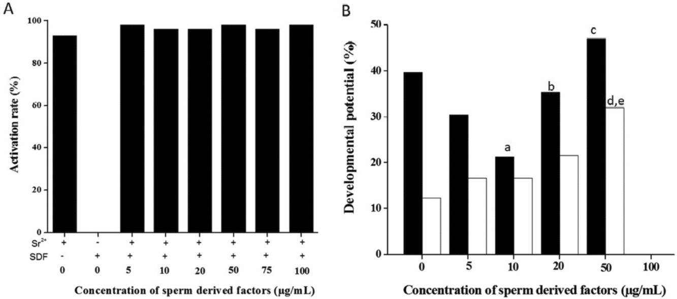
To understand the role played by sperm-derived factors during oocyte activation, varying concentrations of SDF were supplemented in M16 medium with chemical activating agent SrCl2. As shown in Fig. 1(A), the activation potential of oocytes in the control group (SrCl2) was 93%. Incubating the oocytes with SDF alone did not result in oocyte activation. However, supplementation of SDF to the activation medium was found to result in approximately 98–99% activation at all concentrations used (5 to 100 µg/ml). The oocytes activated with SrCl2 were haploid in nature and the presence of SDF during activation did not alter the ploidy status of activated oocytes.
Activation potential and developmental potential of oocytes activated in the presence of sperm-derived factors. (A) Activation potential of mouse oocytes activated with Sr2+ in the presence of varying concentrations of sperm-derived factors (n = 77, 25, 70, 62, 63, 76, 57 and 45 in Sr2+ alone, SDF alone, 5, 10, 20, 50, 75, 100 µg/ml of SDF respectively in three trials). χ2 test was used for statistical analysis of the percentage of oocytes activated after supplementation with sperm-derived factors. (B) Developmental potential of parthenogenetic embryos activated in the presence of sperm-derived factors. 0 µg/ml of SDF corresponds to strontium alone (control). a P < 0.01 vs. C, b P < 0.05, c P < 0.01 vs. 10 µg/ml in morula rate; d P < 0.01 vs. C, e P < 0.05 vs. 5,10 µg/ml in blastocyst rate (n = 77, 25, 70, 62, 63, 57 and 45 in Sr2+ alone, SDF alone, 5, 10, 20, 50, 100 µg/ml of SDF in three trials). χ2 test was used for statistical analysis of the developmental potential of oocytes after activation in presence of sperm-derived factors
 Morula rate;
 Blastocyst rate.

Developmental potential
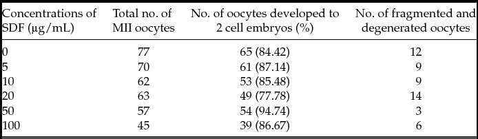
The embryos activated in the presence of varying concentrations of SDF were further cultured in M16 medium and scored at every 24 h intervals. The 2-cell rate in the control group was 84.42% (Table 1), which did not differ much in oocytes activated in the presence of 5, 10 or 20 µg/ml of SDF (87.14, 85.48 and 77.78% respectively). The highest 2-cell rate (94.74%) was observed in the 50 µg/ml group. At 96 h after culture, 39.6% of the control parthenotes underwent compaction (Fig. 1 B), which was marginally lower at the 5 µg/ml (30.5%) and significantly lower (P < 0.01) at the 10 µg/ml concentrations (21.2%). Further increase in SDF concentration to 20 and 50 µg/ml increased the compaction rate to 35.4 and 47.05% respectively. The remaining embryos either arrested at the 2-cell stage or at the 6–8-cell stage. Activation in the presence of 100 µg/ml of SDF resulted in embryonic arrest at the 6–8-cell stage. However, the blastocyst rate was found to be increased in a dose-dependent manner up to 50 µg/ml. The highest blastocyst rate (32%) was obtained in the 50 µg/ml group, which was significantly higher than that of the control (12.31%, P < 0.01).
2 cell rate of oocytes activated in presence and absence of SDF

Influence of SDF on formation of first cleavage axis
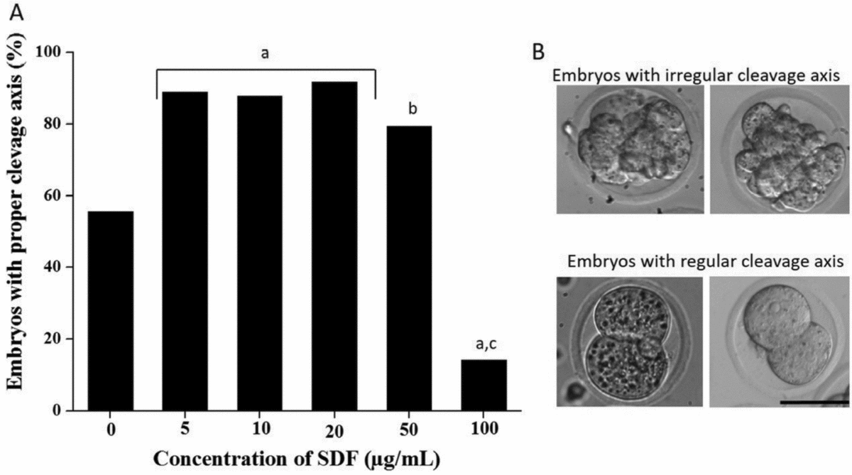
In oocytes activated with SrCl2 at 18 h after activation, only 55.7% of the embryos had regular cleavage axis while the remaining embryos did not have clearly demarcated embryonic boundaries. However, these embryos developed to a ‘proper’ 2-cell stage by 24 to 36 h of culture suggesting delayed cleavage (Fig. 2). Interestingly, we observed that the 2-cell-stage embryos obtained after activation in the presence of SDF up to 20 µg/ml had a significantly increased number of embryos with a normal cleavage axis within 18 h of activation (P < 0.0001). Further increase in SDF concentration to 50 µg/ml decreased the percentage of embryos with a normal cleavage axis to 79.4%, although this percentage was still significantly higher than that of the control group (P < 0.01). At the highest SDF concentration, embryos with a normal cleavage axis at 18 h showed the lowest percentage compared with all other groups (14.2%, P < 0.0001).
Embryos with proper cleavage axis after supplementation with sperm-derived factors. (A) Graph showing the percentage of embryos with proper embryonic axis. a P < 0.0001; b P < 0.01 vs. C, c P < 0.0001 vs. 5, 10, 20, 50 µg/ml (n = 77, 25, 70, 62, 63, 57 and 45 in Sr2+ alone, SDF alone, 5, 10, 20, 50, 100 µg/ml of SDF respectively in three trials). χ2 test was used for statistical analysis. (B) Images of embryos with irregular cleavage pattern at 18 h post activation (top panel), embryos with regular cleavage pattern at 18 h post activation (bottom panel). Bar represents 100 µm.

Influence of SDF on cell cycle
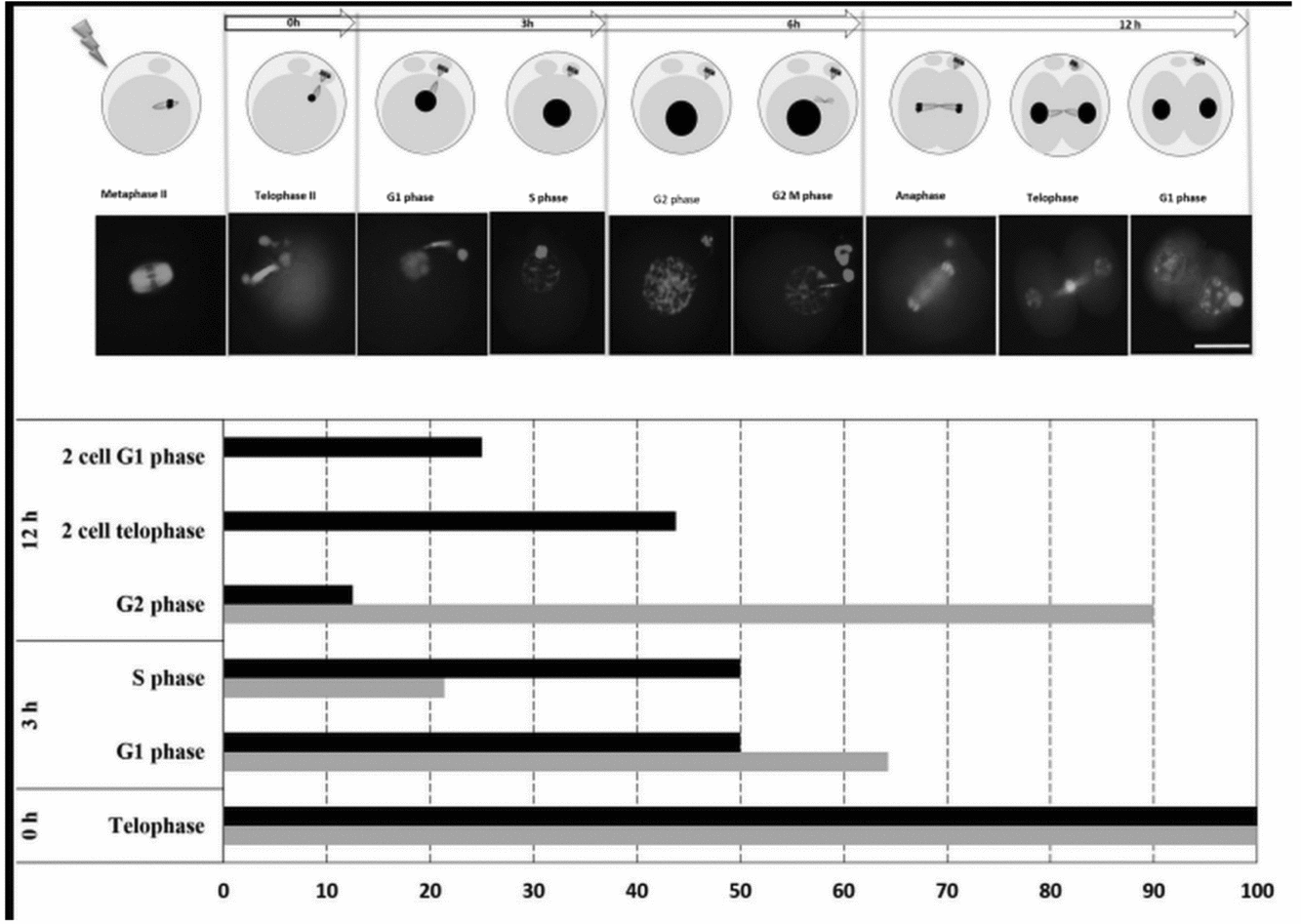
To understand the influence of SDF on early embryonic development, further studies were performed with 50 µg/ml of SDF, which gave the maximum blastocyst rate. To understand whether the presence of SDF changed the cell cycle pattern, nuclear organization along with spindle dynamics was analysed at various time intervals after activation. Oocytes activated in the presence or absence of SDF had all their activated oocytes at telophase at 0 h after activation with chromosomes moved to the spindle poles (Fig. 3). In the control group, at 3 h after oocyte activation, 64.30% of oocytes had their genome covered with a nuclear membrane and 21.40% of oocytes were in S phase. The remaining 14.30% were still in telophase. However, in oocytes activated in the presence of SDF, 50% of zygotes had crossed G1 phase and entered S phase at this interval. At 12 h, a shorter cell cycle was observed in oocytes activated with SDF group compared with the control group. In the SDF group, 68.75% were at early stages of cytokinesis marked by the appearance of a cleavage furrow, while in the control none of the activated oocytes had progressed to the 2-cell stage with 80% of these still in the G2M phase.
Cell cycle stages observed after each interval post artificial oocyte activation in oocytes. Representative images of spindle assembly and nuclear arrangement in oocytes at different cell cycle stages. The nucleus was stained with propidium iodide (red) and α-tubulin immunofluorescence detection was carried out for spindle assembly (green). Activated pronuclear embryos were fixed at specific intervals (0, 3, 6 and 12 h) and stained. The nuclear morphology and appearance of spindle is represented for each cell cycle point in the images. The images (12 h onwards) were pseudocolored with red. Bar represents 100 µm. Cell cycle stages observed in Sr2+ activated oocytes in the presence or absence of sperm-derived factors (50 µg/ml) at 0, 3 or 12 h post activation (n = 15 per group in all the intervals). Oocytes activated in the absence of SDF (control) and activated in the presence of SDF (50 µg/ml SDF) was assessed at different time intervals and the percentage of embryos in each stages of the cell cycle was plotted in the graph. Control;
 SDF;

Cortical granule exocytosis
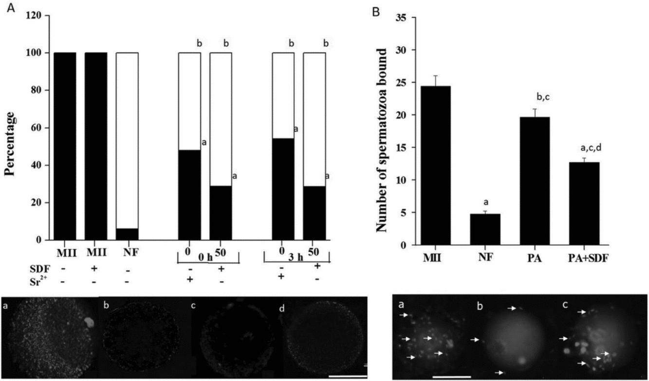
Cortical granule exocytosis was determined by an immunofluorescence method using LCA conjugated with rhodamine. Oocytes at the metaphase II (MII) stage served as the negative control, which had a uniform distribution of cortical granules (Fig. 4 Aa), while the normally fertilized PN stage embryos which served as the positive control, had a cortical distribution pattern (94.12%) (Fig. 4 Ab). MII oocytes incubated in the presence of SDF alone (without activating agent SrCl2) also exhibited a uniform distribution of cortical granules indicating that SDF alone does not have any direct influence on cortical granule exocytosis (Fig. 4 Ac). In artificially activated oocytes only 52% had cortical distribution immediately after activation (0 h) while 48% of oocytes had uniform distribution of cortical granules (P < 0.0001 vs. PN embryos). However, activation of oocytes in the presence of SDF increased the percentage of oocytes with cortical distribution (71.26%) at 0 h (P < 0.0001 vs. PN embryos), which did not change with increase in time (45.83 and 71.43% of oocytes with cortical distribution in oocyte activated in presence and absence of SDF) (Fig. 4 Ad).
(A) Graph showing the percentage of oocytes with uniform and cortical distribution of cortical granules post activation with Sr2+ in the presence or absence of paternal factors. MII, MII oocytes; NF, normally fertilized pronuclear embryos; 0, oocytes activated with strontium in the absence of SDF; 50, oocytes activated in the presence of SDF (50 µg/ml).
Uniform;
Cortical distribution. N = 20 per group in trials. χ2 test was used for statistical analysis. Representative images of cortical granule distribution in (a) MII oocyte; (b) PN stage embryo; (c) parthenotes; (d) parthenotes activated in the presence of SDF (50 µg/ml) by LCA immunofluorescence. Bar represents 100 µm. (B) Sperm bindability assay in oocytes activated in the presence or absence of SDF. MII, MII oocytes; NF, normally fertilized pronuclear embryos; PA, parthenogenetic embryos activated with strontium alone; PA+SDF, parthenogenetic embryos activated with strontium along with 50 µg/ml of SDF a P < 0.001, b P < 0.05 vs. MII, c P < 0.001 vs. PN, d vs. PA N = 42 for MII oocyte; PN for 62, parthenotes for 72, parthenotes with SDF for 26 in three trials. One-way ANOVA test was used for statistical analysis. Images represent the sperm zona binding pattern after staining with propidium iodide a MII oocytes, b PN embryos and c oocytes activated with strontium. The white arrow indicates the spermatozoa bound to zona. Bar represents 100 µm.

Sperm zona binding ability after oocyte activation
Sperm zona binding ability indicates the occurrence of zona reaction, which was assessed in oocytes activated in the presence or absence of SDF. The normally fertilized embryos a had significantly lower number of bound spermatozoa (04.79 ± 0.46) compared with MII oocytes (24.45 ± 01.56, P < 0.001, Fig. 4 Ba). Artificially activated oocytes had significantly higher numbers of sperm bound to the zona (19.65 ± 01.26) when compared with PN stage embryos (P < 0.001, Fig. 4 Bb) and MII oocytes (P < 0.05). The presence of SDF during activation resulted in a decrease in the sperm zona binding ability (12.69 ± 0.71) when compared with the parthenogenetic control (P < 0.01, Fig. 4 Bc), but was significantly higher than that of normally fertilized PN stage embryos (P < 0.001).
Mitochondrial distribution pattern in oocytes activated in presence of SDF
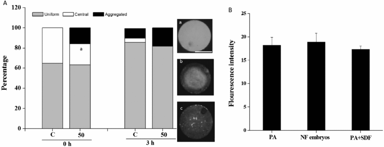
The mitochondrial distribution pattern was analysed at 0 and 3 h post activation. A uniform distribution of mitochondria was observed in approximately 65% of oocytes at 0 h (Fig. 5 Aa). However, the presence of SDF during activation resulted in a significant decrease in the percentage of oocytes with centrally localized mitochondria (P < 0.05, Fig. 5 Ab) while, in 16% of oocytes, aggregation of mitochondria was observed (Fig. 5 Ac). Mitochondrial distribution pattern at 3 h post activation was unaffected with 85% oocytes having uniform distribution, while 10% had aggregated mitochondria. Oocytes activated with SDF had approximately 20% of oocytes with aggregated mitochondria.
(A) Mitochondrial distribution pattern of oocytes activated in the presence or absence of SDF. C, oocytes activate with strontium alone; 50, oocytes activated with strontium along with 50 µg/ml of SDF. a P < 0.05 central distribution at 0 h.
Uniform,
Central,
Aggregated distribution pattern of mitochondria. N = 20 per group in each of three trails. Images represent the distribution pattern of mitochondria post activation after staining with rhodamine 123. Representative images of a uniform, b central and c aggregated mitochondrial distribution patterns in oocytes are shown. Bar represents 100 µm. χ2 test was used for statistical analysis. (B) Oct 4 intensity in activated oocytes after 24 h of culture. PA, parthengenetic embryos activated with strontium alone; NF embryos, normally fertilized embryos; PA+SDF, parthenogenetic embryos activated with strontium along with 50 µg/ml of SDF. N = 15 per group in three biological replicates. One-way analysis of variance (ANOVA) was used for analysis of Oct 4 intensity.

Expression pattern of pluripotency gene oct4
The expression of pluripotency gene Oct4 was analysed by quantification of immunofluorescence expression. Oct4 expression was observed in both nucleus and cytoplasm and the intensity of fluorescence was found to be similar in normally fertilized and parthenogenetic embryos after 24 h of activation (Fig. 5 B). Parthenotes activated in the presence of sperm-derived factors also showed a similar expression pattern, indicating that SDF does not have any significant influence on Oct4 expression.
Role of sperm-derived factors during embryonic development
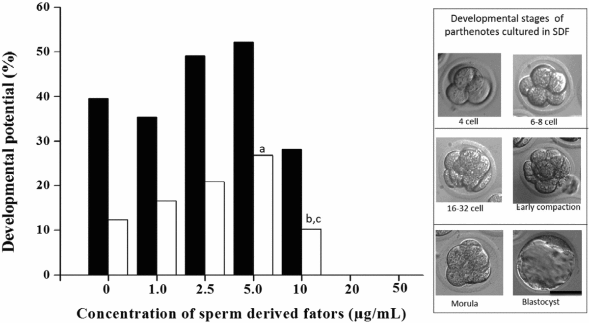
To understand the role played by sperm-derived factors post activation, 2-cell stage parthenogenic embryos were cultured in M16 medium supplemented with varying concentrations of SDF. Embryos were scored at every 24 h and the blastocyst rate was calculated at 120 h of culture. Unlike during activation, the beneficial effect of SDF was observed at much lower concentrations (2.5 and 5.0 μg/ml), while 50 μg/ml concentration was found to be detrimental when supplemented in culture medium for the development of parthenotes. Parthenogenetic embryos cultured at lower SDF concentrations were found to have a marginally increased morula rate when compared with the control (39.60, 35.40, 49.18 and 52.20% in control, 1, 2.5 and 5 μg/ml groups respectively (Fig. 6). The control group (10 mM SrCl2) showed a 12.31% blastocyst rate. While a dose-dependent increase was observed in parthenotes cultured at varying concentrations of SDF up to 5 μg/ml, parthenotes cultured at lower SDF concentrations (1 or 2.5 μg/ml) resulted in a blastocyst rate of 16.60 and 20.80%. A significantly increased (P < 0.05) blastocyst rate (26.80%) was obtained when parthenotes were cultured in M16 medium containing 5 μg/ml of SDF. However, at higher concentration (10 μg/ml), both morula rate and blastocyst rate were lower than that of the control (28.20 and 10.20% respectively). Furthermore, increasing the concentrations of SDF to 20 and 50 μg/ml resulted in failure of the embryos to undergo compaction.
Developmental potential of embryos cultured in varying concentrations of SDF. a P < 0.05 vs. C, b P < 0.05 vs. 2.5, c P < 0.01 vs. 5 µg/ml SDF in blastocyst rate (N = 45 per group in three trials). χ2 test was used for statistical analysis. Images represent different cell stages of embryos during development. Bar represents 100 µm. Morula rate
Blastocyst rate

Quantification and characterization of sperm-derived factors
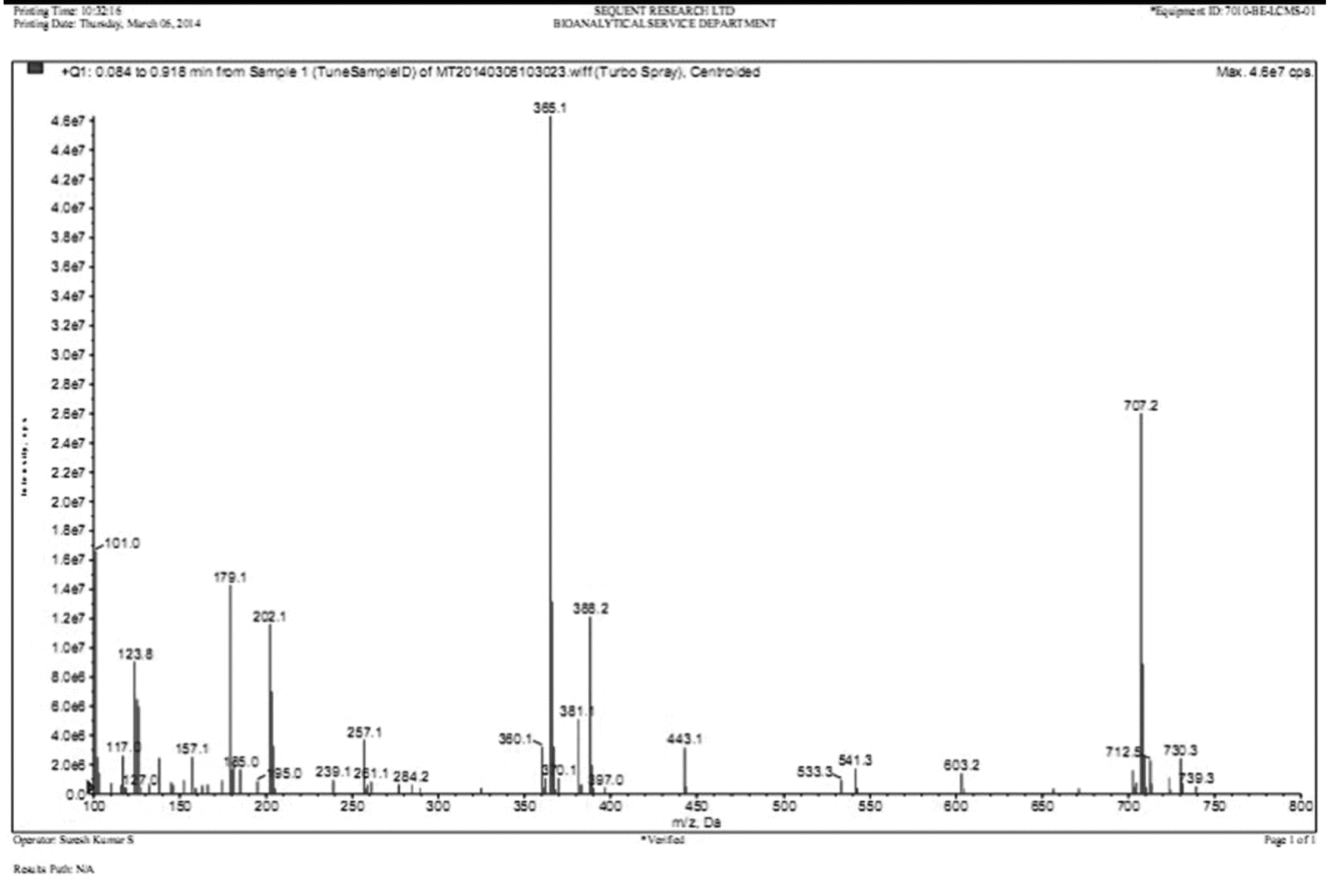
Spectrophotometric analysis was carried out to estimate the amount of protein present in SDF. A standard graph was plotted with bovine serum albumin (BSA) (5 mg/ml) as the standard. The protein concentration of lyophilized product was 98 μg/ml. Further characterization of the obtained sperm-derived components was carried out by mass spectrophotometry. A representative mass spectrum of sperm-derived factor is shown in Fig. 7. The possible fragmentation products obtained indicated the presence of phosphatidylserine (385.30 k), phosphatidylethanolamine (744.03 k), phosphatidylcholine (768.00 k), sphingomyelin (740.867 k) and caveolin (25 k) in the lyophilized product. The analysis of mass spectrometric spectra revealed the presence of about 41 proteins, which were further classified based on their function (Table S1).
Mass spectra of sperm-derived factors.

Discussion
In the present study, we demonstrated that the presence of SDF in the culture medium during artificial activation of oocytes can improve the developmental potential of haploid parthenotes without using any microinjection methods for the delivery of SDF into the oocyte. Previous studies have shown that haploid parthenotes are characterized by delayed embryonic development and decreased blastocyst rate (Kaufman & Sachs, Reference Kaufman and Sachs1975; Henery & Kaufman, Reference Henery and Kaufman1992) which is also observed in the present study (Fig. 3). Beatty & Fischberg (Reference Beatty and Fischberg1951) indicated that ploidy is one of the major factor that regulate the development of preimplantation embryos as both haploidy and polyploidy in mouse embryos can lead to a reduction in cell number. Similarly, Han et al. (Reference Han, Abeydeera, Kim, Moon, Cabot, Day and Prather1999), have reported the abnormal cleavage of polyploid embryos to three or more cells within 36 h of insemination in porcine embryos, while diploid embryos were still at the 2-cell stage.
The lack of paternal contribution in parthenotes is considered to cause poor in vitro development (Surani et al., Reference Surani, Barton and Norris1984; Solter, Reference Solter1988). Along with the sperm nucleus, certain cytoplasmic factors are found to be released into the oocyte during fertilization. Earlier studies have shown that the microinjection of SDF into oocyte results in calcium oscillations with a duration and amplitude similar to that observed during normal fertilization (Homa & Swann, Reference Homa and Swann1994; Wu et al., Reference Wu, He and Fissore1998) indicating that they have a significant role to play in fertilization. However, in these studies, SDF was microinjected into oocyte mainly focussing on understanding its effect on the intracellular calcium metabolism. None of these studies attempted to see whether the microinjection of SDF enhances the further development of activated oocytes. In this report, for the first time, we provide experimental evidence for the role of SDF in oocyte activation and early embryonic development of preimplantation embryos.
The presence of proteins that determines the fertility of spermatozoa has been observed in several species including human (Martınez-Heredia et al., Reference Martínez-Heredia, Estanyol, Ballescà and Oliva2006; Domagala et al., Reference Domagała, Pulido, Kurpisz and Herr2007; Secciani et al., Reference Secciani, Bianchi, Ermini, Cianti, Armini, La Sala, Focarelli, Bini and Rosati2009), mouse (Stein et al., Reference Stein, Go, Lane, Primakoff and Myles2006; Baker et al., Reference Baker, Reeves, Hetherington and Aitken2010; Guo et al., Reference Guo, Shen, Xia, Zhang, Zhang, Zhao, Xing, Chen, Chen, Lin and Huo2010) and bovine species (Newton et al., Reference Newton, Kastelic, Wong, Van der Hoorn and Thundathil2009; Saadi et al., Reference Saadi, van Riemsdijk, Dance, Rajamanickam, Kastelic and Thundathil2013). Mass spectrometric analysis in the present study revealed the presence of certain proteins that have been already reported to have role in fertility (Table S1).
Supplementation of SDF during oocyte activation was found to improve the development of haploid parthenotes. The presence of cytosolic sperm protein PLC zeta1 has been shown to activate the oocytes upon microinjection (Saunders et al., Reference Saunders, Larman, Parrington, Cox, Royse, Blayney, Swann and Lai2002; Rogers et al., Reference Rogers, Hobson, Pickering, Lai, Braude and Swann2004; Yoon et al., Reference Yoon, Eum, Lee, Lee, Kim, Han, Won, Park, Shim, Lee and Fissore2012). In addition to this, microinjection of PLCζ mRNA was found to rescue oocytes with failed sperm oocyte activation in humans (Yoon et al., Reference Yoon, Jellerette, Salicioni, Lee, Yoo, Coward, Parrington, Grow, Cibelli, Visconti, Mager and Fissore2008) and these embryos can progress up to the blastocyst stage (Saunders et al., Reference Saunders, Larman, Parrington, Cox, Royse, Blayney, Swann and Lai2002; Yu et al., Reference Yu, Saunders, Lai and Swann2008). However, we did not detect the presence of PLCζ in SDF in our study. But we observed the presence of proteins that are involved in the calcium signalling pathway such as tyrosine protein kinases SYK, solute carrier family 8 member 1 (Slc8a1), eukaryotic translation initiation factor 5A (Uorf1) and voltage gated calcium channels [calcium channel voltage dependent 1 type subunit (Cacnals), opioid receptor mu1] which might have helped in improving the developmental potential through their action on regulating the intracellular calcium level (Table S1). Further experiments are necessary to understand the exact mechanism of beneficial action by which these soluble factors from SDF induce the beneficial effects by entering into the oocyte or from outside.
To understand whether SDF involvement in fertilization and embryo development is stage specific, we cultured 2-cell-stage parthenotes in medium supplemented with SDF. The presence of SDF in culture medium during development was found to be beneficial, but was required at a much lower concentrations than that during oocyte activation. Following the first cleavage in mouse embryos, zygotic genome activation occurs, making it independent of parental genetic control. There are studies in the literature that indicate that paternal factors have a significant influence on the development of embryo up to blastocyst stage (Janny & Menezo, Reference Janny and Menezo1994; Liu et al., Reference Liu, Pang, Chiu, Wong, Lao, Lee and Yeung2012). SDF supplementation would have helped in regulation of the cell cycle of parthenogenetic embryos, as SDF contains few cell cycle regulators such as host cell factor C1 (HCFC1 protein) and tyrosine protein kinase Fe.
The paternal genome is reported to influence the first cell cycle in mouse (Renard & Babinet, Reference Renard and Babinet1986; Schabronath & Gärtner, Reference Schabronath and Gärtner1988), in bovine (Eid et al., Reference Eid, Lorton and Parrish1994), as well as in humans (Tesarik et al., Reference Tesarik, Mendoza and Greco2002). We used nuclear and spindle dynamics to identify the stage of embryonic cell cycle. By 6 h post activation, parthenotes in the control group were found to enter M phase, which is evident from the formation of nascent spindle fibres. Normally fertilized embryos at the PN stage are reported to have a functional cell cycle check point at M phase (Fulka et al., Reference Fulka, First, Fulka and Moor1999). Oocytes activated in the presence of SDF were found to have decreased duration of G2M phase, as ~70% of embryos had already completed cytokinesis and progressed to the 2-cell stage by 12 h of activation, while oocytes activated in the absence of SDF were found to reach the 2-cell stage only by 24 h (data not given). Eukaryotic initiation factor 4E (Eif4Ebp1) present in SDF has been shown to play a significant role in regulation of G1S transition (Fingar et al., Reference Fingar, Richardson, Tee, Cheatham, Tsou and Blenis2004).
Cytoplasmic streaming along with microtubule polymerization is also found to play an important role in exocytosis of cortical granules immediately after sperm entry into oocytes (Kim et al., Reference Kim, Day, Lee and Chung1996). There are contradictory reports on the stimulus of cortical granule exocytosis. Horvath et al. (Reference Horvath, Kellom, Caulfield and Boldt1993) reported that sperm act as the stimuli, while other studies have indicated that repetitive calcium oscillation triggers cortical granule exocytosis (Kline & Kline, Reference Kline and Kline1992; Abbott & Ducibella, Reference Abbott and Ducibella2001). The increase in the percentage of oocytes with cortical granule exocytosis after activation in the presence of SDF observed in this study could be due to the ability of SDF to increase amplitude and duration of calcium oscillations during artificial activation. However, this aspect needs to be studied further.
The number of sperm bound to parthenotes in the presence of SDF was also found to be significantly lower, indicating the occurrence of a zona reaction. However, it was not efficient as the sperm-induced zona reaction as observed in pronuclear embryos (Fig. 5 B). Tatone et al. (Reference Tatone, Van Eekelen and Colonna1994) have reported the occurrence of a transient phenomenon of sperm block within 30 min of artificial oocyte activation, unlike a permanent sperm-induced block in normally fertilized zygotes. The interval assessed in the present study is 3 h after the addition of strontium, by which time the block to further binding of the spermatozoa must have been reduced.
Apart from the effect of SDF on oocyte activation and early events of fertilization, we were interested to understand the cytoplasmic quality of the embryos developed. Mitochondria, which is the major cytoplasmic organelle and an indicator of developmental potential of embryos (Acton et al., Reference Acton, Jurisicova, Jurisica and Casper2004; Schatten et al., Reference Schatten, Sun and Prather2014) were analysed in oocytes activated in the presence or absence of SDF. SDF supplementation during oocyte activation however did not alter the distribution of mitochondria until after 3 h of activation. The uniform distribution of mitochondria was observed in artificially activated oocytes both in the presence or absence of SDF and was similar to that of earlier reported studies (Katayama et al., Reference Katayama, Zhong, Lai, Sutovsky, Prather and Schatten2006). The aggregation that was observed post activation could be due to reorganisation of the oocyte cortex during cortical granule exocytosis after fertilization, as indicated by Katayama et al. (Reference Katayama, Zhong, Lai, Sutovsky, Prather and Schatten2006).
The expression of the pluripotency gene (POUF51), which regulates early embryogenesis was also analysed to understand whether the presence of SDF during activation had any influence on the expression of pluripotency gene. Quantification of immunofluorescent signals indicated that, at early intervals of fertilization, embryo quality is unaffected even after SDF supplementation in parthenotes. Oct 4 is a maternal transcript protein and is usually found to be downregulated after the 2-cell stage in the mouse (Palmieri et al., Reference Palmieri, Peter, Hess and Schöler1994; Wu & Schöler, Reference Wu and Schöler2014) for proper embryonic development (Zuccotti et al., Reference Zuccotti, Merico, Bellone, Mulas, Sacchi, Rebuzzini, Prigione, Redi, Bellazzi, Adjaye and Garagna2011).
In conclusion, this is the first study that gives experimental evidence for the role of SDF during oocyte activation and early embryonic development. As the supplementation of these factors can improve the blastocyst rate in haploid parthenotes it can have a beneficial role in derivation of haploid stem cells for experimental and therapeutic purposes. We used haploid parthenotes to understand the beneficial role of sperm-derived factors on development. Further studies are necessary to understand whether diploid parthenotes will show a similar response, whether the components present in soluble SDF are able to enter the ooplasm without any microinjection method and how they improve the cleavage pattern in parthenotes.
Acknowledgement
Authors thankfully acknowledge the financial support from the Department of Health Research (DHR), Indian Council of Medical Research (ICMR), Government of India (DHR/HRD/Women scientist/Type-V/8/2014–15) and Department of Biotechnology (DBT), Government of India (BT/PR15130/GBD/27/328/2011).
Author contributions
RN and SA performed the experiments; GK designed the study and wrote the manuscript; SKA and SM helped in statistical analysis and writing the manuscript.
Supporting Information
Additional Supporting Information is available online at the publisher's website.
Table S1
Supplementary material
To view supplementary material for this article, please visit https://doi.org/10.1017/S0967199417000569