Implications
Immunosuppression threatens the swine industry by increasing susceptibility to secondary infections and sub-optimal response to vaccinations. Using nutrition strategies to improve organism immunity is one of the current research directions. Previous studies have demonstrated the immunomodulatory properties of conjugated linoleic acid (CLA). However, studies addressing the relationship between CLA and cellular immunosuppression are still lacking in piglets. The present study demonstrated that dietary CLA effectively ameliorated cellular immunosuppression in piglets treated with immunosuppressant drug.
Introduction
CLA is a mixture of geometrical and positional isomers of linoleic in which the two double bonds are conjugated. CLA has many important properties that regulate physiological and metabolic responses, for example, immune modulation, anti-cancer effects, prevention of cardiovascular disease and diabetes, and weight control (Ip et al., Reference Ip, Chin, Scimeca and Pariza1991; Houseknecht et al., Reference Houseknecht, Vanden-Heuvel, Moya-Camarena, Portocarrero, Peck, Nickel and Belury1998; Islam et al., Reference Islam, Kim, Oh, Kim, Won, Kim, Choi, Kim and Ha2010; Eftekhari et al., Reference Eftekhari, Aliasghari, Babaei-Beigi and Hasanzadeh2013; Kim et al., Reference Kim, Park, Kweon and Han2013). In animal production, studies on the effects of CLA on immune function have thus far been conducted in the normal physiological status or inflammatory challenge status (O’Shea et al., Reference O’Shea, Bassaganya-Riera and Mohede2004; Lai et al., Reference Lai, Yin, Li, Zi, Qiu and Xu2005; Zhang et al., Reference Zhang, Guo and Yuan2005; Renner et al., Reference Renner, Pappritz, Kramer, Kersten, Jahreis and Dänicke2012).
By immunizing pigs against pseudorabies virus (Prv), Bassaganya-Riera et al. (Reference Bassaganya-Riera, Hontecillas, Zimmerman and Wannemuehler2002) found that the magnitude of Prv-specific recall responses of CD8+ T cells was greater in the immunized pigs fed CLA. Furthermore, Bassaganya-Riera et al. (Reference Bassaganya-Riera, Pogranichniy, Jobgen, Halbur, Yoon, O’Shea, Mohede and Hontecillas2003) reported that dietary CLA ameliorated viral infectivity in a pig model of type-2 porcine circovirus (PCV2)-induced immunosuppression. Long et al. (Reference Long, Guo, Wang, Liu, Zhang and Yang2011) discovered that dietary CLA enhanced immune function in broiler chickens, particularly those of the infectious bursal disease virus (IBDV)-induced immunosuppressive status. These findings indicated the protective effects of dietary CLA on animals with a viral-induced immunosuppressive status.
T lymphocytes depletion is a hallmark immunological effect associated with immunosuppressive viral infections such as Prv, PCV2 and IBDV. So we hypothesized that CLA could ameliorate the cellular immunosuppression in piglets. Cyclosporin A (CsA), a cellular immunosuppressive drug, prevents the synthesis of T-cell cytokines by blocking a late stage signaling pathway initiated by the T-cell receptor, which affects the production of interleukin-2 (IL-2); hence, T-cell proliferation is affected (Lillehoj, Reference Lillehoj1987; Hill et al., Reference Hill, Rowland, Latimer and Brown1989; Sigal and Dumont, Reference Sigal and Dumont1992). The objective of this study was to evaluate the effects of 2% dietary CLA on the cellular immune response of piglets intramuscularly injected with CsA.
Material and methods
Experimental design
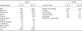
This experiment was conducted according to the protocols approved by the Henan University of Animal Husbandry and Economy Animal Care and Use Committee. A total of 72 Duroc×Landrace×Yorkshire weaned piglets (21 days old) were randomly assigned to four treatments in a design with 2×2 factorial arrangement, that is two levels of CLA (0% or 2% in diets) and with or without CsA challenge. In control diets, 2% CLA was replaced by 2% soybean oil to keep both the CLA-supplemented and the control diets isoenergetic. CLA was provided by the Qingdao Aohai Biology Technology Co. Ltd (Qingdao, Shangdong Province, China) and contained 80.80% conjugated dienes with 39.24% 9c, 11t-CLA and 38.93% 10t, 12c-CLA representing 96.74% of the contained isomers. Each treatment had six replications with three piglets each. The ingredients and nutrient levels of the basal diet were formulated to meet the NRC (1998) nutrient requirements (Table 1). The piglets were fed for 42 days. Piglets were raised in 2×2 m net beds with a stainless steel adjustable chute and nipple-type drinking water apparatus. Temperature was controlled at 25°C to 27°C. Piglets were vaccinated using a routine immunization program and had free access to feed and water.
Ingredients and nutritional values of control diet as-fed basis

1 Supplied per kilogram of diet: VA, 9000 IU; VD3, 2500 IU; vitamin E, 20.0 IU; VK3, 3.0 mg; VB1, 1.5 mg; VB2, 4.0 mg; VB6, 3.0 mg; VB12, 0.012 mg; niacin, 30.0 mg; pantothenic acid, 15.0 mg; folic acid, 0.75 mg; biotin, 0.05 mg; Zn, 75 mg (as ZnSO4); Fe, 75 mg (as FeSO4·H2O); Mn, 20 mg (as MnSO4); Cu, 225 mg (as CuSO4·5H2O); I, 0.04 mg (as ethylene diamine dihydroiodide).
2 Calculated values.
On days 14, 21, 28 and 35 of the study, the challenged group was administrated intramuscularly in the neck with CsA (diluted in olive oil, 10 mg/kg BW). The unchallenged group was injected with 0.9% (w/v) NaCl solution with olive oil. CsA was purchased in a crude state from Fujian Kerui Drug Co., Fujian, China.
Growth performance and the thymus
At the beginning and end of the study, piglets were weighed and feed intake was recorded for each replication to calculate growth performance including the average daily gain (ADG) and feed : gain (F : G). One piglet from each replication was euthanized on day 42 of the experiment. The thymus was excised under sterile condition. The relative weights of the thymus were measured using the formula: thymus relative weight (g/kg)=thymus weight g/per BW of piglet kg.
Separation of peripheral blood and thymus lymphocytes
Six piglets (one per replication) were randomly chosen from each treatment on day 42. Heparinized blood samples were collected by anterior vena cava puncture. Peripheral blood mononuclear cells were isolated from heparinized whole blood by Ficoll-Paque density gradient centrifugation as described by Lai et al. (Reference Lai, Yin, Li, Zi, Qiu and Xu2005). Briefly, 3 ml heparinized whole blood layered over 4 ml lymphocyte Ficoll-Paque density gradient centrifugation medium (density=1.077; HaoYang Biological Manufacture Co. Ltd, Tianjin, China) in 10-ml centrifuge tubes and centrifuged at 2500×g for 30 min. Approximately 1 ml of mononuclear cell layer was collected and washed twice with RPMI-1640 (Gibco, Invitrogen, Grand Island, New York, USA) medium. Subsequently, the cells were re-suspended in complete RPMI-1640 medium supplemented with 10% (v/v) heat-inactivated fetal calf serum, 105 U/l of penicillin, 100 mg/l of streptomycin and 25 mmol/l of 4-(2-hydroxyethyl)-1-piperazineethanesulfonic acid (HEPES) buffer (Sigma Chemical Co., St. Louis, MO, USA). The pH of the medium was adjusted to 7.4 by the addition of a sterile solution of 56 g/l sodium bicarbonate. Then cells were counted on a hemocytometer using a light microscope and viability was determined by trypan blue exclusion method.
The pigs for the thymus investigation were the same as those for peripheral blood collection. These piglets had their thymus removed for thymus lymphocytes subsets analysis. The thymus was trimmed to remove the connective tissue and fat, and washed with cold phosphate buffer saline (PBS). After shearing in Hank’s salt solution (pH 7.4), the fragments were ground with a glass syringe core on a stainless steel cell screen. The collected thymus lymphocytes layered over equivalent lymphocyte Ficoll-Paque density gradient centrifugation medium and centrifuged at 2500×g for 20 min. The cloud-like white cell layer at the interface was collected and treated with hemolysis buffer to remove erythrocytes. Thymocytes were counted and re-suspended in RPMI-1640 medium. The cell concentration was adjusted to 1×10⁷/ml.
Peripheral blood lymphocyte proliferation by Concanavalin A
A 3-[4,5-dimethylthiazol]-2,5-diphenyltetrazolium bromide (MTT, Sigma Chemical Co.) assay was used to determine the peripheral blood lymphocyte proliferation response at day 42 of the experiment. Suspensions of peripheral blood mononuclear cells (1×10⁶/ml) were incubated in 96-well culture plate at 180 μl/well; each sample was seeded in six wells. Exactly 20 μl of RPMI-1640 nutrient solution containing concanavalin A (Con A; 45 μg/ml; Sigma Chemical Co.) was then added into three wells. As the control, the same volume of RPMI-1640 nutrient solution containing no Con A was added into other three wells. Cell cultures were incubated at 37°C in a humidified atmosphere at 5% CO2 for 72 h. At 4 h before the end of incubation, a 10 μl MTT solution (5 mg/ml) was added to each well and the plates were incubated for another 4 h. Subsequently, 100 μl of 10% SDS dissolved in 0.04 mol/l HCl solution was added into each well to lyse the cells and solubilize the MTT crystals. After 30 min, the optical density was measured at a wavelength of 570 nm using a Microplate Reader.
Peripheral blood and thymus lymphocyte subsets analysis
The peripheral blood and thymus lymphocytes were incubated with an optimal concentration of fluorescence-labeled mouse anti-swine CD monoclonal antibody for 30 min at 4°C in the dark, and later with a fluorescein isothiocyanate-conjugated goat anti-mouse IgG. Cells were washed and re-suspended in fluorescence activated cell sorting (FACS) buffer, and analyzed with a Beckman-Coulter flow cytometer. A minimum of 104 viable cells from each experiment was analyzed. Phycoerythrin (PE)-labeled rat anti-swine CD3+ antibody, PE-labeled mouse anti-swine CD4+ and fluorescein isothiocyanate (FITC)-labeled mouse anti-swine CD8+ antibody were obtained from Southern Biotechnology Associates, Inc., Birminghan, AL, USA. For thymus CD4+ CD8+ double-positive lymphocyte analysis, thymus lymphocyte suspensions were incubated with PE-labeled mouse anti-swine CD4+ monoclonal antibody, FITC-labeled mouse anti-swine CD8+ monoclonal antibody and a mix of PE and FITC-labeled mouse IgG1 isotype control.
Interleukin-2-induced activity of peripheral blood lymphocytes
The cells were placed onto a 24-well plate and incubated for 48 h with 45 μg/ml of Con A at 37°C with 5% CO2 in an incubator. The supernatant was collected. IL-2-induced activity was detected as follows: on 96-well cell culture plates, each well contained 50 μl of RPMI-1640 complete medium, tested IL-2 samples and 50 μl of 1×10⁷/ml target cells. RPMI-1640 medium was used as a blank control. Cell cultures were incubated at 37°C in a humidified atmosphere of 5% CO2 for 30 h. A volume of 10 μl of 5 g/ml MTT solution was added to each well and cultured for 6 h. Then, 100 μl of 10% SDS dissolved in 0.04 mol/l HCl solution was added to each well and cultured for 2 h. The optical density was measured with a Microplate Reader at a wavelength of 570 nm.
Statistical analysis
Data were analyzed as a 2×2 factorial arrangement of treatments by GLM procedure of SPSS 16.0 program. The main effects of challenge (CsA or saline injection), CLA levels (0% or 2% CLA in diets) and their interactions were analyzed. The statistical model utilized was Yijk=μ+Infectioni+Dietj+(Infection×Diet)ij+errorij, where μ was the general mean, Infectioni the main effect of the ith level of the challenge effect, Dietj the main effect of the jth level of the dietary effect, (Infection×Diet)ij the interaction effect between infection and diet, and errorij the random errors. Differences among each treatment group were tested by Duncan’s multiple comparison when the significant interaction between the main effects was observed. A level of P<0.05 was set as the criterion for statistical significance. Data were expressed as means±SEM.
Results
Effects of dietary CLA and cyclosporin A injection on the growth performance of piglets
As shown in Table 2, both dietary CLA and CsA injection had no significant effects on the ADG and the average daily feed intake of piglets (P>0.05). CsA treatment significantly increased the F : G of piglets (P<0.05), whereas dietary CLA decreased it (P<0.05). There was significant interaction between CsA treatment and dietary CLA on the F : G of piglets (P<0.05). Dietary CLA prevented the increase of F : G caused by CsA treatment (P<0.05).
Effects of dietary CLA supplementation on growth performance of Duroc×Landrace×Yorkshire piglets after cyclosporin A (CsA) injection

Data were analyzed as a 2×2 factorial arrangement of treatments with CsA challenge (CsA or saline injection), CLA levels (0% or 2% CLA in diets) and their interactions as main effects. Data were expressed as means±SEM (n=18, six replications of three piglets each).
a,b,cDifferences among each treatment group were tested when dietary CLA and CsA injection show a significant interaction (P<0.05). Means in the same line without the same superscript differ significantly (P<0.05).
Effects of dietary CLA and cyclosporin A treatment on peripheral blood lymphocytes subsets and lymphocytes transformation rate
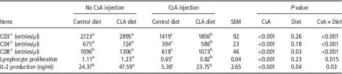
As shown in Table 3, CsA injection significantly decreased the numbers of peripheral blood CD3+, CD4+ and CD8+ T lymphocytes (P<0.01). Dietary CLA did not change the numbers of the peripheral blood CD3+, CD4+ T lymphocytes (P>0.05), but significantly increased the number of peripheral blood CD8+ T lymphocytes (P<0.05). There was significant interaction between CsA treatment and dietary CLA on the numbers of the peripheral blood CD3+, CD4+ and CD8+ T lymphocytes of piglets (P<0.05). Dietary CLA supplementation significantly inhibited the decreases of the peripheral blood CD3+, CD4+ and CD8+ T lymphocytes counts after CsA injection (P<0.01). Piglets treated with CsA showed the lower peripheral blood lymphocyte transformation rate (P<0.01). There was a significant interaction between dietary CLA and CsA injection on the peripheral blood lymphocyte transformation rate of piglets (P<0.05). Dietary CLA partially inhibited the decrease of the peripheral blood lymphocyte transformation rate resulted from CSA treatment (P<0.05).
Effects of dietary CLA supplementation on the numbers, proliferation and interleukin-2 (IL-2) production of peripheral blood T lymphocytes of Duroc×Landrace×Yorkshire piglets after cyclosporin A (CsA) injection

Data were analyzed as a 2×2 factorial arrangement of treatments with CsA challenge (CsA or saline injection), CLA levels (0% or 2% CLA in diets) and their interactions as main effects. Data were expressed as means±SEM (n=6).
a,b,cDifferences among each treatment group were tested when dietary CLA and CsA injection show a significant interaction (P<0.05). Means in the same line without the same superscript differ significantly.
Effects of dietary CLA and cyclosporin A injection on thymus lymphocytes subsets and thymus relative weight
Most of thymus lymphocytes were CD4+ CD8+ double-positive lymphocytes, and there were few single-positive lymphocytes (Table 4). CsA treatment decreased the percentages of thymus CD4+ and CD8+ single-positive lymphocytes (P<0.01). Dietary CLA significantly increased the percentages of thymus CD4+ CD8+ double-positive and CD8+ single-positive T lymphocytes (P<0.05). Dietary CLA had the tendency to block the decrease of thymus CD4+ single-positive lymphocytes resulted from CsA treatment (P=0.07). Both dietary CLA and CsA injection did not change the thymus relative weight of piglets (P>0.05).
Effects of dietary CLA supplementation on T lymphocyte subsets and relative weight in thymus of Duroc×Landrace×Yorkshire piglets after cyclosporin A (CsA) injection

Data were analyzed as a 2×2 factorial arrangement of treatments with CsA challenge (CsA or saline injection), CLA levels (0% or 2% CLA in diets) and their interactions as main effects. Data were expressed as means±SEM (n=6).
a,b,cDifferences among each treatment group were tested when dietary CLA and CsA injection show a trend of interaction on thymus CD4+ single positive (P=0.07). Means in the same line without the same superscript differ significantly.
Effects of dietary CLA and cyclosporin A injection on interleukin-2-induced activity of peripheral blood lymphocytes
As shown in Table 3, CsA treatment decreased IL-2 production of peripheral blood lymphocytes (P<0.01), whereas CLA supplementation resulted in significantly higher IL-2 production (P<0.05). There was significant interaction between CsA challenge and dietary CLA on IL-2 production (P<0.05). Dietary CLA partly inhibited the decrease of IL-2 production of peripheral blood lymphocytes after CsA injection (P<0.05).
Discussion
In the present study, dietary CLA prevented the increase of F : G and the decrease of the peripheral blood lymphocytes transformation rate, and effectively blocked the exhaustion of peripheral blood CD3+, CD4+ and CD8+ T lymphocytes induced by CsA injection (P<0.05). Dietary CLA supplementation also showed the tendency to block the decrease of thymus CD4+ single-positive lymphocytes after CsA treatment (P=0.07). Thus, dietary CLA alleviated cellular immunosuppression resulted from CsA injection in piglets.
Effects of dietary CLA and cyclosporin A injection on the growth performance of piglets
In the current study, CsA injection worsened the feed conversion efficiency of piglets (P<0.05), but had no effect on BW gain (P>0.05). This result indicated that CsA had little effect on swine growth, and thus was an ideal immunosuppressive induction agent. This result is in line with that of Fitzgerald et al. (Reference Fitzgerald, Williams and Reed1996) who reported that CsA specifically altered T lymphocytes activity, but did not damage immune organs, so the side effects of CsA were minor.
Dietary CLA supplementation significantly improved the feed conversion efficiency of piglets (P<0.05) and alleviated the deterioration of feed conversion efficiency after CsA treatment (P<0.05). Michael et al. (Reference Michael, Jennifer and John2004) reviewed the results of 14 relevant studies, 12 of which indicated that dietary CLA improved swine feed conversion efficiency. Lai et al. (Reference Lai, Yin, Li, Zi, Qiu and Xu2005) found that the feed conversion efficiency of swine improved linearly when the additive amount of dietary CLA was from 0% to 5%. Therefore, most previous studies have shown the beneficial effects of dietary CLA on feed conversion efficiency in swine regardless of immunosuppression.
Effects of dietary CLA and cyclosporin A injection on the thymus relative weight and thymus lymphocytes subsets
The thymus is the important organ for lymphocytes maturation, differentiation and proliferation. In this study, CsA injection showed no effect on the relative weight of thymus, indicating that CsA treatment was not destructive to thymus development. However, at pharmacological doses (⩾20 mg/kg), CsA-induced morphological changes and significantly reduced the thymus medulla of rat, and at a virulence dose (⩾50 mg/kg), induced vacuolization and necrosis of the thymus medulla of rats (Blair et al., Reference Blair, Thomson, Whiting, Davidson and Simpson1982; Tanaka et al., Reference Takahashi, Kawamata and Akiba1988). The use of a low dose of CsA in the present study might explain the difference in results compared with previous studies. Alternatively, these differences might be due to different reactions of swine and rats to CsA treatment.
CsA was reported to primarily suppress T lymphocytes, that is, maintains resting lymphocytes in the G0 or G1 phase. In this experiment, CsA injection had little effect on CD4+ CD8+ double-positive lymphocytes (P=0.11), but significantly depleted CD4+ and CD8+ single-positive lymphocytes (P<0.01). Therefore, CsA inhibition of thymocytes differentiation mainly occurred in the CD4+ CD8+ double-positive phase to block the transformation of CD4+ CD8+ double-positive lymphocytes to CD4+ and CD8+ single-positive lymphocytes. Dietary CLA significantly increased the percentages of CD4+ CD8+ double-positive and CD8+ single-positive T lymphocytes in the thymus (P<0.05), and had the trend to inhibit the decrease of CD4+ single-positive T lymphocytes in the thymus after CsA injection (P=0.07). In the absence of exogenous stimulus, dietary CLA increased the number of thymus CD8αα T lymphocytes in piglets (Bassaganya-Riera et al., 2001). These collective findings demonstrate the regulation by dietary CLA on thymus T lymphocytes of swine with or without external immunosuppressive factors.
Effects of dietary CLA and cyclosporin A injection on peripheral blood lymphocyte subsets and interleukin-2-induced activity
In the present study, CsA injection significantly decreased the numbers of peripheral blood CD3+, CD4+ and CD8+ T lymphocytes (P<0.01). The result is consistent with the decreases of thymus CD4+ and CD8+ T lymphocytes because peripheral blood T lymphocytes originate from the thymus. Dietary CLA effectively blocked the decreases of peripheral blood CD3+, CD4+ and CD8+ T lymphocytes induced by CsA injection. This result indicated that dietary CLA could alleviate the exhaustion of peripheral blood lymphocytes of piglets under immunosuppression. In the trials of swine infected with Prv and swine circovirus type-2, dietary CLA effectively maintained CD8+ T lymphocyte profile (Bassaganya-Riera et al., Reference Bassaganya-Riera, Hontecillas, Zimmerman and Wannemuehler2002 and Reference Bassaganya-Riera, Pogranichniy, Jobgen, Halbur, Yoon, O’Shea, Mohede and Hontecillas2003). In the absence of exogenous stimulation, dietary CLA induced the proliferation of CD8+ T lymphocytes such as (TCR)γQCD8αα and TCRαβ CDαβ, and enhanced the immune function mediated by CD8+ T cells (Bassaganya-Riera et al., Reference Bassaganya-Riera, Hontecillas, Zimmerman and Wannemuehler2001). Taken together, these results indicated a proliferative effect of dietary CLA on peripheral blood CD8+ T lymphocytes.
IL-2 is a multi-functional protein, most notably known for the ability to induce the proliferation and maturation of activated T cells (Furue and Ishibashi, Reference Furue and Ishibashi1991). Piglets treated with CsA showed significantly low IL-2 production of peripheral blood lymphocytes. However, dietary CLA increased the IL-2 production of peripheral blood lymphocytes and alleviated the inhibitory effect of CsA injection on IL-2 inductive activity (P<0.01). Several studies in humans, poultry and rats have shown that dietary CLA increased IL-2 activity induced by Con A (Wong et al., Reference Wong, Chew, Wong, Hosick, Boylston and Shultz1997; Nugent et al., Reference Nugent, Roche, Noone, Long, Kelleher and Gibney2005; Takahashi et al., Reference Tanaka, Shinohara, Fukumoto, Tanaka and Kaneko2007). IL-2 messenger RNA expression levels in the CLA diet group were significantly higher than in the control group in a pig model of PCV2-induced immunosuppression (Lee et al., Reference Lee, Kritchevsky and Pariza1994). Therefore, dietary CLA maybe increased IL-2 production by increasing IL-2 gene expression to promote the proliferation and differentiation of T lymphocytes. On the other hand, it was reported that CLA supplementation in diets showed no effects (Kelley et al., Reference Kelley, Warren, Simon, Bartolinin, Mackey and Erickson2002; Yamasaki et al., Reference Yamasaki, Chujo, Hirao, Koyanagi, Okamoto, Tojo, Oishi, Iwata, Yamauchi-Sato, Yamamoto, Tsutsumi, Tachibana and Yamada2003) or adverse effects on IL-2 production (Hayek et al., Reference Hayek, Han, Wu, Watkins, Meydani, Dorsey, Smith and Meydani1999).
In conclusion, the current study demonstrated that (i) dietary CLA partly ameliorated the deterioration of feed conversion rate caused by CsA treatment; (ii) dietary CLA inhibited the decreases of CD4+ and CD8+ T lymphocytes in the thymus after CsA treatment; (iii) dietary CLA markedly inhibited the decreases of peripheral blood CD3+, CD4+, CD8+ T lymphocyte counts and IL-2 production after CsA treatment; (iv) dietary CLA ameliorated CsA-induced cellular immunosuppression in piglets.
It bears pointing out that the immune system of piglets at 21 days old is in development. Significantly different, the immune system of the adult pigs is mature, so it is not clear whether the above-mentioned immunomodulatory effects of dietary CLA on piglets can be extrapolated to adult pigs. To clarify this, further studies are necessary.
Acknowledgment
This research was supported by Henan Province Natural Science Fund Project (Project No. 102102110123).