Introduction
Over the past decades, total body irradiation (TBI) with megavoltage photon beam has become an important component of the patients’ conditioning regimen prior to bone marrow transplant (BMT). TBI is also a treatment technique for some haematological diseases. Reference Giraud and Houle1–Reference Allahverdi, Geraily, Esfehani, Sharafi, Haddad and Shirazi5 The combination of TBI and chemotherapy is the most common pre-treatment conditioning regimen. Reference Allahverdi, Geraily, Esfahani, Sharafi, Haddad and Shirazi3,Reference Sarradin, Simon, Huynh, Gilhodes, Filleron and Izar6–Reference Leer, Broersel, Vroomel, Chinl, Noordijkl and Dutreix8 TBI is usually implemented with two large opposing radiation fields with the aim to cover the whole body with one left and one right lateral single field, requiring the patient to be positioned at an extended distance from the radiation source. Reference Jahnke, Jahnke and Molina-Duran9,Reference Lu, Filippi and Patel10
Although some centres apply a dedicated treatment room and machine for the TBI technique, most centres implement this technique using standard radiotherapy equipment. TBI implementation with standard equipment is a difficult and time-consuming, cumbersome task that requires several dosimetric measurements, careful treatment planning and significant physical effort. Reference Yao, Bernard and Turian11,Reference Shahzadeh, Gholami, Aghamiri, Mahani, Nabavi and Kalantari12 One of the most important dosimetric challenges for TBI commissioning is the fact that the data used for dose calculation algorithms in modern treatment planning systems (TPS) are obtained under standard conditions, and they cannot be applied directly for the TBI technique because this is delivered at an extended treatment distance. In addition, the extended source skin distance (SSD) and large radiation field size make the scattering conditions of TBI more complex than other standard radiotherapy techniques. Reference Yao, Bernard and Turian11
Although TBI techniques differ from one centre to another regarding the dedicated installation of the department, all centres have a common goal of delivering prescribed dose uniformly to the whole body in the range of ±10%. Reference Wills, Cherian, Yousef, Wang and Mackley13–Reference Wolden, Rabinovitch and Bittner16
In TBI and half-body irradiation (HBI) where the SSD distance increases more than the standard distance introduced by the manufacturer (i.e., 100 cm), it is not possible to apply TPS for dose calculations. Precise monitor units (MU) calculation for reducing the amount of error in dose delivery requires measuring basic dosimetric parameters under TBI conditions. Measurements in large fields and at extended distance involve several dosimetric complications, and therefore data measured experimentally at an extended distance have more uncertainties than those determined at the isocentre in normal SSD conditions. In addition, it would be more accurate if the basic data can be calculated from standard measurement values. Therefore, it is necessary to know if we can use dosimetric data calculated from the measurements performed under standard conditions by the TPS or if it is more accurate to use the basic dosimetric parameters measured directly under TBI conditions. Regarding the calculations’ error values, it can then be decided whether to directly measure or calculate these parameters at extended SSDs. Reference Houdek and Pisciotta17,Reference Spunei, Mihai and Mălăescu18 Despite there being similar studies in this field, dosimetric data obtained in one centre cannot be used for another. The purpose of this study is to compare the basic dosimetric parameters including dose rate, percentage depth dose (PDD) measured directly under TBI conditions with those calculated from the measurements performed in the standard situation by the TPS for 6- and 18-MV photon beams produced by Clinac 2100C/D linear accelerator (Varian Medical System, Palo Alto, CA, USA).
Materials and Method
Materials
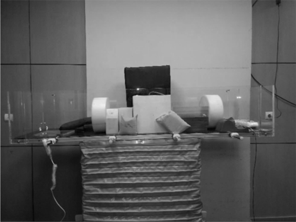
In order to achieve the large radiation field sizes required to deliver TBI, treatment needs to be delivered in a large treatment room so that the source to skin distance can be increased to accommodate the field sizes required to cover the patient’s entire body, and this requires a nonstandard treatment table. Reference Van Dyk, Galvin, Glasgow and Podgorsak19 In this study, a TBI treatment table was designed and fabricated to allow for irradiation at an extended treatment distance (Figure 1). The TBI table has a variable height and is equipped with a Plexiglas frame in which the build-up effect arising from a high-energy photon beam can be accommodated.
TBI treatment table and phantoms arrangements for depth dose measurement.

All absorbed doses were measured with a Markus parallel-plate ion chamber (model TM23343, PTW-Freiburg, Germany) and a Farmer-type ionization chamber (model TM30010, PTW-Freiburg) with a 0·6 cc sensitive volume in conjunction with an Unidos electrometer (model T10001, PTW-Freiburg) for depths in the build-up region and beyond it, respectively. A 30 × 30 × 30 cm3 water phantom and layers of slab phantoms (PTW-Freiburg) with different thicknesses were used for dose rate, PDD and beam profile measurements.
Method
In this study, three parameters including dose rate, PDD and beam flatness were measured in simulated treatment conditions. All irradiations were done under the defined TBI conditions (i.e., field size = 40 × 40 cm2, SSD = 312 cm, collimator rotation = 45° and gantry angle = 90°) with 18- and 6-MV photon beams produced by CLINAC 2100C/D linear accelerator.
Central axis PDD
PDD is defined as the percentage of absorbed dose in any depth (
 $z$
) to the absorbed dose in reference depth (
 ${z_0}$
) along the central axis of the beam.
Reference Osei20,Reference Khan and Gibbons21
PDD can be calculated using Equation (1):
 $${\rm{PDD}} = {z \over z_0} \times 100\%$$
The central axis depth dose is dependent on beam energy, depth, field size, SSD and beam collimation. Reference Khan and Gibbons21 During TBI treatment, both field size and SSD are changed in comparison to standard conditions. The change in PDD due to TBI dosimetry conditions can be obtained in two ways. One way is to multiple Mayneord factors by depth dose obtained under standard situations using Equation (2). Reference Houdek and Pisciotta17
 $${\rm{D}}{{\rm{D}}_{\rm{c}}}\left( {d.f.z} \right) = {\rm{D}}{{\rm{D}}_{\rm{m}}}\left( {100.f.z} \right) \times MF\left( {d.z} \right)$$
where
 ${\rm{D}}{{\rm{D}}_{\rm{c}}}$
is the depth dose at the desired depth (
 $z$
), f is the side of square field at isocentre, d is the extended treatment distance, and
 ${\rm{D}}{{\rm{D}}_{\rm{m}}}\left( {d.f.z} \right)$
is the depth dose measured at a standard distance (
 $d = 100\;cm$
) but for the same field size (
 $f$
) and the same depth (
 $z$
). MF is the Mayneord factor, which is obtained using Equation (3)
Reference Khan and Gibbons21
:
 $${\rm{MF}}\left( d.z \right) = \left( {100 + z \over d + z} \right)^2 \times \left( {d + z_0 \over 100 + z_0} \right)^2$$
where
 $z$
and d have the same significance as in Equation (2), and
 ${z_0}$
is the depth of dose maximum.
Another way to obtain depth dose data for TBI is by direct measurement under TBI conditions.
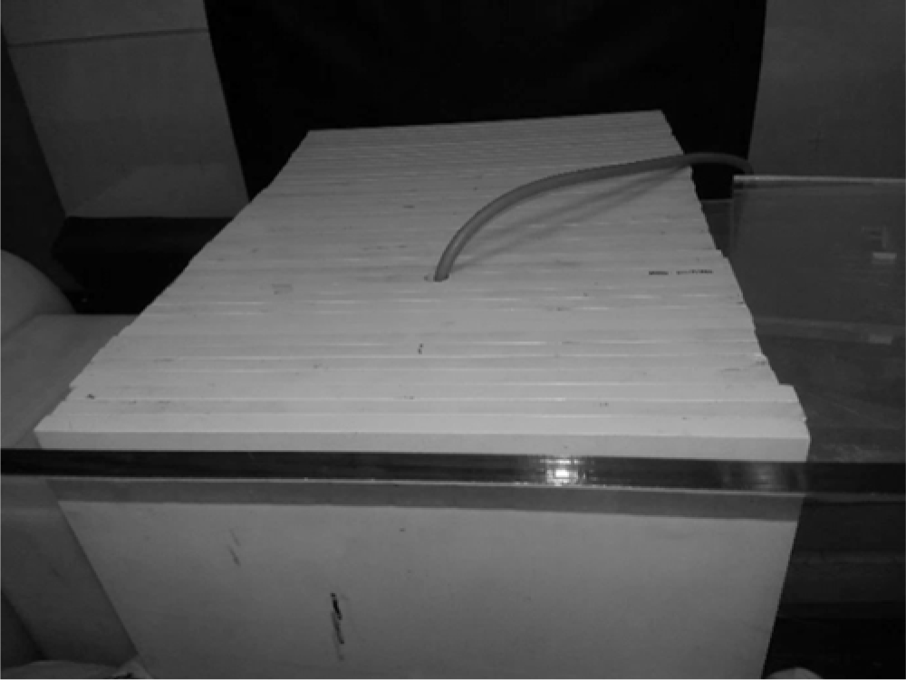
In this study, depth dose data for TBI were directly measured using layers of RW3 slab phantoms (LAP, Luneburg, Germany) with the thicknesses of 1, 2, 5, and 10 mm. These layers were arranged side by side on the TBI-couch at 312 cm SSD while a 5-cm gap maintained between the front surface of slab phantoms and the compensator (Figure 2). Reference Shahzadeh, Gholami, Aghamiri, Mahani, Nabavi and Kalantari12,Reference Van Dyk, Galvin, Glasgow and Podgorsak19 Dose measurements were carried out with Farmer and parallel plane ionization chamber for both 6- and 18-MV radiation beams.
Arrangement of slab phantoms.

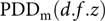
Finally, the measured
 ${\rm{PD}}{{\rm{D}}_{\rm{m}}}\left( {d.f.z} \right)$
at each depth was compared with the calculated
 ${\rm{PD}}{{\rm{D}}_{\rm{c}}}\left( {d.f.z} \right)$
using Equation (4)
Reference Houdek and Pisciotta17
:
 $$R\left( d.f.z \right) = {{\rm{PDD}}_{\rm{c}}\left(d.f.z\right) \over {\rm{PDD}}_{\rm{m}}\left( d.f.z \right)}$$
where
 $R\left( {d.f.z} \right)$
is the ratio of calculated and measured central axis PDD.
Relative dose rate
The dose rate is the amount of absorbed dose per each MU irradiation at reference depth. The measurement unit of dose rate is centigray per MU (
 ${\rm{cGy}}/{\rm{mu}}$
).
Reference Houdek and Pisciotta17,Reference Izewska, Rajan and Podgorsak22
In linear accelerators, for a 10 × 10 cm2 square field at SSD of 100 cm, the transmission ionization chambers are usually set to correspond the beam output to
 $1{\rm{cGy}}/{\rm{mu}}$
at reference depth (usually depth of maximum dose).
Reference Houdek and Pisciotta17,Reference Izewska, Rajan and Podgorsak22
So,
 $${\rm{D}}{{\rm{R}}_{\rm{r}}}\left( {{z_r}.{d_r}.{f_r}} \right) = {\rm{D}}{{\rm{R}}_{\rm{r}}}\left( {{z_0}.100.10} \right) = 1.00{\rm{\;cGy}}.{\rm{m}}{{\rm{u}}^{ - 1}}$$
where
 ${\rm{D}}{{\rm{R}}_{\rm{r}}}\left( {{z_{{\rm{max}}}}.{d_r}.{f_r}} \right)$
is the dose rate in reference depth (
 ${z_{{\rm{max}}}})$
at reference distance
 $({f_r} = 100\;{\rm{cm}})$
) and reference side of the square field (
 ${d_r} = 10{\rm{\;cm}}$
).
If any of the distance, depth and field size values are changed, the dose rate will differ accordingly. Reference Houdek and Pisciotta17,Reference Izewska, Rajan and Podgorsak22
An increase in the SSD is accompanied by the change in PDD and therefore dose rate. The dose rate in a large field size (f) and at extended treatment distance (SSD = d) was calculated using the values measured at isocentre and Equation (6):
 $${\rm{D}}{{\rm{R}}_{\rm{c}}}\left( {d.f} \right) = {\rm{D}}{{\rm{R}}_{\rm{r}}}\left( {100.10} \right) \times {\rm{ISL}}\left( d \right) \times {\rm{SF}}$$
where
 ${\rm{D}}{{\rm{R}}_{\rm{c}}}\left( {d.f} \right)$
is the calculated dose rate in reference depth at any treatment distance (
 $d$
) and field size (
 $f$
).
In this study, the TBI treatment was carried out while the Plexiglass frame on the treatment table was placed as a compensator between the phantom and the head of the treatment unit, and the transmission factor for the compensator had to be considered by applying the compensator (spoiler) transmission factor (
 ${\rm{SF}}$
). The SF values for the 6- and 18-MV radiation beams were 0·952 ± 0·02 and 0·972 ± 0·01, respectively. Inverse square law (ISL) is an inverse square correction that is calculated using Equation (7)
Reference Khan and Gibbons21
:
 $${\rm{ISL}}\left( d \right) = \left[ {\left( 100 + z_0 \right) \over \left( d + z_0 \right)} \right]^2$$
where (
 $d$
) is the therapeutic distance and
 ${z_0}$
is the depth of maximum dose.
The calculated relative dose rate then was compared with the directly measured relative dose rate using Equation (8):
 $${\rm{RD\;}}\left( {312.40} \right) = {{\rm{DR}}_{\rm{c}}\left( {d.f} \right) \over {\rm{DR}}_{\rm{m}}\left( {d.f} \right)}$$
where
 ${\rm{RD}}\left( {{\rm{d}}.{\rm{f}}} \right)$
is the ratio of calculated dose rate
 ${\rm{D}}{{\rm{R}}_{\rm{c}}}\left( {d.f} \right)$
and measured dose rate
 ${\rm{D}}{{\rm{R}}_{\rm{m}}}\left( {d.f} \right)$
at an extended distance which here is 312 cm and TBI field size (40 × 40 cm2).
In addition to the method for calculating the dose rate, another way to obtain the dose rate in TBI is the direct measurement method. The geometry of dose rate measurement in TBI is similar to that implemented for PDD measurement. The dose rate at depth of maximum dose for both 6- and 6-MV photon beams was measured using a Farmer-type ionization chamber. Phantom and chamber were irradiated under TBI condition while a 5-cm gap remained between the front surfaces of the phantom to the compensator. The objectives of this part were to validate the results of the dose rate calculated using Equation (6) and compare these results with measured dose rate under TBI conditions.
Beam flatness
TBI requires a large radiation field to cover the entire body of the patient. To this end, the collimator is usually rotated 45° and the patient lays along the diagonal of the field. Therefore, the beam profile, which may also affect the beam flatness, must be measured in the diagonal of the field. In addition, the uniformity of the beam decreases with an increase in the SSD. Thus, the beam profile and flatness must be measured under TBI conditions. Reference Li, Kong and Sun23
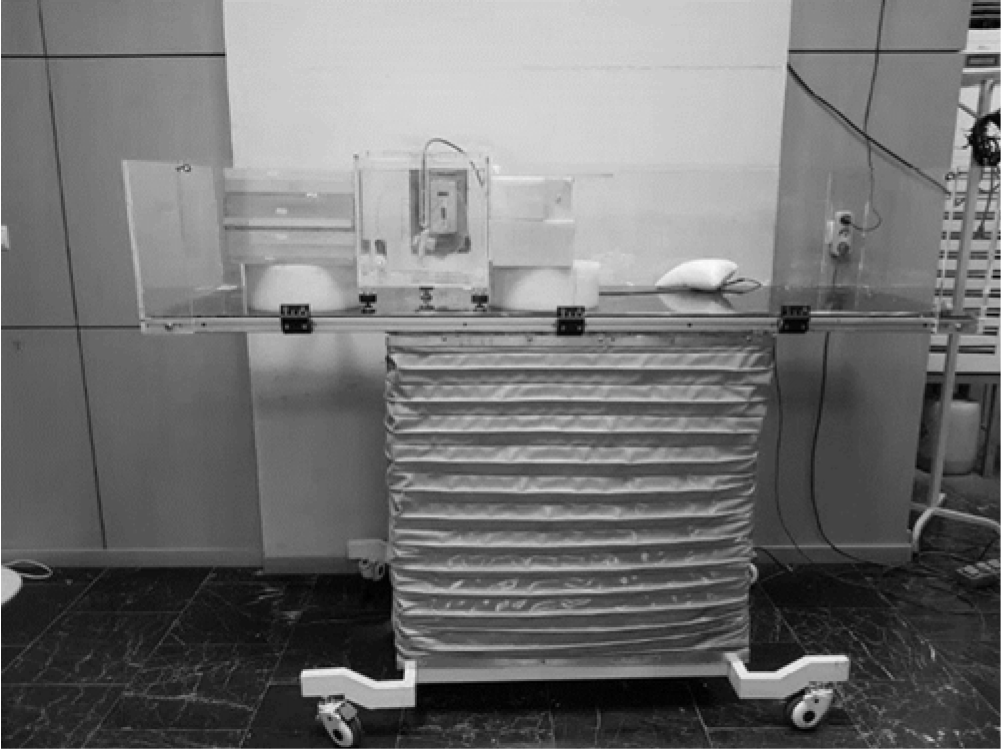
To measure the beam profile, a 30 × 30 × 30 cm3 water phantom was placed on a TBI-couch at SSD = 312 cm while a 5 cm gap was maintained between the front surface of the water phantom and the compensator. The Farmer ionization chamber was placed at a depth of 10 cm in the water phantom. Slab phantoms with different sizes were placed nearby the water phantom to produce full lateral scatter conditions (Figure 3). First, the phantom and chamber were irradiated under TBI conditions, and the central axis of the radiation field was placed on the phantom’s center and chamber. For each of next irradiations, the phantom was moved to the edge of the radiation field and irradiated. In the last step, all measurements were normalised to the central axis absorbed dose. After beam profile measurements, the flatness of the radiation beam was obtained using the maximum dose (
 ${D_{{\rm{max}}}}$
) and minimum dose (
 ${D_{{\rm{min}}}}$
) across 80% of the central width of the beam profile and Equation (9)
Reference Izewska, Rajan and Podgorsak22
:
 $$F = {D_{\rm{max}} - D_{\rm{min}} \over D_{\rm{max}} + D_{\rm{min}}} \times 100$$
Phantoms arrangement for beam profile measurement at SSD = 312 cm.

The beam flatness was calculated along the axis by measuring the profile for both the 6- and 18-MV energies.
Results
Central axis PDD results
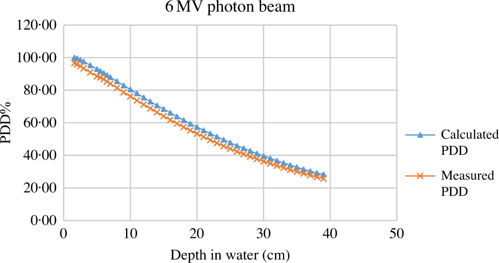
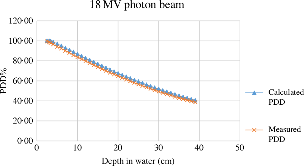
Figure 4 and Figure 5 show the measured and calculated PDD curves at SSD = 312 cm for a 40 × 40 cm2 (at isoplane)radiation field for the 6- and 18-MV photon beams, respectively.
Measured and calculated PDD at SSD = 312 cm and 40 × 40 cm2 field in the water. Line with multiplication sign markers presents calculated PDD and line with triangle markers presents measured PDD.

Measured and calculated PDD along curve central axis in a water phantom for SSD = 312 cm, field size = 40 × 40 cm2, and 18-MV photon beam. Line with multiplication sign markers is presenting calculated PDD and line with triangle markers demonstrate measured PDD.

Relative dose rate results
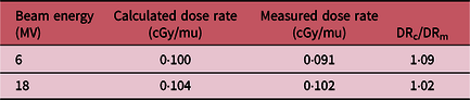
The calculated relative dose rate was compared with the directly measured relative dose rate under TBI conditions using Equation (8). The results of the calculated and measured dose rate and the ratio of calculated to measured dose rate are presented in Table 1.
The ratio of calculated and measured dose rate at in water phantom at SSD = 312 cm, depth of dose maximum and field size = 40 × 40 cm2

Beam flatness results
The beam profile was directly measured under TBI conditions (i.e., the field size of 40 × 40 cm2 at isoplane, SSD = 312 cm, 45° collimator rotation and 90° gantry angle), and then the flatness of the beam was calculated using Equation (9).
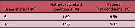
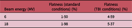
Table 2 presents the beam flatness under standard conditions versus the beam flatness under TBI conditions for both 6- and 18-MV photon beams.
Beam flatness under standard conditions and beam flatness under TBI conditions
Discussion
TBI is a significant and complex radiation therapy technique that is implemented in numerous radiation oncology departments around the world. The use of this technique requires a series of complex dosimetry measurement procedures. Some of these parameters could either be derived from standard conditions measured data or can be measured directly under TBI conditions. In the current study, PDD and dose rate were measured directly under TBI conditions and then were compared with those calculated from measured data under standard conditions. Furthermore, the flatness of the radiation beam was obtained under both TBI and standard treatment conditions for 6- and 18-MV photon beams.
Figure 4 shows the measured and calculated PDD curves for the 6-MV photon beam. The results indicate that the values of the calculated depth dose using Equation (2) are greater than the directly measured ones. The average of the calculated-measured ratio (R-ratio) for all depths is equal to 1·07. Percentage differences of calculated and measured values were also calculated for each depth, and the average of these values is equal to −6·97%. This difference is due to the fact that the Mayneord factor does not consider the scatter component in the absorbed dose estimation and thus this factor overestimates the PDD increment as a result of an increase in the SSD. Reference Khan and Gibbons21 This difference between the calculated and measured depth doses can also be realised in the 18-MV photon beam, which is presented in Figure 5. However, the agreement between the calculated and measured values is improved. In other words, with energy increment from 6- to 18-MV, the difference between calculated-measured depth dose was decreased such that the R-ratio and percentage difference of calculated and measured values were 1·04 and −4·14%, respectively. R factor decrement with energy increment can be explained by the fact that the amount of scatters decreases while energy increases. Reference Khan and Gibbons21 According to the results, since PDD calculation error for 6-MV photon beam is more than 5%, it is necessary to measure depth dose directly under TBI conditions, while it is not essential for the 18-MV photon beam direct measurement. Our results are in agreement with those of other studies. Reference Spunei, Mihai and Mălăescu18
The results in Table 1 indicate a good consistency between the calculated and measured dose rate for the 18-MV photon beam; however, for the 6-MV photon beam, the calculated-measured ratio was a considerable amount. Although these data are consistent with the results obtained by Houdek and Pisciotta, there is a slight difference between the results, which is due to the difference in radiation beam energy and SSD. Reference Houdek and Pisciotta17 The difference between the measured and calculated values can be due to the fact that using the ISL, it is assumed that we only deal with primary photon beams; therefore, with an increase in the scatter radiations, the ISL may generate some errors. Reference Khan and Gibbons21 This difference is more pronounced for 6-MV energy in comparison with the 18-MV energy. The explanation for this result is the higher contribution of the scattered component in the absorbed dose for a 6-MV photon beam than the 18-MV one. For the 18-MV photon beam, the dose rate can be calculated instead of direct measurement but for 6-MV due to the high ratio of a calculation error, direct measurement is required.
The results of the beam flatness measured under standard and TBI conditions (Table 2) show that with SSD increment, the beam flatness deteriorates; this is consistent with other studies, Reference Khan and Gibbons21,Reference Izewska, Rajan and Podgorsak22 and is more severe for an 18-MV photon beam than 6-MV. To improve the beam flatness, a filter can be designed and mounted to the head of the linear accelerator.
Basic dosimetric parameter measurements are an important and valuable step toward extended treatment distance radiation therapy techniques such as TBI and HBI. The obtained data can also be used for TPS commissioning. For future studies, measurement of other basic dosimetric parameters such as tissue maximum ratio (TMR), tray factor (TF), collimator and phantom dispersion factor (
 ${{\rm{S}}_{{\rm{cp}}}}$
) under TBI conditions is recommended.
In TBI, dose uniformity improves with SSD, but because of treatment room size limitation, SSD could not exceed 312 cm. Due to the time-consuming procedure of direct measurements and the high workload of the treatment centre, direct measurements at other extended SSDs and field sizes were not possible. In addition, it is recommended undertaking studies using measurements using other photon energies, which are not accessible with Clinac 2100C/D.
Conclusion
Based on the results of this study, although the depth dose and PDD values under TBI conditions can be derived by calculations from the standard measurement data, the results have −4·14 and −6·97% error values for 6-MV and 18-MV photon beams, respectively. Dose rate calculation errors were −9·89 and −1·96% for the 6- and 18-MV radiation beams, respectively. The beam flatness was also measured under standard and TBI conditions separately. The results indicated that with an increase in SSD at a constant field size, the flatness of the radiation beam becomes deteriorates. This problem was strongly observed for 18-MV photon beams.
Acknowledgements
The authors wish to thank the contribution of staff at the Cancer Institute of Imam Khomeini hospital for their collaboration in this project. This study was funded and supported by Tehran University of Medical Sciences (TUMS); Grant no. 96-03-30-36299.