Introduction
Nodular fasciitis is a rare, benign, pseudoneoplastic process occurring on mucosal, fascial and tendinous surfaces. Its overall incidence has been cited as 0.025 per cent, of which 15 per cent localise to the head and neck region.Reference Batsakis, Rice and Howard1 Although rare, it is the most common soft tissue pseudosarcoma.Reference Dahl and Angervall2 Given its rarity, varied histological presentation and pseudosarcomatous appearance, nodular fasciitis is frequently misdiagnosed on pre-operative, intra-operative and final analyses.Reference Montgomery and Meis3 More importantly, it is often mistaken for a sarcoma, due to its rapid growth, increased cellularity, prolific mitotic activity and infiltrative borders.Reference Silva, Bruce, Malik, Homer and Banerjee4
Although there have been recent reports of nodular fasciitis in the literature, there has been no discussion of the difficulty in diagnosing nodular fasciitis pre-operatively.Reference Silva, Bruce, Malik, Homer and Banerjee4–Reference Thompson, Fanburg-Smith and Wenig7 The purpose of this study is to report the clinical, radiological, cytological and intra-operative findings made prior to the determination of nodular fasciitis on final histopathological analysis.
Materials and methods
From 1999 to 2007, four female patients aged between 39 and 71 years with a final diagnosis of nodular fasciitis were reviewed at a tertiary care centre in New York City. Patients underwent pre-operative investigation for a head or neck mass, which included contrast-enhanced computed tomography (CT) of the neck and fine needle aspiration cytology. A frozen section was reviewed intra-operatively. The final diagnosis was based on pathological examination of the mass in its entirety.
Results
Case one
A 52-year-old woman presented with a mass in the left parapharyngeal space. Pre-operative radiological examination revealed a left parapharyngeal space tumour, non-vascular in origin. Fine needle aspiration cytology was consistent with either a possible sarcoma or a neurogenic tumour. The patient underwent resection of the mass. The frozen section diagnosis suggested a malignant process of a spindle cell type.
Case two
A 44-year-old woman presented with a left, level four neck mass. The CT scan suggested lymphoma. Fine needle aspiration cytology was consistent with a pseudo-inflammatory process, reported as a possible undifferentiated malignancy. Frozen section diagnosis was consistent with a neoplasm, possibly spindle cell type in origin.
Case three
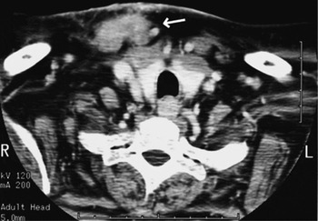
A 71-year-old woman with a history of breast cancer seven years previously presented with a mass at the right sternoclavicular joint (Figure 1). The presumed clinical and radiological diagnosis was a metastatic breast cancer lesion. Fine needle aspiration cytology suggested a diagnosis of either neurofibroma or fibromatosis. Intra-operatively, frozen section analysis revealed a neurogenic tumour, probably benign, with an inflammatory component.
Nodular fasciitis. Axial, contrast-enhanced neck computed tomography scan demonstrating a heterogenous, soft tissue mass (arrow) at the right sternoclavicular joint.

Case four
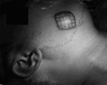
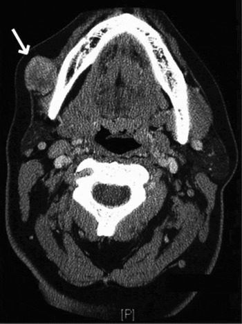
A 39-year-old woman presented with a right cheek mass (Figure 2). Radiologically, the mass appeared to be an accessory parotid tumour (Figure 3). Fine needle aspiration cytology revealed scattered epithelial cells with focal nuclear atypia, and spindle cell features embedded in a mucoid and myxoid matrix. The differential diagnosis ranged from a salivary gland adenoma to a low-grade salivary gland carcinoma. The patient underwent resection of the lesion. Intra-operatively, biopsy results were consistent with either a spindle cell neoplasm or a schwannoma.
Nodular fasciitis. A 39-year-old woman who presented with a right cheek mass.

Nodular fasciitis. Axial, contrast-enhanced neck computed tomography scan demonstrating a heterogenous, soft tissue mass (arrow) located anterior to the right parotid gland.

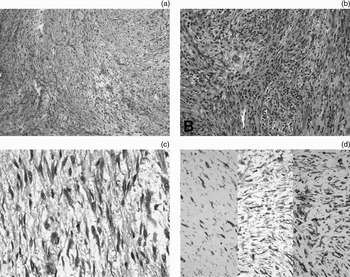
Grossly, the lesion in case four comprised an irregular, nodular structure measuring 2.7 cm in its greatest dimension. The cut surface was pale pink, glistening and rubbery, with intermingling fascicles of firm, fibrous tissue. Microscopically, the tissue showed a fascicular arrangement of spindle cells mainly comprising fibroblasts and myofibroblasts. In some areas, these were more tightly packed and intermingled with scanty bundles of mature collagen, whereas in others they were loose and surrounded by a moderate amount of stromal oedema and microhaemorrhages (Figure 4a). There was an appreciable variation in cellularity, with hypercellular spindle cell areas with lymphoid infiltrate (Figure 4b), together with less cellular areas showing a characteristic loosely textured, ‘feathery’ appearance. Lesional cells showed bland cytological features, with an absence of hyperchromasia or variably sized nuclei (Figure 4c). Less than three mitotic figures were observed per 10 high power fields, and none were atypical. Immunohistochemical testing using antisera to smooth muscle actin, vimentin and CD68 was positive in lesional cells (Figure 4d). However, immunostaining for cytokeratin and S-100 was negative, as expected.
All patients underwent resection of their masses without peri-operative complication. In all patients, the final diagnosis of nodular fasciitis was made only after permanent section and immunohistological analysis.
Photomicrographs of nodular fasciitis, demonstrating: (a) cellular areas and scanty collagen bundles (left), plus less cellular areas with oedema and extravasation of erythrocytes (right) (HPS, hematoxylin-phloxine-saffron; ×100); (b) hypercellular areas with abundant lymphocytic infiltrate and rare plasma cells (HPS, hematoxylin-phloxine-saffron; ×200); (c) bland cytological features, absence of hyperchromasia and pleomorphism, and occasional mitotic figures (centre) (HPS, hematoxylin-phloxine-saffron; ×400); and (d) reactivity with antisera to CD68, smooth muscle actin and vimentin (×40).

Discussion
Nodular fasciitis presents clinically as a rapidly enlarging, subcutaneous mass, which may be tender or painful and can extend into surrounding tissues. The peak incidence is in the fourth decade, and men and women are equally affected. Seven to 12 per cent of all cases occur in children.Reference Batsakis, Rice and Howard1, Reference Silva, Bruce, Malik, Homer and Banerjee4, Reference Matusik, Wiberg, Sloboda and Andersson8 On CT and magnetic resonance imaging, nodular fasciitis has a varied presentation; lesions appear as discrete masses, either cystic or solid in appearance, with enhancement.Reference Kim, Kim, Park, Baek, Byun and Kim9 On cytology, nodular fasciitis appears as a cellular smear composed primarily of various-sized spindle cells, occurring as single cells or as cohesive tissue particles in a myxoid background. Ganglion-like cells have also been described. In these cells, there is no atypia or atypical mitoses.Reference Matusik, Wiberg, Sloboda and Andersson8
Microscopically, nodular fasciitis is mainly composed of immature fibroblasts arranged in bundles and interweaving fascicles, with a small amount of collagen, within a loose, mucopolysaccharide-rich stroma. The vasculature of the lesion is fairly constant, consisting of a fine capillary network arranged in a radial pattern. Other cells that may be found include foamy histiocytes and multinucleate giant cells.Reference Batsakis, Rice and Howard1, Reference Montgomery and Meis3, Reference Matusik, Wiberg, Sloboda and Andersson8 Nodular fasciitis is often confused with neurofibroma, fibromatosis, fibrous histiocytoma, fibrosarcoma and leiomyosarcoma.Reference Han, Hu, Yang, Wang and Huang6, Reference Matusik, Wiberg, Sloboda and Andersson8
Due to the nonspecific nature of nodular fasciitis, its diagnosis is aided by specialised staining, including that for smooth muscle, muscle-specific actins, vimentin, CD68 and KP1 (a histiocyte marker). Staining for cytokeratin, S-100 protein, HMB45, CD34, factor VIII related antigens and desmin is negative.Reference Montgomery and Meis3, Reference Nishi, Brey and Sanchez5, Reference Matusik, Wiberg, Sloboda and Andersson8, Reference Kayaselcuk, Demirhan, Kayaselcuk, Ozerdem and Tuncer10 Unfortunately, special staining and multiple immunohistochemical studies may be difficult to perform on a small needle aspirate, and a surgical tissue biopsy may be required.
• Nodular fasciitis is a rare, benign, pseudoneoplastic process occurring on mucosal, fascial and tendinous surfaces
• Although rare, it is the most common soft tissue pseudosarcoma
• This paper describes four cases of nodular fasciitis presenting in the head and neck
• In a patient with nonspecific results following investigation of a head or neck mass, nodular fasciitis should be considered. Use of appropriate immunohistochemical markers will aid the final diagnosis
Nodular fasciitis, although rare, can be mistaken for other benign or malignant head or neck lesions. Radiographical and pathological studies can lead to equivocal diagnoses. Nodular fasciitis is a benign process that is self-limited; it never metastasises. Often, lesions can be followed clinically as they may regress spontaneously.Reference Matusik, Wiberg, Sloboda and Andersson8 If the diagnosis is unclear, local excision of the tumour is the preferred treatment.Reference Dahl and Akerman11 Recurrence is rare; if it does occur, the initial diagnosis should be reconsidered.Reference DiNardo, Wetmore and Potsic12 Pre-operative findings suggesting a malignant process can have a negative effect on surgical planning, with unnecessary risk of injury to adjacent structures. In a patient with nonspecific results after investigation of a head or neck mass, nodular fasciitis should be considered.