


Highlights
-
• Rising EEG demand and technologist shortages highlight the need for appropriate test utilization.
-
• This study identified clinical scenarios with low diagnostic yield and revealed substantial gaps in requisition completeness across physician specialties.
-
• Findings directly informed the redesign of the KHSC EEG requisition to improve clarity, appropriateness and resource allocation.
Introduction
The electroencephalogram (EEG) is a widely used diagnostic tool that measures electrical activity in the brain through electrodes placed on the scalp. It is an essential supportive tool in evaluating neurological conditions, particularly epilepsy. EEG findings are broadly categorized as being either normal, demonstrating epileptiform activity or containing non-epileptiform abnormality. Epileptiform abnormalities, characterized by features like spikes and sharp waves, can be supportive of a diagnosis of epilepsy. Reference Britton, Frey and Hopp1 In contrast, non-epileptiform abnormalities, such as focal or generalized slowing, may reflect underlying brain dysfunction that may not be directly related to epilepsy or seizures. Reference Britton, Frey and Hopp1
Clinical utility and limitations of short-duration EEGs
Short-duration EEGs are widely employed in clinical practice due to their accessibility and efficiency. These tests, which include routine, portable and sleep-deprived EEGs, are typically recorded for about 30–60 minutes and have a total session duration of around 1.5 hours including electrode application and removal. Video recording is integrated as the gold standard for these tests to correlate observable events with electrical brain activity. Reference Peltola, Leitinger and Halford2 Studies have shown that prolonged inpatient video EEGs are able to provide more comprehensive data compared to short-duration EEGs. Reference Aslan and Gungor3,Reference Chen, Atallah, Pauldurai, Becker and Koubeissi4 However, these are limited by their cost and resource demands, making short-duration EEGs a more accessible first-line investigation. Short-duration EEGs also have limitations, such as reduced likelihood of capturing abnormal activity due to the length of recording. Reference Baldin, Hauser, Buchhalter, Hesdorffer and Ottman5 Additionally, it is also more difficult to record activity in specific states, such as during natural sleep or exposure to environmental triggers of seizure activity. Reference Baldin, Hauser, Buchhalter, Hesdorffer and Ottman5
The demand for EEGs may be constrained by available resources, reflecting pressure on healthcare systems. In Canada, there is a nationwide shortage of trained EEG technologists, which may delay the scheduling and performance of tests. Reference Van Winssen, Andrade and Andrade6 In Ontario, the increasing prevalence of epilepsy and related neurological disorders intensifies the demand for EEG services, as the Canadian government reported a crude epilepsy prevalence of 168,800 in the 2023–2024 fiscal year. 7 However, all neurodiagnostic centers that perform EEGs across Ontario have been affected by labor shortages, especially for EEG services. Reference Van Winssen, Andrade and Andrade6 This resource limitation affects patient care and puts additional pressure on staff.
Diagnostic yield and its variability
The diagnostic yield of EEGs, defined as the proportion of tests detecting abnormalities, varies significantly across studies, with reported rates ranging from 10% to 50%. Reference Angus-Leppan8–Reference Gilbert and Gartside14 This variability is influenced by cohort characteristics, clinical settings and methodological differences. Several clinical and demographic factors have been associated with increased diagnostic yield, though findings have been inconsistent across studies.
Challenges in previous studies
Despite the widespread use of EEGs, significant gaps remain in understanding the factors influencing diagnostic yield. One major limitation in previous studies is the exclusion or selective inclusion of pediatric populations. Reference Bozorg, Lacayo and Benbadis9–Reference Phamnguyen, Szekely and Swinburn11,Reference Watemberg, Tziperman, Dabby, Hasan, Zehavi and Lerman-Sagie13–Reference DeRoos, Chillag, Keeler and Gilbert15 Pediatric EEGs are often indicated for conditions distinct from those in adults, such as febrile seizures, developmental delay or genetic epilepsy syndromes. By excluding or focusing solely on pediatric patients, many studies fail to capture the full spectrum of diagnostic yield across age groups.
Another limitation is the focus on specific clinical settings, such as inpatient or outpatient populations, without accounting for differences between them. Reference Bozorg, Lacayo and Benbadis9,Reference Monif and Seneviratne10,Reference Gilbert and Gartside14,Reference Angus-Leppan16–Reference Sundaram, Hogan, Hiscock and Pillay19 Inpatients, often evaluated for acute or severe conditions, are more likely to yield abnormalities on EEG than outpatients, who are typically assessed for more stable or less urgent conditions. Reference Phamnguyen, Szekely and Swinburn11 By addressing multiple settings, including different types of inpatient settings, a more comprehensive understanding of the factors influencing yield can be achieved.
Guidelines for EEG utilization
There are two established United Kingdom guidelines to outline appropriate reasons for requesting an EEG for the investigation and diagnosis of epilepsy. These include the National Institute of Clinical Excellence (NICE) and the Scottish Intercollegiate Guidelines Network (SIGN). The Canadian Neurological Society (CNS) formed a Choosing Wisely task force in August 2018 with the American Board of Internal Medicine and produced guidelines recommending strategies to reduce unnecessary testing and treatments. Reference Beyak, Costello, Burton and Couillard20
EEG requisition forms
A requisition form is important in facilitating effective communication during the referral process. Reference Hartveit, Vanhaecht, Thorsen, Biringer, Haug and Aslaksen21 It serves as the primary tool through which referring clinicians convey essential information, the rationale for the test and specific diagnostic concerns to the interpreting team. However, requisition forms may be poorly completed.
Study rationale
With the growing demand for EEGs in Ontario and existing literature emphasizing the need to minimize unnecessary diagnostic testing, this study addressed two complementary objectives: the characterization of EEG findings across clinical factors and the assessment of requisition quality as a component of efficient clinical communication. Our ultimate goal was to inform improvements in EEG referral processes and support the development of a more effective requisition form that promotes optimal use of healthcare resources.
Methods
Experimental design
This single-center study used a retrospective cohort design to examine EEGs performed over a 6-month period between March and August 2024 at Kingston General Hospital, a Canadian, tertiary-level, acute-care university teaching hospital and District Epilepsy Centre. The EEG department provides adult and pediatric EEG services across South-Eastern Ontario. At the time of the study, there were 4 adults and 2 pediatric neurologists interpreting EEG studies, 5–6 EEG technologists, and 2 trainees. Ethics approval for the study was obtained from the Queen’s University Health Sciences and Affiliated Teaching Hospitals Research Ethics Board (HSREB, reference 6041410).
The inclusion criteria for this study were all routine, portable and sleep-deprived EEGs conducted during this period. A portable EEG is a bedside recording performed outside the standard EEG lab for inpatients and emergency department patients. Exclusion criteria included long-duration EEGs (i.e., continuous, ambulatory, epilepsy unit monitoring). For patient confidentiality and data security, all collected data were anonymized by using sequential identification numbers, and patient assent for retrospective use of anonymized data for research was verified.
Data collection and variables
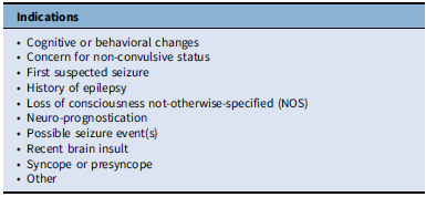
Demographic and clinical data were extracted from paper and digital requisition forms, as well as the EEG reports retrieved from the electronic patient medical records. Variables of interest included age, setting where the EEG was performed (inpatient – critical care, inpatient – non-critical care, emergency department or outpatient), requesting physician specialty, whether the patient was receiving anti-seizure or sedating medications (yes/no), whether the patient has a stated prior history of epilepsy (yes/no) and the type of EEG (routine, portable or sleep-deprived). The indication for the EEG was classified into categories informed by previous studies Reference Bozorg, Lacayo and Benbadis9–Reference Phamnguyen, Szekely and Swinburn11 and expert clinical opinion. The indication categories are outlined in Table 1.
Classification for EEG indications

Table 1. Long description
A textbox titled ʼIndicationsʼ contains a bulleted list of reasons for performing an EEG. The list includes: Cognitive or behavioral changes, Concern for non-convulsive status, First suspected seizure, History of epilepsy, Loss of consciousness not-otherwise-specified (NOS), Neuro-prognostication, Possible seizure event(s), Recent brain insult, Syncope or presyncope, and Other.
EEG = electroencephalography.
Requisition form completion data was collected by examining each requisition section for any pen stroke or text: “Medications, Patient History, Neurological & Psychiatric Signs, Clinical Diagnosis, and Type of Information Sought.” If one or more requisition sections were not filled in correctly, the entire requisition was marked incomplete. A sample KHSC EEG requisition form can be found in Supplementary Figure 1.
Additional data were collected for patients who underwent a sleep-deprived EEG to determine whether stage 2 (N2) sleep was achieved. These patients were then reviewed to identify if they had an EEG within the prior two years, and data regarding the findings of that previous EEG were extracted.
Classification of EEG findings
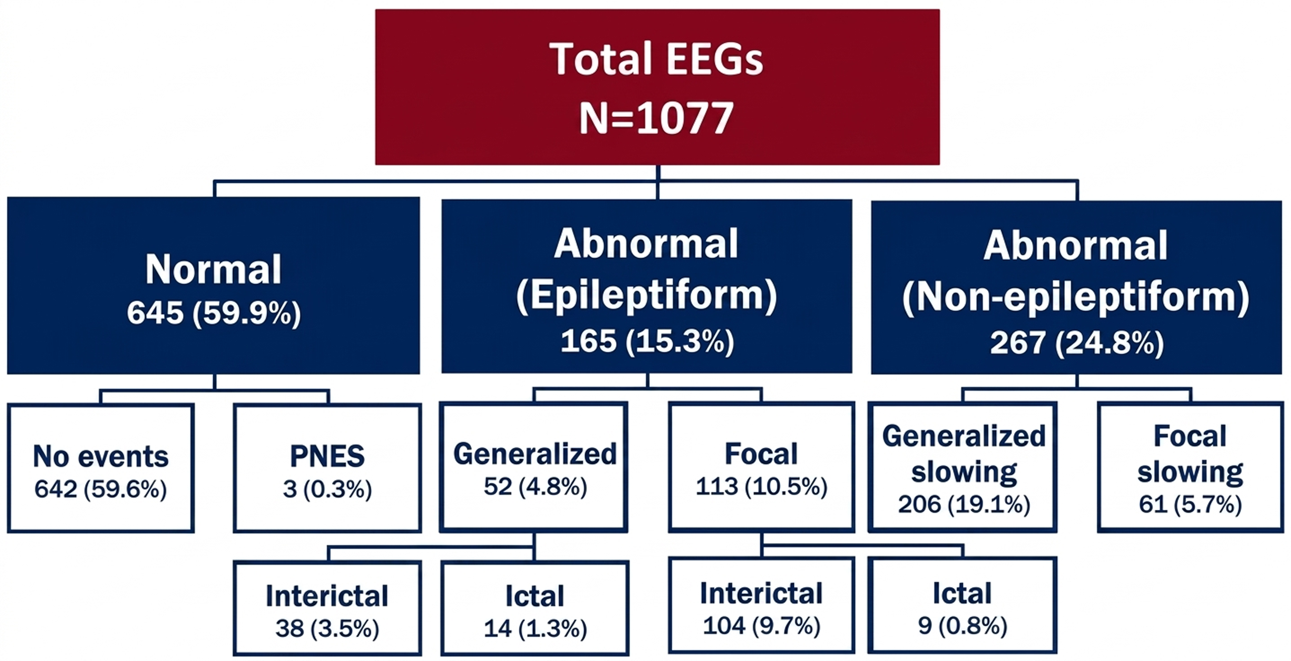
The findings of the EEGs were categorized into three broad classifications: normal, abnormal non-epileptiform and abnormal epileptiform. These classifications each have further subclassifications, outlined in Figure 1.
Yield by EEG finding classification for Cohort 1. This figure outlines the classification categories for EEG findings chosen for this study, as well as the proportion of EEGs under each classification. PNES = psychogenic non-epileptic seizures; EEG = electroencephalography.

Outcome measures
The primary outcome of this study was the diagnostic yield, defined in two ways:
-
1. Total diagnostic yield: the proportion of all EEGs demonstrating any abnormality, whether epileptiform or non-epileptiform.
-
2. Epileptiform diagnostic yield: the proportion of EEGs showing epileptiform abnormalities.
Additional analyses focused on identifying associations between clinical and demographic variables, requisition completeness and the likelihood of detecting epileptiform abnormalities.
Statistical analysis
Descriptive statistics (frequencies and percentages) were used to report the demographic and clinical data of the cohort. Yield was reported as the proportion of abnormal EEGs within each result classification and for each event per variable.
Univariate analyses were conducted to examine associations between individual variables and the binary outcome of epileptiform abnormalities using chi-squared tests. Those with a significant association were included in two multivariate logistic regression models with a forward stepwise approach to identify predictors of all abnormalities and epileptiform abnormalities. Variables with a p-value of <0.05 were included in the final regression models. The results of these analyses were reported as odds ratios (ORs) with corresponding 95% confidence intervals (CIs). A McNemar’s test was used to assess paired samples of patients who underwent both a prior EEG outside the study period and a subsequent sleep-deprived EEG. Statistical significance was defined as a p-value of less than 0.05. All analyses were conducted using IBM SPSS Statistics for MacOS, version 29.0.2.
Results
Data cohorts
In the 6-month period, a total of 1215 EEGs were performed. These were divided into two cohorts for analysis. In Cohort 1, 132 repeat EEGs were removed, retaining only the first for each patient within the time period, and 6 that were incomplete or with missing reports were removed, resulting in a sample of 1077 EEGs.
For Cohort 2, the repeated EEGs were retained, and only EEGs with associated paper requisitions or digitally scanned copies found were included as we discovered that some requisitions were inadvertently lost. This cohort was used in the analysis of requisition completeness (N = 960).
Demographics and diagnostic yield of EEG findings
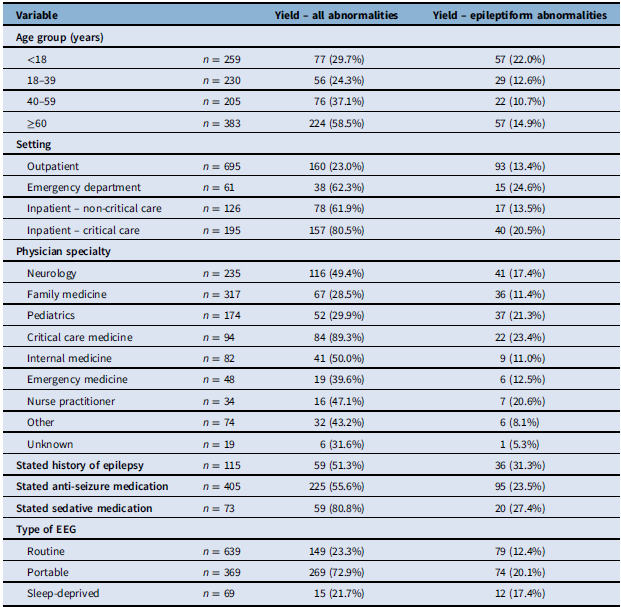
Clinical data for Cohort 1 are summarized in Table 2. Patients’ ages ranged from 0 to 101 years (median 45, IQR 48). Nearly two-thirds of patients (64.5%) were outpatients, and just over one-third (37.6%) were stated to be taking anti-seizure medications (ASMs) at the time of EEG. Recordings were most commonly ordered by Family Medicine physicians (29.4%), followed by Neurology (21.8%), and Pediatrics (16.5%). Most EEGs performed were routine (59.3%), followed by portable (34.3%) and sleep-deprived EEGs (6.4%).
Relationship of diagnostic yield of EEG in Cohort 1 to demographic and clinical factors

Table 2. Long description
A table comparing the diagnostic yield of EEG for all abnormalities and epileptiform abnormalities across different variables. The table has 11 rows and 3 columns. Column headers are Variable, Yield – all abnormalities, and Yield – epileptiform abnormalities. Row labels include Age group, Setting, Physician specialty, Stated history of epilepsy, Stated anti-seizure medication, Stated sedative medication, and Type of EEG. Each row provides the number of patients (n) and the yield percentages for all abnormalities and epileptiform abnormalities. Notable trends include higher yields in the inpatient critical care setting, among patients with a stated history of epilepsy, and those taking anti-seizure or sedative medications. Routine EEGs show lower yields compared to portable and sleep-deprived EEGs.
EEG = electroencephalography.
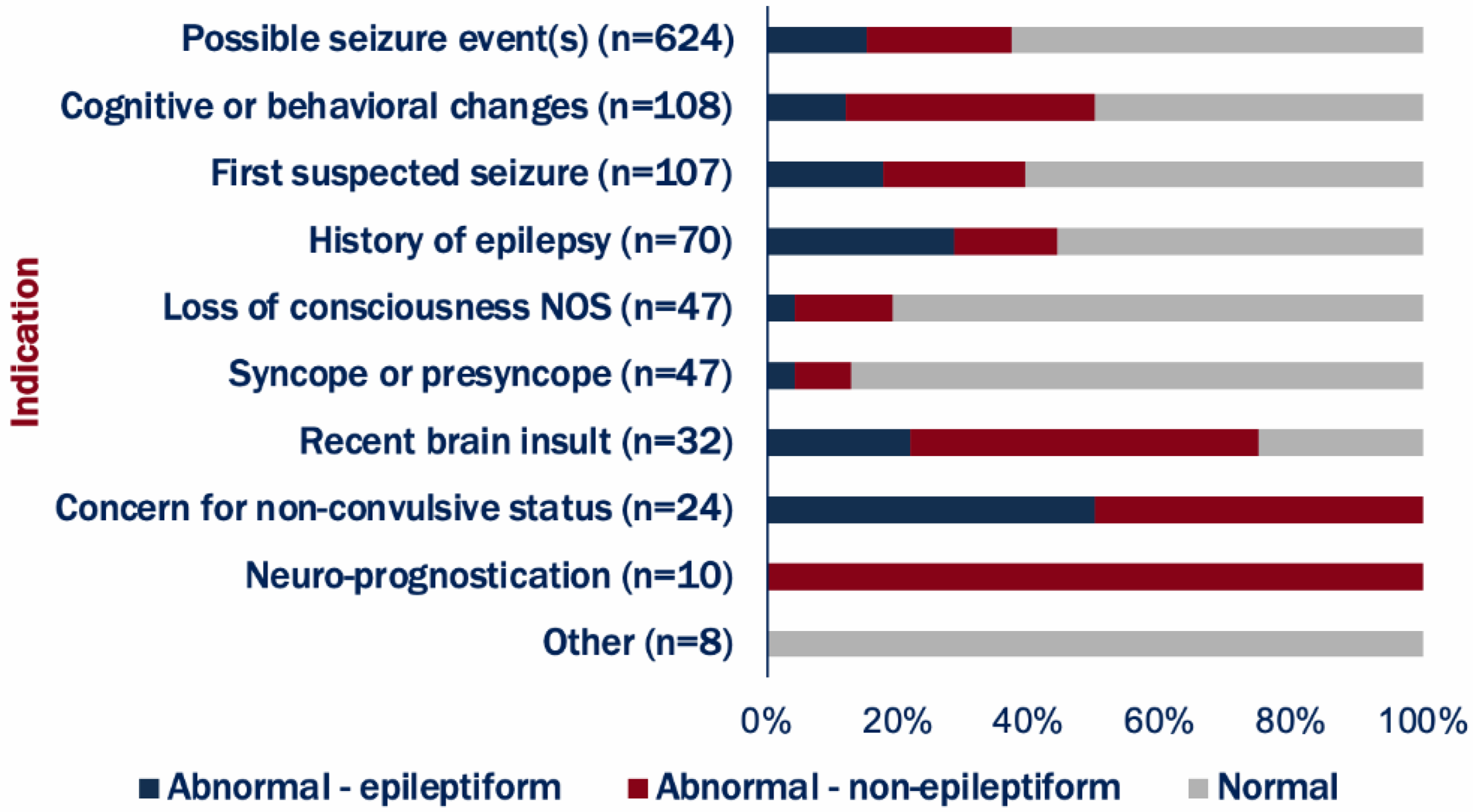
Of these EEGs, 645 (59.9%) were normal, while 432 (40.1%) showed abnormalities – 165 (15.3%) were epileptiform and 267 (24.8%) were non-epileptiform (Figure 1). Yield varied by clinical indication (Figure 2). The most common indication by far was possible seizure events (n = 624). Indications such as “concern for non-convulsive status” and “neuro-prognostication” had the highest yields, with abnormalities present in 100% of cases. Notably, while neuro-prognostication cases showed abnormalities in all recordings, these were all non-epileptiform but important for patient management (e.g., global suppression suggesting poor prognosis). In contrast, “syncope or presyncope” had the lowest yield besides “other.” Of the 47 referred for syncope or presyncope, 2 (4.3%) had epileptiform abnormalities, 4 (8.5%) had non-epileptiform abnormalities and 41 (87.2%) were normal.
EEG findings by clinical indication. Indications for EEG requisitions, categorized by abnormal (epileptiform) (blue), abnormal (non-epileptiform) (red) and normal (gray) findings. Indications are ranked in descending order based on the number of requests, with the most common indication at the top. NOS = not-otherwise-specified; EEG = electroencephalography.

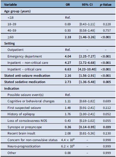
Using multivariate logistic regression, five variables proved significant in predicting an EEG with any abnormality (Table 3). Older age was significantly associated with an EEG with abnormalities; those aged ≥60 years had increased odds compared to those aged under 18 years (OR 2.18; 95% CI 1.46–3.26). Compared with outpatients, those who had an EEG conducted in the emergency department, as an inpatient in non-critical care, or as an inpatient in critical care were more likely to show an abnormality, with ORs of 4.04 (95% CI 2.25–7.27), 4.27 (95% CI 2.72–6.68) and 6.63 (95% CI 4.23–10.40), respectively. Patients also had increased odds if they were on a stated ASM or sedative medication, compared to those who were not, with ORs of 2.16 (95% CI 1.56–2.91) and 2.73 (95% CI 1.36–5.48), respectively. Presyncope or syncope as an indication was associated with decreased odds of having an EEG with abnormalities (OR 0.36; 95% CI 0.14–0.95).
Multivariate logistic regression model for predictive factors of any EEG abnormality in Cohort 1

Table 3. Long description
A table with 15 rows and 4 columns. The columns are labeled Variable, OR, 95% CI, and p-Value. The table presents data on the odds ratios (OR), 95% confidence intervals (CI), and p-values for various predictive factors of any EEG abnormality. The variables are grouped into age group, setting, stated anti-seizure medication, stated sedative medication, and indication. Row 1: Age group (years), <18, Ref., -. Row 2: Age group (years), 18-39, 0.69, [0.43-1.11], 0.128. Row 3: Age group (years), 40-59, 0.93, [0.58-1.48], 0.757. Row 4: Age group (years), >=60, 2.18, [1.46-3.26], <0.001. Row 5: Setting, Outpatient, Ref., -. Row 6: Setting, Emergency department, 4.04, [2.25-7.27], <0.001. Row 7: Setting, Inpatient - non-critical care, 4.27, [2.72-6.68], <0.001. Row 8: Setting, Inpatient - critical care, 6.63, [4.23-10.40], <0.001. Row 9: Stated anti-seizure medication, 2.16, [1.56-2.91], <0.001. Row 10: Stated sedative medication, 2.73, [1.36-5.48], 0.005. Row 11: Indication, Possible seizure event(s), Ref., -. Row 12: Indication, Cognitive or behavioral changes, 1.11, [0.68-1.82], 0.689. Row 13: Indication, First suspected seizure, 1.48, [0.91-2.41], 0.112. Row 14: Indication, History of epilepsy, 1.76, [1.00-2.41], 0.052. Row 15: Indication, Loss of consciousness NOS, 0.43, [0.19-1.02], 0.055. Row 16: Indication, Syncope or presyncope, 0.36, [0.14-0.95], 0.039. Row 17: Indication, Recent brain insult, 2.08, [0.81-5.36], 0.130. Row 18: Indication, Concern for non-convulsive status, 4.4 x 10^8, -, 0.998. Row 19: Indication, Neuro-prognostication, 6.2 x 10^8, -, 0.999. Row 20: Indication, Other, 0.00, -, 0.999.
EEG = electroencephalography; NOS = not-otherwise-specified.
A second logistic regression conducted to identify predictors of only epileptiform abnormalities identified four significant variables (Supplementary Table 1). Younger age was significantly associated with an increased likelihood of abnormalities; those aged 18–39, 40–60 and ≥60 years all had reduced odds compared to the pediatric (<18) group (OR 0.37; 95% CI 0.22–0.63, OR 0.28; 95% CI 0.16–0.49, OR 0.45; 95% CI 0.29–0.70). Patients also had increased odds if they had a previous history of epilepsy, were on a stated ASM or were on a sedative medication, compared to those who were not, with ORs of 1.97 (95% CI 1.20–3.22), 2.71 (95% CI 1.83–4.03) and 2.29 (95% CI 1.29–4.04), respectively.
Analysis of sleep-deprived EEGs
A chi-squared test for independence showed no significant association between undergoing a sleep-deprived EEG and the likelihood of detecting an epileptiform abnormality in Cohort 2 (χ2(1) = 1.447, p = 0.229, OR 1.503; 95% CI 0.77–2.93). Similarly, no significant association was found between achieving N2 sleep during recording and the likelihood of an epileptiform abnormality (χ2(1) = 0.023, p = 0.88, OR 0.91; 95% CI 0.26–3.22).
Among the 68 patients with sleep-deprived EEGs, 51 (75%) had at least one previous EEG. Of those with a prior EEG, 39 (76.5%) had it within the previous 2 years, of which 34 (87.2%) were routine, 3 (7.7%) were portable and 2 (5.1%) were sleep-deprived. Regarding the findings of these prior EEGs, 2 showed non-epileptiform abnormalities, 3 showed epileptiform abnormalities, 8 had minor findings and 26 were normal. In 13 cases (33.3%), a sleep-deprived EEG was recommended by the reporting neurologist: 3 due to abnormal findings, 7 to confirm minor findings and 3 despite normal results due to strong clinical suspicion. Of these, three (23.1%) showed epileptiform abnormalities on the subsequent sleep-deprived EEG.
A McNemar’s test for paired data was used to compare initial and subsequent sleep-deprived EEGs in the 39 patients to assess whether sleep deprivation increased the likelihood of abnormalities. The test revealed no significant effect (p = 0.219). However, the data suggest a potential trend toward increased abnormalities with sleep deprivation, but the sample size may have been too small to confirm this (Supplementary Table 2).
EEG requisition forms
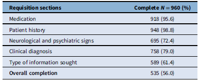
For the 1215 EEGs conducted, a total of 960 requisition forms were available (Cohort 2). This cohort included repeat EEGs, defined as multiple EEG recordings performed on the same patient at different time points, to allow evaluation of how requisition completeness varied by patient setting or ordering physician specialty. Online EEG reports (N = 1215) were utilized for patient clinical and demographic variable collection. Out of the total requisitions (N = 960), 535 requisitions (56%) were incomplete and 425 requisitions (44%) were complete. Table 4 outlines the different requisition sections and the completeness per section. A chi-squared test indicated no significant association between requisition completeness and EEG findings (χ2 = 2.022, p = 0.155). Therefore, requisition completion and EEG findings were analyzed separately.
EEG requisition completeness

Table 4. Long description
A table titled ʼRequisition sectionsʼ with two columns: ʼRequisition sectionsʼ and ʼComplete N = 960 (%)ʼ. The table has six rows, including a header row. The rows are as follows: Row 1: Medication, 918 (95.6 percent); Row 2: Patient history, 948 (98.8 percent); Row 3: Neurological and psychiatric signs, 695 (72.4 percent); Row 4: Clinical diagnosis, 758 (79.0 percent); Row 5: Type of information sought, 589 (61.4 percent); Row 6: Overall completion, 535 (56.0 percent). The table shows the number and percentage of complete requisition sections for each category, with patient history having the highest completeness and type of information sought having the lowest.
EEG = electroencephalography.
In the analysis of requisition completeness by patient setting, outpatients served as the reference group for the logistic regression analysis due to the highest proportion of requests (Supplementary Table 3). Emergency department and critical care requests were found to be significantly less likely to have complete requisitions than outpatients (OR 0.464; 95% CI 0.24–0.89, OR 0.586; 95% CI 0.42–0.82).
In the logistic regression analysis of requisition completeness by patient setting, neurologists served as the reference group (Supplementary Table 4). The analysis revealed that neurologists are less likely to complete their requisitions compared to other specialties. Emergency medicine (OR 2.484; 95% CI 1.21–5.10), family medicine (OR 2.616; 95% CI 1.80–3.81), pediatrics (OR 3.812; 95% CI 2.46–5.90), nurse practitioners (OR 3.372; 95% CI 1.59–7.16) and other (OR 2.108; 95% CI 1.18–3.77) were all significantly more likely to have complete requests.
Discussion
This study revealed several important trends regarding EEG utilization and diagnostic yield. The proportion of EEG showing abnormalities (∼40%) falls within the ranges previously reported in the literature. Reference Angus-Leppan8–Reference Gilbert and Gartside14 As observed in a previous study, Reference Phamnguyen, Szekely and Swinburn11 abnormal findings were more likely in patients undergoing EEGs in inpatient and emergency settings compared to outpatients, likely reflecting greater illness severity and a higher pretest probability of neurological pathology.
Clinical indication was strongly associated with diagnostic yield. Indications such as neuro-prognostication and suspected non-convulsive status epilepticus were highly predictive of EEG abnormalities, which is expected given that patients referred for these indications were acutely ill or in critical care. Conversely, the indication of “syncope or presyncope” was associated with a significantly reduced likelihood of abnormalities (Table 3), supported by literature Reference Mecarelli, Pulitano, Vicenzini, Vanacore, Accornero and De Marinis22–Reference Poliquin-Lasnier and Moore24 and NICE/SIGN guidelines, 25,26 showing low yield in EEGs referred for syncope. These findings reinforce the need for careful patient selection and indication-based requisition to optimize resource use.
The NICE guidelines recommend that EEGs be primarily requested to support the diagnosis of epilepsy and differentiate between seizure types. 25 Additionally, EEG is not recommended to investigate transient loss of consciousness. 27 NICE guidelines should be strictly adhered to maximize EEG potential and decrease inappropriate referrals. 25 Similarly, the SIGN guidelines state that an EEG is recommended when there is a high clinical suspicion of epilepsy and should not be performed if a patient’s symptoms point to non-epileptic events, such as syncope. 26 Furthermore, EEG should not be the only tool used to diagnose epilepsy. As for Choosing Wisely Canada, there are five neurology recommendations, one of which states, “Don’t order neuroimaging or EEG in asymptomatic patients in the emergency department with syncope and a normal neurological evaluation.” 28
Guidelines for EEG utilization vary internationally, reflecting differences in scope and level of specificity. For example, NICE and SIGN guidelines recommend EEG primarily to support the diagnosis of epilepsy and to help differentiate between seizure types. 25,26 In contrast, Choosing Wisely Canada provides broader recommendations focused on reducing unnecessary testing in situations such as syncope. NICE and SIGN guidelines offer more detailed, indication-specific guidance, while Choosing Wisely Canada emphasizes resource stewardship.
These variations influence diagnostic approaches, test indications and overall management strategies involving EEGs. Countries urgently need to develop and establish clinical guidelines for EEG utilization to decrease the number of referral requests, as previous research has suggested that 50–60% of requests are unnecessary. Reference Smith, Bartolo, Pickles and Tedman29 Given the increasing demand and labor shortage, guidelines can help promote the optimal use of EEG services to decrease the burden on the healthcare system.
Multivariate regression analyses provided further insight into predictors of EEG abnormalities. Older age, inpatient status and use of anti-seizure or sedative medications significantly increased the odds of detecting any abnormality. In contrast, predictors of epileptiform abnormalities included some distinct predictors like younger age and history of epilepsy. This age-dependent pattern aligns with known developmental differences in seizure susceptibility and EEG findings. Reference Monif and Seneviratne10,Reference Gilbert and Gartside14 Several studies have identified younger age as a factor associated with higher diagnostic yield in EEG, even when analyzing predominantly adult populations. Reference Monif and Seneviratne10,Reference Marsan and Zivin18 However, the significance of age as a predictor differs from other studies conducted with varying cohorts that found no effect, including one with exclusively an inpatient population and one with patients having a known history of seizures. Reference Phamnguyen, Szekely and Swinburn11,Reference Betjemann, Nguyen, Santos-Sanchez, Douglas and Josephson17,Reference Sundaram, Hogan, Hiscock and Pillay19 This presents the possibility that the correlation of age with the likelihood of abnormalities may be impacted by other confounding factors, such that inpatients are usually older.
Inpatient status is another variable associated with higher diagnostic yield. Reference Phamnguyen, Szekely and Swinburn11 Inpatient EEGs are frequently performed for acute conditions, such as possible seizures or cognitive/behavioral changes, where abnormalities are more likely. Sedating medications, commonly used in inpatient intensive care settings, may also influence diagnostic yield but have not yet been explored in this context. Research suggests that sedatives – such as propofol – alter EEG patterns by inducing changes in frequency and waveform characteristics, causing burst suppression and generalized slowing of EEG. Reference San-juan, Chiappa and Cole30,Reference Sonkajarvi, Puumala and Erola31 These effects can mask or mimic pathological findings, potentially complicating EEG interpretation.
A prior history of epilepsy has also shown to be a predictor of higher diagnostic yield. Reference Monif and Seneviratne10,Reference Phamnguyen, Szekely and Swinburn11 Patients with known epilepsy are more likely to exhibit epileptiform activity on EEG, as their condition involves recurrent abnormal brain activity, which can persist despite treatment. However, ASMs can complicate this relationship. While ASMs are designed to suppress seizures and epileptiform activity, their effects on EEG yield are variable. Some studies suggest that ASMs significantly reduce the likelihood of detecting abnormalities, whereas others have found no correlation. Reference Monif and Seneviratne10,Reference Marsan and Zivin18,Reference Sundaram, Hogan, Hiscock and Pillay19 One study in a pediatric cohort even showed that odds of detecting an abnormality were greater when the patient was on ASMs, which may reflect that ASM use correlates with a more severe or established pathology. Reference Gilbert and Gartside14
Importantly, yield should not be interpreted as a proxy for clinical utility. In this study, “diagnostic yield” reflects the presence of abnormalities detected by the test. It does not directly indicate whether these findings meaningfully influence diagnosis, treatment decisions or patient outcomes. For instance, an abnormal EEG in a patient with known epilepsy already established on treatment would contribute to diagnostic yield irrespective of whether it leads to a change in treatment. In a study evaluating the utility of the routine EEG in the emergency department, an EEG was considered “useful” only when it led to changes in clinical management. Reference Rodriguez Quintana, Bueno, Zuleta-Motta, Ramos, Velez-van-Meerbeke and tNR32 Future prospective studies are therefore needed to assess the extent to which EEG findings influence management across clinical settings.
We compared distinct patient groups who had either routine or sleep-deprived EEGs and found no significant difference in epileptiform yield. The impact of sleep deprivation as an activation procedure has been inconsistently reported. Some studies demonstrate a clear benefit, particularly in patients with suspected epilepsy. Reference DeRoos, Chillag, Keeler and Gilbert15,Reference Giorgi, Perini and Maestri33 However, others find negligible differences compared to routine EEGs, which may reflect variations in protocols or patient populations. Reference Daniel, Dennis, Anita and Roy34,Reference Guerrero-Aranda, Enriquez-Zaragoza, Lopez-Jimenez and Gonzalez-Garrido35
As sleep-deprived EEGs are often performed following a routine EEG in the same patient, we also conducted a within-subject analysis. This follow-up similarly showed no significant increase in epileptiform abnormalities, although the small sample size may limit the strength of this finding. Still, the clinical practice of using routine EEGs to guide decisions for subsequent sleep-deprived studies appears justified in select cases, especially given that a third of these follow-up tests were neurologist-recommended based on prior results. Interestingly, the lack of association between achieving N2 sleep and the detection of epileptiform abnormalities is consistent with literature suggesting that sleep-deprived EEGs may increase interictal epileptiform discharges even during wakefulness, independent of capturing sleep stages. Reference Degen36,Reference Fountain, Kim and Lee37
Over half (56%) of the EEG requisition forms in this study were found to be incomplete. The “Medication” and “Patient History” sections show the highest completion rates, 95.6% and 98.8%, respectively. In contrast, the remaining three sections have comparatively low completion rates, with many requisitions containing only one out of these three sections completed. This pattern highlights inconsistencies in how the form is filled out and suggests the current requisition form may lack a clear structure. If information is missing from the requisition form, healthcare professionals reviewing the forms and reports are often required to seek out additional clinical details, which is time-consuming and inefficient. Reference Laing, Jarmain and Elliott38 Consequently, the data points to a broader issue regarding the overall poor quality of the form. Clinicians completing these requests may be uncertain about what information is required in each section due to ambiguous and overlapping section titles of the current requisition form. This analysis also identified important differences in requisition completeness across patient settings and ordering specialties. Compared with outpatient requests, requisitions originating from the emergency department and critical care were significantly less likely to be complete, consistent with research indicating that under time pressure, physicians’ documentation accuracy declines. Reference Mashoufi, Ayatollahi, Khorasani-Zavareh and Talebi Azad Boni39,Reference Soucek, Reggiani and Kairies-Schwarz40
When examining ordering physicians’ specialties, neurologists were chosen as the reference group in the logistic regression because research has shown neurologists tend to submit a high proportion of “appropriate referrals.” Reference Pearce and Cock41 However, in the present study, neurologists were less likely to submit complete requisitions compared with physicians in all other specialties. This pattern may reflect other forms of inter-departmental communication or variability in how neurologists balance clinical knowledge with form completion. As well, verbal discussion between physicians about the patient may partially substitute written communication on the paper requisition. Physicians with less knowledge of neurology may provide additional less important details or be less aware of what information an EEG can provide.
A clear, effective and well-documented requisition form improves the accuracy and efficiency of test interpretation and ensures that diagnostic tests are appropriately tailored. Reference Hartveit, Vanhaecht, Thorsen, Biringer, Haug and Aslaksen21 However, previous literature has found that referral guidelines lack consistency and specificity, therefore reducing the number of quality referrals in Canada. Reference Laing, Jarmain and Elliott38 A review of Canadian referral guidelines identified 42 distinct types of referral data recommended for inclusion in referral documentation. However, there was substantial variability in how consistently these data elements were included across referral guidelines, with individual items appearing in only 8 to 77% of recommendations. Although these findings were derived from a variety of referral processes and not specific to a requisition form for a single diagnostic test, the results highlight broader systemic inconsistencies. This variation highlights a notable discrepancy between standardized guideline recommendations and referral forms, suggesting inconsistency in referral processes and potential barriers to efficient communication. Reference Laing, Jarmain and Elliott38 Therefore, high-quality requisition forms remain important for enhancing communication within healthcare systems.
Incomplete requisitions are a persistent issue in broader healthcare settings and are associated with a range of adverse consequences. Studies in other diagnostic settings, including a study of incomplete laboratory request forms, have shown that incomplete request forms can contribute to errors in patient scheduling, miscommunication and increased risk of diagnostic errors. Reference Nutt, Zemlin and Erasmus42 Inadequate information on referral forms can also lead to repeated testing, unnecessary procedures or missed diagnoses compromising patient outcomes and highlighting the importance of accuracy and thorough documentation. Reference Savoy, Khazvand and Mathew43–Reference Kotze and Parrish45 In this context, the inclusion of a clearly defined clinical question is particularly important, as it helps ensure that diagnostic testing is appropriately targeted and that results are interpreted within the correct clinical framework. While some research suggests that initial EEG interpretation may be performed without clinical information to reduce history bias, this step is typically followed by integration with clinical context to ensure meaningful interpretation. Reference Amin and Benbadis46,Reference Nascimento, Jing and Beniczky47 Standardized reporting guidelines from the American Clinical Neurophysiology Society emphasize that clinical correlation is an integral component of EEG interpretation. Reference Tatum, Olga and Ochoa48 Therefore, EEG requisition forms should explicitly include a specific clinical question to improve the contextual framework in which the test is interpreted and reported. The results of this study supported the updating of KHSC’s EEG requisition form (Supplementary Figure 2) to address unnecessary duplication in the sections and provide clear indications for the investigation.
On February 25, 2025, the provincial health authority Shared Health notified all Manitoba physicians that an incomplete MRI requisition would be rejected 49 as they had noticed a trend in incomplete physician requisitions. Although this policy pertains to MRI rather than EEG, it serves as an example of a system-level intervention aimed at improving requisition completeness. A similar approach could be considered for EEG requisitions as a potential future strategy to improve the quality of clinical information provided.
The low diagnostic yield seen in outpatient settings and from family physicians and pediatricians may suggest potential overuse or inappropriate use of EEG. Targeted educational interventions may help address this issue by enhancing clinicians’ understanding of appropriate referral. Reference Smith, Bartolo, Pickles and Tedman29 Other factors, including variability in EEG acquisition practices, laboratory resources and clinician expertise, may further moderate the extent to which requisition quality translates into improved diagnostic outcomes. As such, optimizing requisition forms should be considered as one component within a wider framework of improving EEG service delivery and resource utilization. By focusing on these areas of weakness, it is possible to increase diagnostic efficiency, reduce unnecessary testing and ultimately improve patient outcomes.
This study should be interpreted in the context of several limitations. First, the data were collected over a limited sampling period from a single tertiary-care center, which may restrict the generalizability of the findings to other institutions with different referral practices, patient populations or requisition formats. Second, the high proportion of incomplete requisitions limits the generalizability of our findings regarding the quality of clinical information conveyed through requisition forms, as this may reflect site-specific documentation practices or form design. Additionally, several variables may not have been entirely independent, introducing collinearity. However, these were accounted for through multivariate regression analysis to minimize confounding effects.
Another limitation relates to the source of clinical information. Clinical variables, including ASM use and epilepsy diagnosis, were obtained solely from EEG requisition forms and reports rather than comprehensive medical charts, which may have introduced under-ascertainment bias. Furthermore, we did not have access to full clinical charts or longitudinal follow-up data to determine whether EEG results influenced diagnosis or patient management, as our referrals come from all specialists and family doctors in the region. As a result, the impact of EEG findings on clinical decision-making could not be assessed.
Yield may also have been affected by the time between the event and the test being performed. Evidence suggests that EEG timing after a first unprovoked seizure affects its diagnostic and prognostic value. In one study evaluating EEG timing following a first unprovoked seizure, both early and late EEGs were found to be valuable but served different purposes: early EEGs (within 2 days) were more useful for diagnostic evaluation, whereas later EEGs provided greater prognostic value in predicting seizure recurrence. Reference Gilboa, Hanuka, Shifman and Ekstein50 In our institution, most inpatient requests are performed on the same day, whereas the wait time for routine outpatients was under 2 months at the end of the study period. Urgent outpatient requests (e.g., first seizure, possible infantile spasms) were prioritized. However, the interval between the clinical event and EEG acquisition was not consistently recorded in requisitions, and therefore its impact on diagnostic yield could not be systematically analyzed in this study.
Another limitation to this study is that, in Cohort 1, only the first EEG within the study period was retained for analysis to maintain independence of observations. However, this approach may not capture longitudinal diagnostic information. Patients may have undergone additional EEGs prior to, within or following the study period, and it is possible that abnormalities detected on these recordings were not reflected in the analysis. As a result, the reported diagnostic yield reflects the first recorded EEG within the study time frame rather than the first lifetime EEG or the full spectrum of EEG results across repeated testing within individuals.
During the study period, all EEGs required paper-based free-text requisitions. However, the widespread adoption of electronic medical records could limit the quality/quantity of information provided in the future if free-text boxes are replaced by a drop-down box with limited options. Lastly, out of the 1215 total completed EEGs, only 960 requisition forms were available, limiting the cohort size. Future research may focus on optimizing EEG indications to improve diagnostic efficiency and yield, as well as refining clinical guidelines to best suit specific populations. Prospective studies incorporating longitudinal patient follow-up may also be valuable to better evaluate how EEG results influence diagnostic outcomes and patient management.
Conclusion
This study provides valuable insights into the optimal use of EEG in various clinical scenarios. By identifying factors associated with a higher diagnostic yield, such as patient age, clinical setting and specific indications, our findings can guide more efficient resource allocation in healthcare settings. Limitations in the requisition form identified in this study led to revision of the form to improve communication.
Supplementary material
The supplementary material for this article can be found at https://doi.org/10.1017/cjn.2026.10657.
Acknowledgments
The authors specially thank the members of the WiNTER Lab at Queen’s University and the EEG Laboratory at Kingston General Hospital, in particular Janelle Bailey, for their valuable support and contributions. The authors declare no competing financial interests.
Author contributions
S.P. and A.S. contributed equally to formal analysis, investigation, data curation and writing – original draft. C.W. and S.L. contributed to investigation, data curation and writing – review & editing. L.B.L. and G.S. contributed to investigation and writing – review & editing. C.H. contributed to methodology, formal analysis and writing – review & editing. G.P.W. contributed to conceptualization, methodology, writing – review & editing, supervision and project administration.
Funding statement
The authors received no external funding for this study.
Competing interests
The authors declare no competing interests.


 Open access


Target article
Evaluating EEG Requisition Quality and Clinical Factors Influencing Outcomes: A Retrospective Audit
Related commentaries (1)
Reviewer Comment on Patrick et al. “Evaluating EEG Requisition Quality and Clinical Factors Influencing Outcomes: A Retrospective Audit”