The staged Fontan palliation is a surgical approach to treat patients with functionally univentricular heart defects.Reference Jaquiss and Imamura 1 In Fontan circulation, the ventricle(s) supports the systemic arterial circulation. The pulmonary circulation is driven by the residual non-pulsatile superior caval vein pressure. The second stage of the Fontan palliation is the creation of a bidirectional cavopulmonary anastomosis and is usually performed between the ages of 4 and 6 months.Reference Barron, Kilby, Davies, Wright, Jones and Brawn 2 The superior caval vein is connected to the pulmonary artery in order to channel venous blood from the upper part of the body directly to the lungs. After this procedure, the mean pulmonary arterial pressure equals the superior caval vein pressure, which is also the central venous pressure, in the upper half of the body, and thus the pressure of the veins draining the brain.
An association of superior caval vein – central venous – hypertension, communicating hydrocephalus, and macrocephaly has been suggested in various case reports.Reference Dillon, Berman, Yabek, Seigel, Akl and Wernly 3 – Reference Sweeney, Bell, Doty and Schieken 9 Compression,Reference Kollar, Johnston and Sholler 5 – Reference McLaughlin, Loeser and Roberts 7 , Reference Sweeney, Bell, Doty and Schieken 9 stenosis,Reference Karmazyn, Dagan, Vidne, Horev and Kornreich 4 – Reference McLaughlin, Loeser and Roberts 7 , Reference Sweeney, Bell, Doty and Schieken 9 or obstruction after cardiac surgeryReference Dillon, Berman, Yabek, Seigel, Akl and Wernly 3 , Reference Markowitz, Kleinman, Hellenbrand, Kopf and Ment 6 , Reference Rosman and Shands 8 of the superior caval vein can cause venous hypertension.
The mechanism leading to a communicating hydrocephalus in central venous hypertension is not completely understood. A common theory says that high venous pressure may obstruct the re-absorption of cerebrospinal fluid into the major dural venous sinuses by the Pacchioni’s granulations, subsequently leading to ventriculomegaly and macrocephaly.Reference Dillon, Berman, Yabek, Seigel, Akl and Wernly 3 – Reference Rosman and Shands 8 , Reference Williams 10 – Reference Rekate 12 The development of ventriculomegaly and macrocephaly along with high superior caval vein pressure does not depend on age and has even been reported in children older than 3 years of age.Reference Sweeney, Bell, Doty and Schieken 9
The aim of this study was to correlate superior caval vein pressure – that is, pulmonary artery pressure – and head circumference values in children who have undergone bidirectional cavopulmonary anastomosis and Fontan completion. We hypothesised that higher superior caval vein pressure would cause an increased head circumference growth as the superior caval vein pressure might interfere with cerebral venous drainage.
Materials and methods
Study population and assessment
The inclusion criteria for this study were a single ventricle physiology with bidirectional cavopulmonary anastomosis and a previous complete invasive haemodynamic assessment. Children with dysmorphic features, morphological abnormalities of the skull or brain, or macrocephaly (head circumference>97th percentile) at birth were excluded from the study. Data from eligible patients were obtained through a search of the database of the Division of Paediatric Cardiology, Children’s Hospital of Zurich, Switzerland, covering the period between January, 2007 and April, 2013. Demographic data and detailed information about growth, surgical procedures, and haemodynamic data were collected by a review of the clinical histories.
Cardiac catheterisation: In our institution, children underwent haemodynamic evaluation by cardiac catheterisation, including direct measurements of atrial, superior caval vein, and pulmonary artery pressures before proceeding to the second and final third stage of the Fontan pathway. We retrospectively reviewed haemodynamic measurements from the routine catheterisation performed under general anaesthesia before bidirectional cavopulmonary anastomosis and Fontan completion.
Head circumference: The occipital–frontal circumference was measured using a flexible non-stretchable measuring tape, at least at birth, at pre-bidirectional cavopulmonary anastomosis and pre-Fontan catheterisations, at bidirectional cavopulmonary anastomosis and Fontan operations, and at outpatient clinic visits. Age-dependent percentiles for head circumference were calculated according to the current local longitudinal growth charts recommended by the Swiss Society of Pediatrics.Reference Braegger, Jenni, Konrad and Molinari 13
Statistical analysis
All data were analysed using SPSS, 22.0. (IBM Corporation, Armonk, United States of America). Results are presented as frequencies, mean±standard deviation, or median and range as appropriate. The Wilcoxon signed-rank test was used to compare measurements from the catheterisations and the head circumferences. Correlation was calculated using the Spearman rank correlation coefficient test. Linear regression was performed to analyse the effect of the venous pressure on the head circumference. A p-value <0.05 was considered to be statistically significant.
Ethics
The local and institutional ethics committee approved the study.
Results
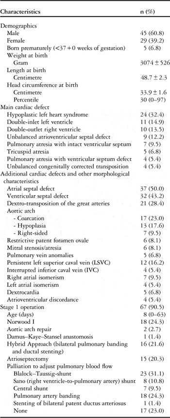
A total of 74 consecutive children fulfilled the inclusion criteria. Demographic data, diagnoses, and stage 1 surgery are summarised in Table 1.
Clinical characteristics of the study cohort (n=74).

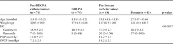
Median age, head circumferences, mean superior caval vein – corresponding to central venous – pressures and mean pulmonary artery pressures at pre-operative catheterisations, bidirectional cavopulmonary anastomosis, and Fontan completion are summarised in Table 2.
Evolution during catheterisations, bidirectional cavopulmonary anastomosis, and Fontan operation.

BDCPA=bidirectional cavopulmonary anastomosis; HC=head circumference; PAP=pulmonary arterial vascular pressure; SVCP=superior caval vein pressure
* pre-BDCPA versus pre-Fontan catheterisation
At pre-bidirectional cavopulmonary anastomosis catheterisation, 29 (39.2%) children had a mean superior caval vein pressure >7 mmHg and 40 (54.1%) had a mean pulmonary artery pressure >14 mmHg. The median head circumference at this stage was at the 7th (0–100th) percentile, with 28 (37.8%) children fulfilling criteria for microcephaly (head circumference <3rd percentile) and one (1.4%) fulfilling criteria for macrocephaly (head circumference >97th percentile).
The superior caval vein pressure of the patient with macrocephaly was 11 mmHg before bidirectional cavopulmonary anastomosis and 15 mmHg before Fontan. His head circumference at birth was at the 88th percentile, increased to the 100th percentile before cavopulmonary anastomosis, and stayed unchanged until pre-Fontan catheterisation.
The median head circumference percentile of children with hypoplastic left heart syndrome did not differ from the head circumference percentiles of children with other heart defects, 6th (0–100th) percentile versus 11th (0–87th) percentile (p=0.33).
In total, 13 patients underwent bilateral bidirectional cavopulmonary anastomosis. Their head circumferences and superior caval vein pressures did not differ from the whole population.
Unfortunately, one child died after bidirectional cavopulmonary anastomosis due to low cardiac output after Glenn takedown, subsequent dilatation of pulmonary artery stenosis, and need for extracorporeal membrane oxygenation.
In 48 (64.9%) patients, a pre-Fontan catheterisation was performed. The remaining patients had not reached the pre-Fontan evaluation at the time of this study. At this stage, 14 (29.2%) children had a superior caval vein pressure – corresponding to pulmonary artery pressure – >14 mmHg.
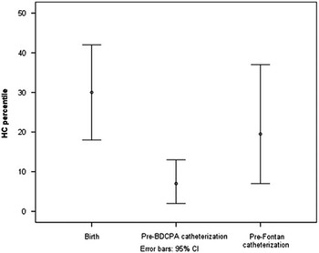
The head circumference percentiles showed a significant decrease after birth and reached the lowest values at the catheterisation before the bidirectional cavopulmonary anastomosis (30th percentile versus 7th percentile, p<0.001). At the time of bidirectional cavopulmonary anastomosis, median head circumference was more than one standard deviation below the 50th percentile. Head circumference increased significantly the following months and reached levels within lower range for age at Fontan operation (Table 2, Fig 1).
Median head circumference percentile.

To test our hypothesis, we correlated the superior caval vein pressure with the head circumference percentiles of each patient using a correlation coefficient regression model (n=74).
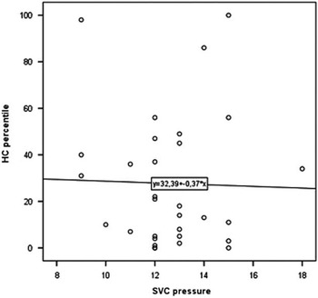
No correlation was found between the superior caval vein pressure at pre-bidirectional cavopulmonary anastomosis catheterisation and the difference in head circumference percentile between pre-bidirectional cavopulmonary anastomosis and pre-Fontan catheterisations (R2=0.08, p=0.122). The superior caval vein pressure did not correlate with head circumference percentile at the time of pre-Fontan catheterisation (R2=0.001, p=0.833, Fig 2).
Correlation between the superior caval vein pressure (SVC), corresponding to pulmonary artery pressure (PAP), and head circumference (HC) percentile at pre-Fontan catheterisation r=−0.032, R2=0.001, p=0.833.

Selective analyses of children with superior caval vein pressure >7 mmHg (n=29) at pre-bidirectional cavopulmonary anastomosis catheterisation and children with superior caval vein pressure >14 mmHg (n=14) at pre-Fontan catheterisation did not demonstrate correlation between head circumference and superior caval vein pressure (R2=0.03, p=0.549, resp. R2=0.02, p=0.805).
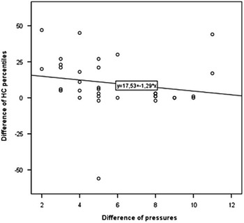
To evaluate the effect of increase in superior caval vein pressure to which the brain was exposed by the bidirectional cavopulmonary anastomosis operation, we performed a regression analysis between the difference in superior caval vein pressures at pre-bidirectional cavopulmonary anastomosis and at pre-Fontan catheterisations and the growth in head circumference percentile between pre-bidirectional cavopulmonary anastomosis and pre-Fontan catheterisations. It revealed a weak negative correlation (R2=0.19, p=0.012, Fig 3).
Correlation between the difference of superior caval vein pressure (SVC) at pre-bidirectional cavopulmonary anastomosis catheterisation and at pre-Fontan catheterisation and the difference of the head circumference (HC) percentile between pre-bidirectional cavopulmonary anastomosis and pre-Fontan catheterisations r=−0.431, R2=0.19, p=0.012.

Discussion
In the present study, we evaluated whether the superior caval vein pressure has an influence on head circumference in children with single ventricle physiology. Patients with moderately elevated superior caval vein pressure associated with single ventricle physiology did not have a tendency to an increase of the head circumference or to develop macrocephaly. There was no positive correlation between venous pressure and the growth of the head circumference.
We found a weak but significant negative correlation between the difference in superior caval vein pressures at pre-bidirectional cavopulmonary anastomosis and at pre-Fontan catheterisations and the difference in head circumference percentiles at the same time. A larger increase in superior caval vein pressure, and thus in the venous pressure to which the cerebral vessels were exposed, was associated with a slower growth of the head circumference. These findings argue against an association between moderately increased superior caval vein pressures and the development of hydrocephalus in children with single ventricle defects. Furthermore, children with lower superior caval vein pressure, reflecting potentially better haemodynamics, showed a more favourable growth of the head circumference from the low levels seen for the entire patient population at the time of bidirectional cavopulmonary anastomosis.
In children with single ventricle physiology, the brain might be exposed to higher superior caval vein pressure after completion of a bidirectional cavopulmonary anastomosis. Rosman et alReference Rosman and Shands 8 reported about an infant who underwent the Fontan procedure and developed communicating hydrocephalus after bidirectional cavopulmonary anastomosis at the age of 3 weeks. To our knowledge, this is the first report where neuropathological investigations showed no abnormality, except an increased venous pressure as cause of the hydrocephalus. It may be explained by the very young age at which bidirectional cavopulmonary anastomosis was performed with still physiologically elevated venous pressure and resistance.
In our population, the increase in superior caval vein pressure was only moderate in the majority of children. None of our patients had a superior caval vein pressure >19 mmHg. Thus, it is possible that higher superior caval vein pressures than those seen in the present study are required to cause frank hydrocephalus. This would be in accordance with the study of Dillon et al.Reference Dillon, Berman, Yabek, Seigel, Akl and Wernly 3 They hypothesised that only a substantial elevation of the mean superior caval vein pressure impairs cerebrospinal fluid absorption. In their cohort, patients with mean superior caval vein pressures of at least 24 mmHg developed macrocephaly, whereas in children with pressures of 13–15 mmHg head circumference did not increase.
Gil et alReference Gil, Specter-Himberg and Gomori 11 could not detect a correlation between head circumference, not adjusted for age, and superior caval vein pressure in a prospective study with 37 patients mostly with biventricular physiology and a mean superior caval vein pressure of 10 mmHg. In the present study, the children were exposed to slightly higher superior caval vein pressures as they underwent the different stages in the single ventricle pathway. Nevertheless, none of our patients had a superior caval vein pressure above 13 mmHg at pre-bidirectional cavopulmonary anastomosis catheterisation or a superior caval vein pressure – corresponding to pulmonary artery pressure – above 19 mmHg at pre-Fontan catheterisation. This supports the hypothesis that higher venous pressures are needed for an effect on the head circumference similar to the aforementioned case reports.
In our study, the median head circumference percentile at pre-bidirectional cavopulmonary anastomosis catheterisation was more than one standard deviation below the 50th percentile for age. At birth, the head circumferences of our cohort were within the norm but then growth was reduced. The drop in head circumference percentile after birth falls in the frail inter-stage period between the first palliative operation and the bidirectional cavopulmonary anastomosis. This stage is associated with considerable mortality and morbidity.Reference Jaquiss and Imamura 1 , Reference Vogt, Manlhiot, van Arsdell, Russell, Mital and McCrindle 14 The low head circumference percentiles at pre-bidirectional cavopulmonary anastomosis catheterisation might reflect poor growth during this delicate early period, where the systemic ventricle has to support both the systemic and the pulmonary circulations, and thus is exposed to a significant volume overload.Reference Ravishankar, Zak and Williams 15 , Reference Burnham, Ittenbach and Stallings 16 After bidirectional cavopulmonary anastomosis, haemodynamics are more stable and can promote better growth. Therefore, increase in head circumference after bidirectional cavopulmonary anastomosis might represent a catch-up growth from the low levels of head circumference percentiles seen at the time of pre-bidirectional cavopulmonary anastomosis catheterisation compared with the levels seen at birth and at Fontan completion. Ravishankar et alReference Ravishankar, Zak and Williams 15 found a similar increase in low head circumference percentiles after bidirectional cavopulmonary anastomosis as was seen in the present study. They termed the phenomenon pseudo-normalisation of the head circumference due to increased superior caval vein pressure with bidirectional cavopulmonary anastomosis. As we did not carry out routine follow-up cerebral imaging in the present study, we cannot differentiate between catch-up growth of head circumference due to an improved circulatory and nutritional state or a pseudo-normalisation due to increased superior caval vein pressure after the bidirectional cavopulmonary anastomosis; however, as pointed out above, the increased growth of head circumference in children with better haemodynamics suggests at least a component of true normalisation of head circumference in these children.
Limitations
Although the method to measure head circumference using a measuring tape is standardised, there is wide intra-observer variability; however, this limitation would not be expected to occur systematically more often at any given time point, but would rather induce a greater variation in the measurements, making smaller differences in head circumference over time harder to detect.
Methodologically, it would have been advantageous to measure the ventricular volume – with ultrasound or magnetic resonance imaging – instead of the head circumference, because macrocephaly may be caused not only by ventriculomegaly but also by megalencephaly; however, in children with congenital heart defects, this can be assumed to be a very rare cause of macrocephaly and may, therefore, be neglected.
The retrospectively collected data did not allow us to measure superior caval vein pressure during the months after bidirectional cavopulmonary anastomosis, and therefore we probably missed cases with high superior caval vein pressure in the direct post-operative phase after bidirectional cavopulmonary anastomosis.
Conclusion
Patients with moderately elevated superior caval vein pressure associated with single ventricle physiology did not have a tendency to develop macrocephaly. After birth, head circumference decreases with the lowest values being reached around the time of the bidirectional cavopulmonary anastomosis operation. Subsequently, until Fontan completion, head circumference increases significantly. The lack of direct correlation between superior caval vein pressure before Fontan and head circumference may be explained by catch-up growth of head circumference in patients with more favourable haemodynamics and concomitant venous pressures in the lower range. Further studies with focus on high superior caval vein pressure are needed to exclude or prove a direct correlation between high superior caval vein pressure and head circumference in children with single ventricle physiology.
Acknowledgements
None.
Financial Support
This research received no specific grant from any funding agency, commercial, or not-for-profit sectors.
Conflicts of Interest
None.