INTRODUCTION
Observations of deep Mediterranean sponge grounds were firstly given in the 1960s, thanks to pioneer scientific explorations through manned submarines (Vaissière & Carpine, Reference Vaissière and Carpine1964; Vacelet, Reference Vacelet1969), but the quantity and quality of the images were too poor to carry out ecological studies, and the impossibility of making direct samplings confined taxonomic investigations to only dredged material. The use of remotely operated vehicles (ROVs) has greatly enhanced the possibility of obtaining information on living deep sponge communities (Longo et al., Reference Longo, Mastrototaro and Corriero2005; Freiwald et al., Reference Freiwald, Beuck, Rüggeberg, Taviani and Hebbeln2009; Taviani & Angeletti, Reference Taviani and Angeletti2009; Beuck et al., Reference Beuck, Freiwald and Taviani2010; Mastrototaro et al., 2010; Vertino et al., Reference Vertino, Savini, Rosso, Di Geronimo, Mastrototaro, Sanfilippo, Gay and Etiope2010; Bo et al., Reference Bo, Bertolino, Borghini, Castellano, Covazzi, Di Camillo, Gasparini, Misic, Povero, Pusceddu, Schroeder and Bavestrello2011, Reference Bo, Bertolino, Bavestrello, Canese, Giusti, Angiolillo, Pansini and Taviani2012a). An outstanding example is the rich sponge assemblage associated with the white coral reefs of St Maria di Leuca (Ionian Sea) described between 400 and 600 m depth by Longo et al. (Reference Longo, Mastrototaro and Corriero2005).
In the last five years various oceanographic campaigns have been conducted along the Tyrrhenian and Ionian coast of Italy with the aim of characterizing the population structure of the megabenthic assemblages found in deep coralligenous habitats (50–200 m depth). They revealed the important role played by sponges in the deep ecosystems and the structuring character displayed by some massive demosponges (Bo et al., Reference Bo, Bertolino, Bavestrello, Canese, Giusti, Angiolillo, Pansini and Taviani2012a).
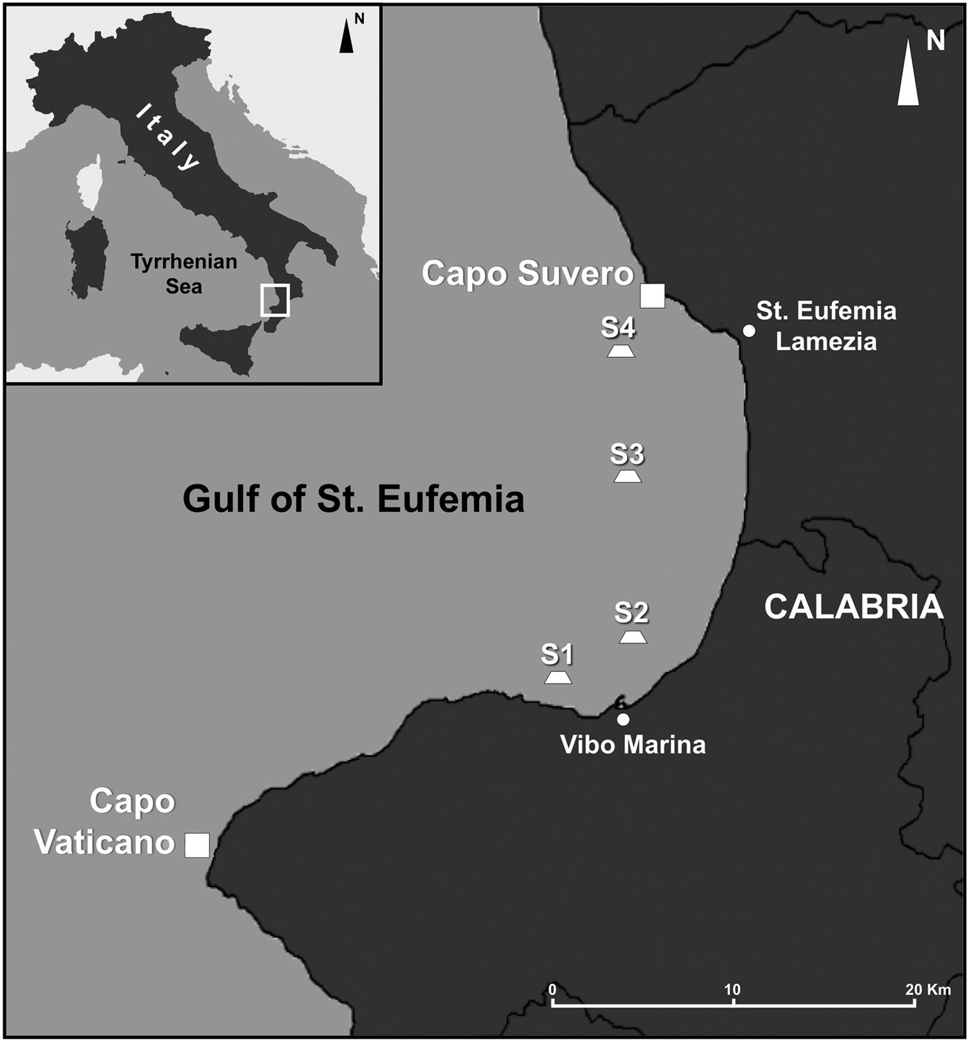
One of the most interesting sites that has been surveyed in the southern Tyrrhenian Sea is the Gulf of St Eufemia (Figure 1). The gulf is a wide arm of sea spotted with small and sparse rocky outcrops emerging from an almost flat muddy bottom, characterized by a slightly turbulent environment (Le Pera & Critelli, Reference Le Pera and Critelli1997; Bo et al., Reference Bo, Canese, Spaggiari, Pusceddu, Bertolino, Angiolillo, Giusti, Loreto, Salvati, Greco and Bavestrello2012b). In these habitats dwell a wide range of arborescent anthozoan colonies which have been recently studied in detail by means of ROV-imaging (Bo et al., Reference Bo, Canese, Spaggiari, Pusceddu, Bertolino, Angiolillo, Giusti, Loreto, Salvati, Greco and Bavestrello2012b).
Map of the studied area. Gulf of St Eufemia (southern Tyrrhenian Sea, Italy), location of the sampling sites: S1, S2, S3 S4 and Capo Vaticano.

Sponges are among the most abundant organisms associated with the St Eufemia's coral gardens (Bo et al., Reference Bo, Canese, Spaggiari, Pusceddu, Bertolino, Angiolillo, Giusti, Loreto, Salvati, Greco and Bavestrello2012b). They largely cover the substrate under the cnidarian ramifications or encrust the dead portions of their skeletons.
The aim of this paper is the description of the sponge communities living on the rocky bottoms of the Gulf of St Eufemia, together with the assemblage found at Capo Vaticano, the cape delimiting the southern boundary of the gulf. Special attention has been dedicated to the description of two new species, and additional information on the identified species and uncommon field images are also given.
MATERIALS AND METHODS
The video and photographic material used for this study was collected by the ROV ‘Pollux’ during five surveys performed between 2007 and 2009 on-board the RV ‘Astrea’. Samplings were carried out at five sites of the Gulf of St Eufemia area (Figure 1): four of them located inside the inlet and a fifth one, namely Capo Vaticano, on the southern boundary of the gulf. The ROV dives were performed to between 70 and 130 m depth, and observations were made mainly on rocky bottoms. The four locations investigated, namely S1, S2, S3 and S4, correspond to the sites studied by Bo et al. (Reference Bo, Canese, Spaggiari, Pusceddu, Bertolino, Angiolillo, Giusti, Loreto, Salvati, Greco and Bavestrello2012b). They consist of rocky outcrops, sometimes with piled boulders, a few kilometres distant from each other, subject to moderate currents and high levels of silting. In contrast, the area of Capo Vaticano is subject to strong currents and the rocky boulders are very poorly silted and almost entirely covered by encrusting coralline algae.
Taxonomic studies were restricted to the collected species. Samples were fixed on-board in 4% formaldehyde in filtered seawater and then preserved in 70% ethanol. Spicule complement and skeletal architecture were examined under light microscopy following Hooper (2000). Length and width of at least 30 spicules per type were measured. Minimum, mean (in parentheses) and maximum values are reported. Dissociated spicules and tissues for scanning electron microscope (SEM) analysis were transferred onto stubs, sputter coated with gold and observed under a SEM Vega3 _TESCAN Microscope type LMU. The type material was entrusted to the Museo Civico di Storia Naturale ‘G. Doria’ of Genova (MSNG).
Over 1100 high resolution photographs were examined for the ecological study. The average abundance (expressed as specimens m−2 ± SE) of some discrete and identifiable target sponge species (Suberites syringella, Haliclona (Haliclona) implexa, Haliclona (Soestella) fimbriata sp. nov., Axinella spp., Topsentia calabrisellae sp. nov., Topsentia vaceleti) was estimated at each study site. The three species of Axinella (damicornis, polypoides and verrucosa) were considered as a single taxon, due to the difficulty of discriminating between them in the photographs. Field pictures, supplied with a scale reference, were also used for acquiring morphometric data of the observed organisms and to draw a general outline of the sponge habitat.
RESULTS
SYSTEMATICS
Order HADROMERIDA Topsent, 1894
Family SUBERITIDAE Schmidt, 1870
Genus Rhizaxinella Keller, 1880
Rhizaxinella pyrifera (Delle Chiaje, 1828)
(Figure 2A)
Several specimens of this well known species were collected at 130 m depth from Site S1. A field photograph is included.
Genus Suberites Nardo, 1833
Suberites syringella (Schmidt, Reference Schmidt1868)
(Figures 2B, C & 3A–C)
Remotely operated vehicle photographs of: (A) Rhizaxinella pyrifera; (B, C) Suberites syringella; (D) Raspailia (Raspailia) viminalis; (E) Haliclona (Gellius) lacazei; (F) Haliclona (Halichoclona) magna covered by a film of cyanobacteria; (G) Haliclona (Soestella) implexa; (H) Haliclona pachastrelloides.
Sponge habitat of the Gulf of St Eufemia: (A–C) patches of Suberites syringella and some specimens of Haliclona (Soestella) fimbriata sp. nov. on the rocky outcrops; (D) Topsentia calabrisellae sp. nov. (white arrow) surrounded by tubular specimens of Haliclona (Soestella) fimbriata sp. nov.; (E) Topsentia vaceleti on the flat and inclined rocky boulders of Capo Vaticano covered by coralline algae. Note the presence of patches of an unidentified Haliclona (?) species mixed with T. vaceleti; (F, G), various morphotypes of H. (Soestella) fimbriata sp. nov. showing alternatively lateral, thin ramifications (F) or a compact, tubular aspect (G); (H) specimens of Haliclona (Soestella) implexa; (I) view of the bottom at Site S1 covered by digitiform and fan-like Axinella spp. sponges; (J, K) lost or discarded trawling nets covering the rocks of Capo Vaticano and impacting T. vaceleti specimens; (L) patches of Aplysina cavernicola at St Eufemia; (M) the silted rocky bottom of Site S3 showing no sponge coverage; (N) patch of Petrosia (Petrosia) ficiformis on the rocks of Capo Vaticano. Scale bars: A–N, 10 cm.
MATERIAL EXAMINED
19, S1 Gulf of St Eufemia, southern Tyrrhenian Sea, rocky outcrop, August 2009, 125 m depth.
DESCRIPTION
The numerous specimens observed and photographed by ROV have an erect habit, with anastomosing cylindrical branches rather variable in diameter (Figures 2B–C, 3A–C). Specimen 19 has branches 3–4 mm in diameter. Colour is pale yellow (Figure 2B, C). Oscules are round or oval, slightly elevated and bordered by an oscular rim, apparently arranged on one side of the branch (Figure 2C). The ectosomal skeleton clearly shows the bouquets of smaller tylostyles with inner bundles of larger tylostyles. The choanosome is rather disordered but not very dense. Spicules: gently curved tylostyles with well formed heads showing almost always a vesicle. They measure 115 (417) 600 µm × 5 (7.5) 10 µm.
DISTRIBUTION AND ECOLOGY
Gulf of Lions, Gulf of Naples, Aegean Sea, Algerian coast, from 60 to 147 m depth. Schmidt (Reference Schmidt1868) did not report the depth of the type locality at La Calle.
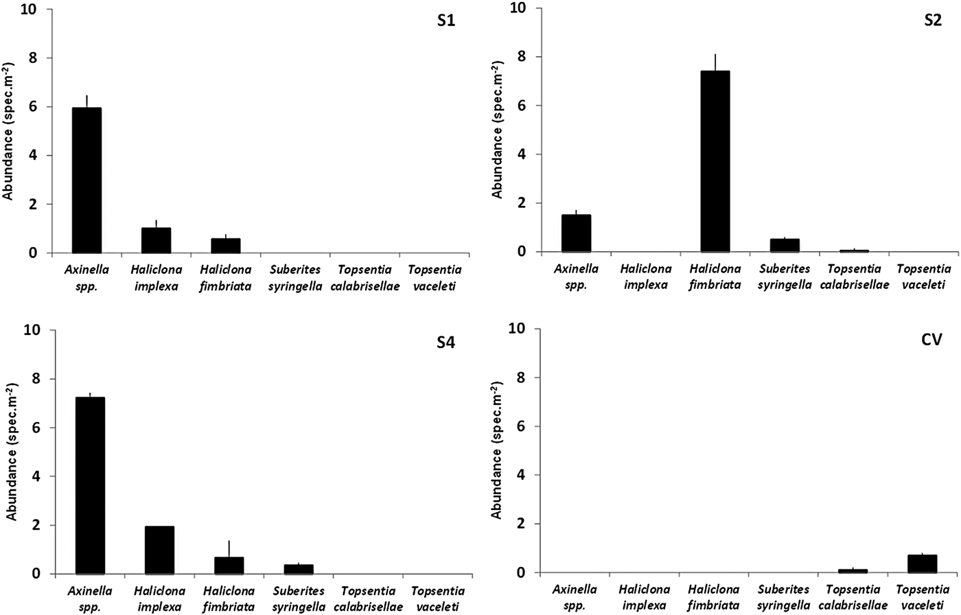
In the Gulf of St Eufemia this species is found only on Sites S1, S2 and S4. Only in the latter two areas does it occur with significant abundance, respectively 0.5 ± 0.1 and 0.4 ± 0.2 specimens m−2 (Figure 4). Most specimens show numerous branches, up to 20 cm long, sometimes bent towards the substrate. Specimens may be found both isolated or in close groups (Figure 3A–C).
Species abundance in the Gulf of St Eufemia and Capo Vaticano sites (No. specimens m−2 ± SE).
REMARKS
Topsent (Reference Topsent1938) considering synonyms its Suberites carnosus var. ramosus (Topsent, 1900) with Suberites carnosus var. syringella of Schmidt (Reference Schmidt1868) recognized that the two taxa, nowadays accepted as separate species (S. carnosus and S. syringella), are very close. Some cylindrical, elongated specimens of S. carnosus observed by one of the authors on the detritic bottom at the base of coralligenous cliffs (Portofino Promontory) differ from S. syringella in habit (repent instead of erect), larger diameter of the body, a darker yellow colour. Tylostyles normally overlap in size, but those of S. carnosus are straight instead of curved and have a bigger vesicle in the tyle.
Order CHONDROSIDA Boury-Esnault & Lopès, 1985
Family CHONDRILLIDAE Gray, 1872
Genus Chondrosia Nardo, 1847
Chondrosia reniformis Nardo, 1847
The species, quite common in the littoral zone, has a wide depth range in the Mediterranean Sea. Topsent (Reference Topsent1934) and Vacelet (Reference Vacelet1961) reported its occurrence around 100 m depth, while the deepest record (450 m) is from trawling the bottom of the Ligurian Sea (Pansini & Musso, Reference Pansini and Musso1991).
Order POECILOSCLERIDA
Family RASPAILIIDAE Hentschel, 1923
Genus Raspailia Nardo, 1833
Raspailia (Raspailia) viminalis Schmidt, 1862
(Figure 2D)
Several, uniramose or ramified specimens, observed and collected at Sites S1 and S4 around 100 m depth.
Order HALICHONDRIDA Gray, 1867
Family AXINELLIDAE Carter, 1875
Genus Axinella Schmidt, 1862
Axinella cannabina (Esper, 1794)
Axinella damicornis (Esper, 1794)
Axinella polypoides Schmidt, 1862
Axinella verrucosa (Esper, 1794)
These four species have been collected in several sites (see Table 1) estimating their abundance by photographic observations.
Family HALICHONDRIIDAE Gray, 1867
Genus Topsentia Berg, 1899
Topsentia calabrisellae sp. nov. Bertolino & Pansini
(Figures 3D, 5–6)
Topsentia calabrisellae sp. nov.: (A) specimen living on the surface of the coralligenous concretion; (B) detail of the ectosomal dermal membrane that does not cover uniformly the sponge body; (C) skeleton of the dermal membrane; (D) ectosomal skeleton with (a) and without (b) dermal membrane; (E) choanosomal skeleton; (F) magnification of the choanosomal skeleton.
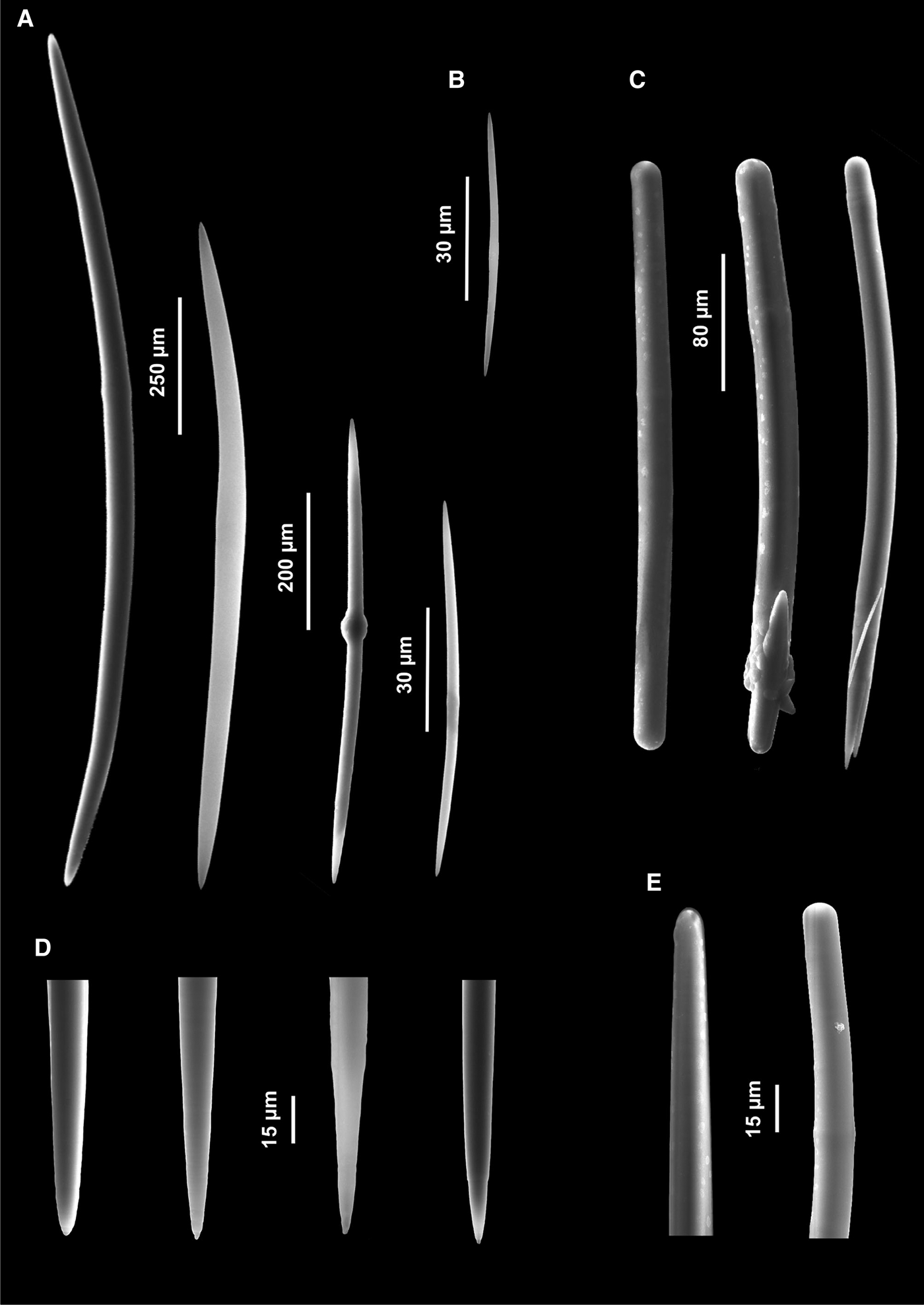
Topsentia calabrisellae sp. nov.: (A) choanosomal large and small oxeas; (B) oxea of the ectosomal dermal membrane; (C) oxeas modified into styles and strongyles, with malformations; (D) magnification of the extremities of oxeas; (E) magnification of the extremities of styles and strongyles.
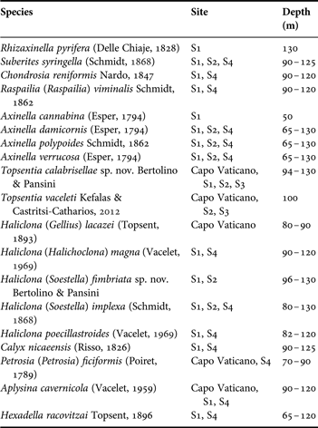
The sponge assemblages of the Gulf of St Eufemia—list of collected species.
TYPE MATERIAL
Holotype: ROV 26, Capo Vaticano, southern Tyrrhenian Sea, sub-vertical rocky shoal, July 2008, 94 m depth (MSNG 57254).
DESCRIPTION
The specimen photographed and collected by ROV is massive lobate, with a furrow along all its length. Oscules are round or oval, 1–2.5 mm across, on top of rather irregular rounded lobes. The overall size of the specimen is 13 × 6 cm. The holotype, sampled by the ROV grabber, consists in three fragments, the largest of which is 3 cm long and 2 cm high. The dermal membrane bearing the ostia does not uniformly cover the sponge body (Figure 5B). Therefore the colour of the living sponge varies from pale yellow to light brown at the surface and from dark yellow to orange in the inner part (Figure 5A, B). It becomes dark orange after formaldehyde preservation and almost black if exposed to air. The surface looks smooth when observed under the light microscope, slightly velvety (Figure 5E) under the SEM. Consistency is stony hard in the dry state (Figure 5A, B).
Skeleton. The ectosomal skeleton—not clearly separable from the choanosome—is covered by an easily detachable dermal membrane that comes off in flakes in the dry specimen (Figure 5C). Where this covering is scraped off (Figure 5D) the ectosome becomes apparent and is characterized by a para-tangential layer of large and small oxeas disorderly arranged. The choanosomal skeleton is dense, halichondroid, with spicules of all sizes directionless intercrossing. No fibres or tracts are detectable but spicules are densely packed (Figure 5). Spongin is absent both in the ectosome and choanosome.
Spicules. Fusiform oxeas are the basic spicule type (Figure 6A, B) but they modify into styles, strongyles and sometimes subtylostyles (Figure 6C, E). They measure 40.8 (464.5) 1632 µm × 2 (12.5) 25 µm in width. Two size categories of small and large oxeas are clearly observable, but they can hardly be separated into classes because intermediate spicules are abundant. Most oxeas are curved, but straight, bent or doubly-bent forms are present. Centrotylotysm is frequent both in small and large spicules, however, the tyle is irregular in the small oxeas and round or elongated in the larger forms (Figure 6A). Tips are acerate and sometimes slightly stepped (Figure 6D). Malformations are frequent. The other spicule types (styles, subtylostyles, strongyles) are clearly derived from oxeas. Styles and strongyles are common, subtylostyles rare. These modified oxeas are rare at the extremities of the size range because the smallest and biggest spicules are generally pure oxeas.
ETYMOLOGY
The name refers to a young girl, celebrated in a typical love song of Calabria, the region where the new species was found.
DISTRIBUTION AND ECOLOGY
The species has been observed only in the area of St Eufemia, at Capo Vaticano and on the rocky outcrops S1, S2 and S3. Its bathymetric range goes from 90 to 130 m depth. It dwells on the rocky elevations of the deep coralligenous community, particularly those devoid of heavy siltation. It is significantly abundant only at Capo Vaticano (0.1 ± 0.1 specimens m−2) and at Site S2 (0.04 ± 0.1 specimens m−2) (Figure 4). Usually it occurs in form of isolated, encrusting or massive specimens with a maximum diameter of 20 cm (Figure 3D).
REMARKS
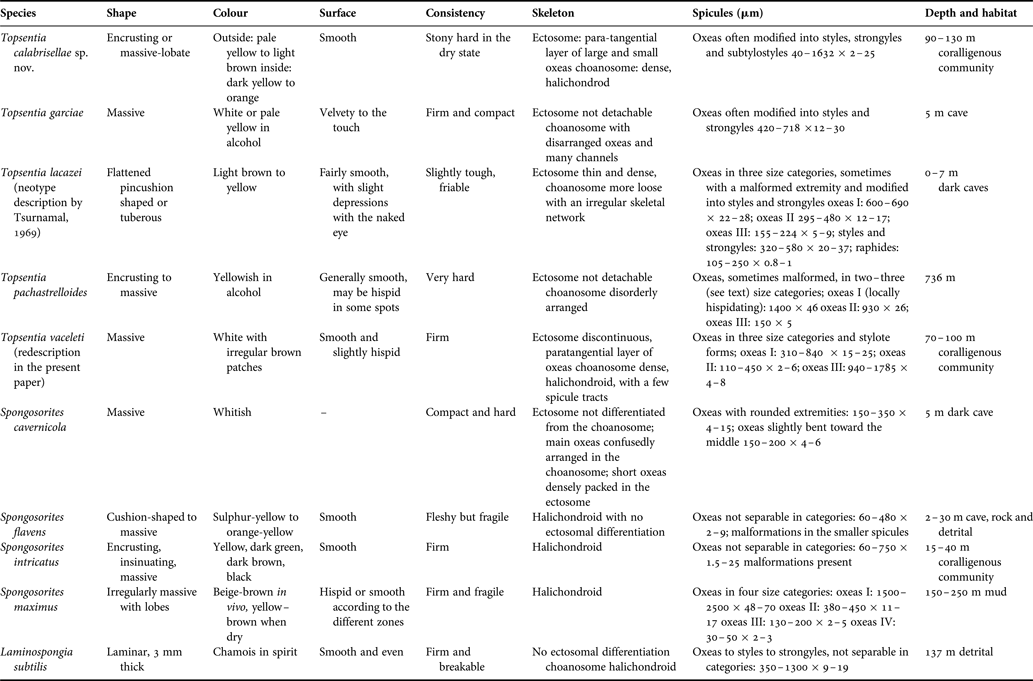
According to the presence of a thin, detachable ectosome with a para-tangential layer of smaller spicules, of a confused choanosomal skeleton almost devoid of collagen and of a wide variability in the spicule shape and size, with frequent malformations, the species is assigned to the genus Topsentia Berg, 1899. However, since several diagnostic characters of Topsentia are shared by Spongosorites Topsent, 1896 and Laminospongia Pulitzer-Finali, Reference Pulitzer-Finali1983, the taxonomic study has been extended to the Mediterranean species of these three genera. A comparison of the morphological characters and the ecology of these species is reported in Table 2. The comparison is useful because the authors still share Lévi's (Reference Lévi1957) statement on the close likeness between Spongosorites and Topsentia: ‘Leur ancienne distinction basée sur la présence ou l'absence d'une membrane dermique est difficile …’.
Morphological characters and ecology of the species of Topsentia, Spongosorites and Laminospongia present in the Mediterranean Sea. Unless further specified, spicule measures refer to the original description.
Four species of the genus Topsentia are present in the Mediterranean Sea: T. garciae Bibiloni, Reference Bibiloni, Uriz and Rützler1993, T. lacazei (Schmidt, Reference Schmidt1868), T. pachastrelloides (Topsent, Reference Topsent1892) and T. vaceleti Kefalas & Castritsi-Catharios, Reference Kefalas and Castritsi-Catharios2012 that has been fully treated in the present paper. In addition, a fifth species recorded from La Calle (Algerian coast) was identified by Topsent (Reference Topsent1901) as T. glabra (Topsent, 1898) and considered by Lévi (Reference Lévi1957) as belonging to Halichondria (Halichondria) genitrix (Schmidt, 1870) and by Pulitzer-Finali (Reference Pulitzer-Finali1983) to S. intricatus (Topsent, Reference Topsent1892).
Topsentia garciae differs from the new species in the ectosome being non-detachable and in the shape and size of the spicules. Topsentia lacazei, carefully redescribed by Tsurnamal (Reference Tsurnamal1969), clearly differs in the thickness of the ectosome, in the presence of an irregular choanosomal skeletal network, in the shape and size of spicules (which are not longer than 680 µm) and in the presence of raphids. Topsentia lacazei, however, has styles, strongyles and frequent malformed spicules, as does the new species and other members of the genus. The closest species—in relation to the large size of spicules—to T. calabrisellae sp. nov. is T. pachastrelloides. This is an Atlantic species recorded twice in the Mediterranean Sea, from the Bonifacio region (Vacelet, Reference Vacelet1961) and from the Adriatic Sea (Pansini, Reference Pansini, Vacelet and Boury-Esnault1987). In both these records were found two categories of oxeas, with a maximum size of 1500 × 45 µm in Bonifacio's specimens. Both specimens have malformed oxeas, as the holotype, but are devoid of styles, sub-tylostyles and strongyles. Vacelet's specimens turn black after preservation, whereas the Adriatic specimen and the holotype maintain their colour in alcohol (Topsent, Reference Topsent1892; Pansini, Reference Pansini, Vacelet and Boury-Esnault1987). The new species shows generally thinner spicules than T. pachastrelloides, not exceeding 25 µm in the biggest forms, and being as thin as 2 µm in the smallest. In addition, in the new species, the great number of intermediate spicules between the two size categories of oxeas is noteworthy. Topsentia calabrisellae sp. nov., in conclusion, differs from T. pachastrelloides in the external morphology (the holotype of the latter is thickly encrusting), in the presence of a detachable ectosome which is absent in T. pachastrelloides and in the spicule characters.
As to spicules, a certain similarity was observed with Laminospongia subtilis Pulitzer-Finali, Reference Pulitzer-Finali1983. All the variations of the oxeas described for Topsentia calabrisellae sp. nov. are present in the type of Laminospongia. However the two species differ in spicule size. Laminospongia subtilis completely lacks the smaller oxeas (less than 350 µm long) and has thinner spicules (maximum thickness 19 µm). In addition it shows long thin oxeas which are absent in the new species.
The four species of Spongosorites Topsent, 1896 present in the Mediterranean Sea (that is S. cavernicola Bibiloni, Reference Bibiloni, Uriz and Rützler1993, S. flavens Pulitzer-Finali, Reference Pulitzer-Finali1983, S. intricatus (Topsent, Reference Topsent1892), S. maximus Uriz, Reference Uriz1978) differ remarkably from the new species in spicule size and other characters.
Topsentia vaceleti Kefalas & Castritsi-Catharios, Reference Kefalas and Castritsi-Catharios2012
(Figures 3E, J, K & 7)
Topsentia vaceleti: (A) two specimens living on the rocky bottom; (B) choanosomal skeleton; (C) undamaged ectosomal skeleton; (D) area devoid of ectosomal cover; (E) oxeas I; (F) oxeas II; (G) oxeas III; (H) magnification of the extremities of oxeas III.
MATERIAL EXAMINED
ROV 25, Capo Vaticano, southern Tyrrhenian Sea, rocky boulders, July 2008, 100 m depth.
DESCRIPTION
The different specimens of this massive species, observed by ROV, show a massive basal part, up to 2 cm high, from which arise cylindrical papillae bearing round or oval apical oscules. Papillae are 12 mm high with walls about 1 mm thick in the only specimen we could collect. However, according to other field photographs, papillae can be much more variable in shape and size. Other flush oscules, about 1 mm in diameter, are dispersed on the sponge surface. The surface is almost smooth and slightly hispid due to the extremities of oxeas protruding through the ectosome. The consistency is firm. The colour is white with irregular brown patches, generally not extended to the papillae (Figures 7A & 3E, J, K). At the dissection microscope the brown patches appear as superficial excavations disseminated by sand grains. These areas, devoid of ectosomal cover, look particularly hispid, due to the extremities of the long, slender, choanosomal oxeas (Figure 7D). It seems highly probable that they are scars left by predators such as sea urchins or molluscs. Their brownish colour may be due to sediment that the sponge is unable to remove from these pinacoderm free areas or to the proliferation of unicellular algae (Figure 7A).
Skeleton. In the ectosome a discontinuous paratangential layer of small, thick and short oxeas (I) is detectable (Figure 7C). The choanosomal skeleton is rather dense, confused, halichondroid, with a few discrete spicule tracts. The extremities of the stout choanosomal oxeas (I) raise the sponge surface whereas the long, slender oxeas (III) cause the hispidation protruding through it (Figure 7B).
Spicules. Spicules are oxeas in three size categories but stylote forms, probably oxea derived, also occur.
Oxeas I: straight, bent, curved and sometimes flexuous; always stout and rarely malformed. The extremities are acerate or slightly mucronate. One of them can be rarely blunt. They measure 310 (645) 840 × 15 (21.5) 25 µm (Figure 7E). The axial canal is often evident, even in the slender, juvenile forms. Stylote and substylote forms, straight or gently curved, probably derived from the oxea (I) category but with a more restricted size range—190–350 × 13–22 µm—are to be found. They seem to be more abundant in the papillae.
Oxeas II: straight or curved, always thin, with pointed ends, not abundant: 110 (345.5) 450 × 2 (4) 6 µm (Figure 7F).
Oxeas III: gently curved or straight, always slender, with pointed extremities: 940–1785 × 4 (6.5) 8 µm (Figure 7G).
In spite of the overlapping in length with oxeas (I), the smaller oxeas (II) may be distinguished by their thinness, the absence of a detectable axial canal and their tangential/paratangential arrangement in the ectosomal skeleton. Kefalas & Castritsi-Catharios (Reference Kefalas and Castritsi-Catharios2012), in the original description of the species, divided the oxeas of ‘wide range of size’ in two categories only. We think that a third category of small oxeas, characterized by their thinness, should be separated.
DISTRIBUTION AND ECOLOGY
The observed specimens lived on a rather flat rocky bottom in exposed positions, apparently well adapted to the light intensity present at 100 m depth as demonstrated by the almost total coverage of the rocky substrate by coralline algae (Figure 3E). The type specimen came from a coralligenous bottom, 70 m deep, south-west of Fourni Island in the Aegean Sea (Kefalas & Castritsi-Catharios, Reference Kefalas and Castritsi-Catharios2012). This species forms relatively abundant patches at Capo Vaticano accounting for 0.7 ± 0.1 specimens m−2 (Figure 4). Rare specimens were found also on the rocky outcrops S2 and S3 of St Eufemia. Moreover, another specimen was observed in the Sicily Channel at about 100 m depth (M. Bo, personal communication). The species generally shows a roundish phenotype (up to 10 cm in diameter) with few erect papillae, however also some massive, large (20 cm across) specimens have been observed, characterized by dozens of papillae. Some small, juvenile specimens have been observed, bearing only 1–2 apical papillae. They generally live on flat boulders, but occasionally also on inclined ridges (Figure 3E, J, K).
REMARKS
The study of the collected material and of the field images allows completion of the recent description of Topsentia vaceleti by Kefalas & Castritsi-Catharios from a specimen dredged in the Aegean Sea (2012). The papillae, variable in shape and size, characterizing the species, were probably broken and removed by the sampling gear in the holotype.
Order HAPLOSCLERIDA Topsent, 1928
Suborder HAPLOSCLERINA Topsent, 1928
Family CHALINIDAE Gray, 1867
Genus Haliclona Grant, 1836
Subgenus Gellius Gray, 1867
Haliclona (Gellius) lacazei (Topsent, 1893)
(Figure 2E)
MATERIAL EXAMINED
13, Capo Vaticano, 80–90 m, rocky cliff, August 2009, alcohol preserved.
DESCRIPTION
Cushion shaped specimen, 3 cm long and 1.8 cm high. Surface rather irregular with slightly elevated round oscules. Colour dark yellow in life. The consistency of the dry specimen is hard, not friable. It was slightly mucous when alive. Skeleton fairly confused, both in the ectosome and choanosome.
Spicules. Oxeas curved, bent or doubly bent: 180 (320) 400 µm × 3 (1) 5 µm. Raphids straight or gently curved, very abundant, 35 (47) 62.5 µm.
DISTRIBUTION AND ECOLOGY
The species is known from the Mediterranean coast of France at depths ranging from 40 to 150 m on hard bottoms, or epibiotic on other sponges. Vacelet (Reference Vacelet1969), according to a personal communication of Griessinger, reports its presence at 15 m depth in a semi-dark cave near Marseille. A specimen has been collected also in a deep coralligenous rocky habitat from Portofino Promontory at about 70 m depth (M. Bo, personal communication).
REMARKS
The species is attributed to Haliclona (Gellius) lacazei rather than to H. (Gellius) laxa (Topsent, Reference Topsent1892) because raphids—which are well visible in the ectosome preparation—are never grouped in trichodragmata. We agree with Vacelet (Reference Vacelet1969) and de Weerdt & van Soest (Reference de Weerdt and van Soest1986) who considered the two species very close.
Subgenus Halichoclona De Laubenfels, 1932
Haliclona (Halichoclona) magna (Vacelet, Reference Vacelet1969)
(Figure 2F)
Several large specimens (up to 30 cm high), partially covered by a film of Cyanobacteria, observed and collected at Sites S1 and S4 around 100 m depth.
REMARKS
This species, described by Vacelet (Reference Vacelet1969) from the Canyon de la Cassidaigne, between 130 and 280 m depth, is here recorded for the second time, but seems to be abundant in the mesophotic zone, not only on steep substrates, but also on flat bottoms. We observed it during ROV surveys between 60 and 150 m deep along the south-eastern Sardinian coast, at the Egadi Islands and in the Ionian Sea (Amendolara Bank). Vacelet (personal communication) observed it during the ROV surveys of the Agence des Aires Marines Protégées (AAMP) in the canyons of the north-western Mediterranean (Toulon) and Corsica, as deep as 243 m.
Subgenus Soestella De Weerdt, 2000
Haliclona (Soestella) fimbriata sp. nov. Bertolino & Pansini
(Figures 8–10 & 3A–D, F, G)
Haliclona (Soestella) fimbriata sp. nov.: (A, B) specimens showing different morphotypes; (C) inner part of an open tube; (D) outer part of a tube.
Haliclona (Soestella) fimbriata sp. nov.: (A) ectosome with roundish meshes at light microscopy; (B) choanosomal skeleton at light microscopy; (C) ectosome with ostia; (D) oblique section through the sponge body showing the ectosome (a) and the choanosome (b); (E) choanosomal skeleton with a stout spicule tract (bottom, centre); (F) detail of the stout choanosomal spicule tracts.
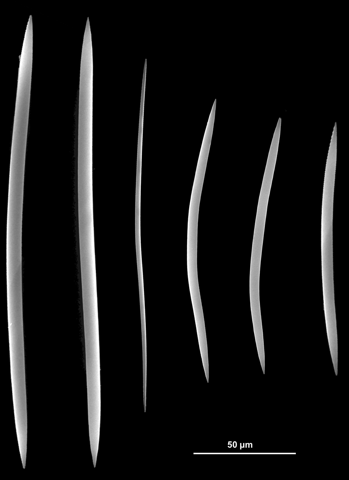
Haliclona (Soestella) fimbriata sp. nov.: oxeas.
TYPE MATERIAL
Holotype: ROV 16, S2 Gulf of St Eufemia, 130 m, rocky shoal, July 2008, fixed in alcohol 95°, then dry preserved (MSNG 57255).
Paratype 1: ROV 9, S2 Gulf of St Eufemia, 96 m, rocky shoal July 2008, fixed in formaldehyde, then dry preserved (MSNG 57256).
Paratype 2: 18, S2 Gulf of St Eufemia, 125 m, rocky shoal, August 2009, fixed in formaldehyde, then dry preserved.
COMPARATIVE MATERIAL EXAMINED
Haliclona (Soestella) mamillata (Griessinger, Reference Griessinger1971), BT4, BTX, Strait of Sicily, 1981, 42 m; Haliclona (Soestella) implexa (Schmidt, Reference Schmidt1868), ROV2, Vibo Marina, 2008, 80 m; Haliclona (Soestella) mucosa (Griessinger, Reference Griessinger1971), AT48, Montecristo Island, 1983, 40 m; PTF37, Portofino, 1981, 20 m; Haliclona (Soestella) valliculata (Griessinger, Reference Griessinger1971), BG10, Strait of Sicily, 1981, 25 m. All these specimens from the collection of the authors at DISTAV (University of Genoa).
DESCRIPTION
The sponge is erect, branching, sometimes with a massive, enlarged base. It forms irregular, tubular processes which are almost always open forming a sort of lamina about 2.5 mm thick. The holotype consists in two fragments of open tubular processes measuring 3 × 1 cm and 2 × 1.5 cm, respectively; paratype 1 consists in several fragments of tubes about 2 mm in diameter; paratype 2 consists in a flattened portion, 3 × 1.8 cm, of an open tube and several smaller fragments.
The biggest specimen observed and photographed by ROV is more than 20 cm high (Figures 8E & 3A–D, F, G). The upper part of the tubular processes is characteristically fringed, lacerated. In other specimens (e.g. ROV 9) the growth form is digitate and most tubes—from 2 to 8 mm in diameter—remain closed. The colour of the sponge in the field is whitish in the well lighted, foreground, specimens and light brown in the more distant ones. Preserved specimens are pale orange-yellow (bisque). The surface is smooth with the naked eye but may also show rounded ridges and grooves. It looks very slightly hispid at the microscope. Oscules are round, 1–4 mm in diameter, almost flush with the surface. They open both on the inner and outer side of the lamina (Figure 8C, D). Living specimens are soft and fragile; they become very friable when dried. The ectosome is not detachable.
Skeleton. The ectosomal skeleton is a tangential paucispicular (1–2) reticulation forming roundish or polygonal meshes (Figure 9A, C, Da). The choanosomal skeleton is a rather dense sub-anisotropic reticulation, with paucispicular (1–2) ascending tracts irregularly connected by single spicules (Figures 9B, E) alternated with stouter tracts cored by 8–10 spicules bound by spongin (Figure 9F). Spongin is never abundant, both in the ectosome and choanosome.
Spicules. Oxeas with acerate extremities, straight, gently curved and sometimes doubly curved or slightly sinuous (Figure 10). Most spicules are pure oxeas: blunt extremities are rare. They measure 102.5 (164.4) 221.4 × 2.6 (5.6) 7.8 µm. Thin, juvenile forms (not considered in the measures) are common in the specimens collected in July (ROV 9 and ROV 16) and rare in specimen 18 collected in August.
DISTRIBUTION AND ECOLOGY
The species is very common on the horizontal and sub-vertical substrates of deep coralligenous hard grounds of the Gulf of St Eufemia (Figure 3A–D, F, G). It has been recorded only at S1, S2 and S4 sites inside the gulf, where it can reach very high abundance values, especially at Site S2 where H. (S.) fimbriata represents the dominant sponge species (7.4 ± 0.7 specimens m−2) (Figure 4). Its abundance decreases of one order of magnitude at S1 and S4 (0.6 ± 0.1 and 0.7 ± 0.2 specimens m−2, respectively) (Figure 4). The species is probably particularly adapted to silted environments, being erect and up to 25 cm high. In St Eufemia this species forms large, dense patches (Figures 8A–B & 3D, F, G). At times, tubes may show a repent behaviour, possibly leading to forms of asexual reproduction (Figure 3A, F).
ETYMOLOGY
The specific name refers to the upper margin of the sponge body, characteristically fringed in most specimens.
REMARKS
The new species of Haliclona is attributed to the subgenus Soestella according to the presence of paucispicular primary lines and thinner tracts in the choanosomal skeleton and to the tendency of spicules to form rounded meshes, especially in the ectosome. A specimen of the type species of the subgenus—Reniera mamillata Griessinger, 1971 = Haliclona (Soestella) mamillata (Griessinger, Reference Griessinger1971)—has been examined for comparison.
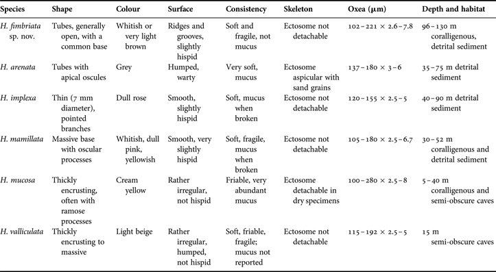
Twenty species belonging to Haliclona (Soestella) are known worldwide (van Soest et al., Reference Soest, Boury-Esnault, Hooper, Rützler, de Voogd, Alvarez de Glasby, Hajdu, Pisera, Manconi, Schoenberg, Janussen, Tabachnick, Klautau, Picton, Kelly, Vacelet, Dohrmann and Cristina Díaz2013). Five of them are present in the Mediterranean Sea: H. arenata Griessinger, Reference Griessinger1971, H. implexa (Schmidt, Reference Schmidt1868), H. mamillata (Griessinger, Reference Griessinger1971), H. mucosa (Griessinger, Reference Griessinger1971) and H. valliculata (Griessinger, Reference Griessinger1971), but none of them is endemic to this area. Their main distinctive characters and preferred habitat are reported in Table 3.
Morphological characters and ecology of the species of Haliclona (Soestella) recorded from the Mediterranean Sea.
All these species of Haliclona (Soestella) (Table 3) are well characterized by their morphology, consistency, and presence/absence of mucus. Most of them share a not detachable ectosome whereas the oxea size, as usual in the genus, is not a discriminating character. Haliclona (Soestella) fimbriata sp. nov. is considered new being characterized by a combination of original characters critically evaluated through the comparison with specimens in the authors’ collection from the Mediterranean Sea and the western Atlantic. The only species compared only by literature data—H. (Soestella) arenata—differs in shape, colour, surface, presence of mucus and spicule size from the new species.
Haliclona (Soestella) implexa (Schmidt, Reference Schmidt1868)
(Figures 2G & 3H)
MATERIAL EXAMINED
ROV2, S2 Gulf of St Eufemia, rocky shoal, July 2008, 80 m.
DESCRIPTION
The collected material and all the specimens photographed by ROV (e.g. Figures 2G & 3H) match with the detailed description reported by Grissinger (1971, p. 133) of an erect sponge, dull rose in colour, with thin branches frequently anastomosing. Ectosome not detachable, with a tangential reticulation of single or paucispicular (2–3) lines tending to form roundish meshes. Choansomal skeleton with paucispicular (3–5) primary lines connected by single spicules. Spongin scarce. Spicules: oxeas 100 (215) 240 µm × 1, 5 (3) 5 µm.
DISTRIBUTION AND ECOLOGY
In the Mediterranean Sea the species is present in the Aegean Sea, Adriatic Sea, Gulf of Lions and the Balearic Sea, often on bottoms with an algal cover (Vidalia, Peyssonnelia) at depths from 5 to 100 m. Haliclona (Soestella) implexa is regarded as cosmopolitan, but de Weerdt & van Soest (Reference de Weerdt and van Soest1986) report more precise information. According to the results of our ROV surveys the species is a very common species in the deep circa-littoral rocky zone throughout the Tyrrhenian Sea. In the Gulf of St Eufemia this species shows a distribution opposite to that of H. (Soestella fimbriata) sp. nov., being present with high abundances at S1 and S4 sites (1.0 ± 0.3 and 1.9 ± 0.6 specimens m−2, respectively), while it is almost completely absent at Site S2 (Figure 4). Also in this case, the different rate of sedimentation appears to be the putative factor driving the species distribution. Haliclona implexa may occur in the form of very large (more than 30 cm), branched specimens extending both vertically and horizontally over the substrate; however, it is possible that adjacent specimens partially hide the real size of each individual (Figure 2G). Juvenile specimens with a single short branch have often been reported.
REMARKS
De Weerdt & van Soest (Reference de Weerdt and van Soest1986) reported some skeletal differences between the Mediterranean specimens and those of the Azorean region. Our specimens strictly conform to the description of the skeletal arrangement illustrated by these authors in their figure 10. On the other hand, the bushy specimen that they show at figure 9 has a dense arrangement of the tubes and a pale yellow colour which are rather different from those of our specimens (Figures 2G & 3H).
Suborder PETROSINA Boury-Esnault & Van Beveren, 1982
Family PHLOEODICTYIDAE Carter, 1882
Genus Calyx Vosmaer, 1885
Calyx nicaeensis (Risso, 1826)
Several specimens observed at Sites S1, S4 between 90 and 125 m depth. This species, which almost disappeared within 50 m depth along the eastern Ligurian coast after mass mortality events (unpublished data), seems rather common in the investigated area.
Family PETROSIIDAE van Soest, 1980
Genus Petrosia Vosmaer, 1885
Sub genus Petrosia Vosmaer, 1885
Petrosia (Petrosia) ficiformis (Poiret, 1789)
(Figure 3N)
Specimens with the typical repent–ramose habit displayed by the species in dim light conditions were observed at Capo Vaticano between 70 and 90 m depth.
Order VERONGIDA Bergquist, 1978
Family APLYSINIDAE Carter, 1875
Genus Aplysina Nardo, 1834
Aplysina cavernicola (Vacelet, 1959)
(Figure 3L)
Several patches of this gregarious species were observed at Sites S1 and S4 between 90 and 120 m depth.
Family IANTHELLIDAE Hyatt, 1875
Genus Hexadella Topsent, 1896
Hexadella racovitzai Topsent, 1896
Frequently observed by ROV between 65 and 120 m depth both on vertical and sub-vertical substrata.
SPONGE COMMUNITY OF THE GULF OF ST EUFEMIA
On the basis of the taxonomic and photographic analyses, 19 species of sponges have been identified in the St Eufemia area (Table 1). However, according to the ROV footage, there may likely be at least 80 discrete, putative species identified from their external morphology. Sixty-six of them are present in the St Eufemia internal area, while 21 have been recorded at Capo Vaticano, with an overlap of only seven species.
The sponge community of the rocky outcrops of St Eufemia shows marked differences that partially emerge from the visual census data of the target species (Figure 4). Sponge abundance is very similar at Sites S1 and S4. The species of the genus Axinella are the most common, with average values of 5.9 ± 0.5 and 7.2 ± 0.8 specimens m−2, respectively, for S1 and S4 (Figures 4 & 3I). Also, the two target species of Haliclona were recorded at both these sites with similar values of abundance (Figure 4). On the other hand, Suberites syringella was repeatedly recorded at S2 and S4 (Figure 4). The relative abundance of the species is significantly different in S2, where Haliclona (Soestella) fimbriata sp. nov. is the most common sponge. At Site S3 no target sponge species with significant abundance have been recorded. The soft bottoms surrounding the rocky outcrops host a rich community of species adapted to live in an homogeneous muddy or sandy environment, including numerous cnidarians (Bo et al., Reference Bo, Canese, Spaggiari, Pusceddu, Bertolino, Angiolillo, Giusti, Loreto, Salvati, Greco and Bavestrello2012b) and the stalked sponge Rhizaxinella pyrifera (Figure 3D). No significant abundance data are available for this species, which is usually found as scattered specimens. Occasionally, however, up to three specimens were observed close to each other.
The situation at Capo Vaticano is completely different; at this site the only target sponge species recorded are the two Topsentia, but T. calabrisellae sp. nov. is only occasional, while T. vaceleti is definitely dominant (Figure 4). Apart from the Topsentia species, at this site there are also dense patches of massive non-target species, such as Petrosia (Petrosia) ficiformis and another branched Haliclona (?) species (Figure 3E). A low coverage of encrusting sponges is reported for this site, probably as a result of competition with coralline algae (Figure 3E).
Evident traces of fishery impact have been recorded in this site, mainly in the form of lost trawling nets (Figure 3J, K) which are entrapped in large portions on hard bottoms and directly affect the sponge community (Figure 3J, K).
The identified trends between the sites are visible also when considering the number of putative discrete species. Biodiversity levels are higher for the most external sites (33 and 29 for S1 and S4, respectively) and lower for the internal sites (20 and 9 for S2 and S3, respectively). Sociophilous species such as Haliclona poecillastroides, Raspailia (Raspailia) viminalis and Calyx nicaeensis do occur at S1 and S4 (Figure 4), together with patches of species with a wider depth range, such as Aplysina cavernicola (Figure 3L), Hexadella racovitzai and Chondrosia reniformis. The largest sponge species (up to 30 cm high), occurring in the form of isolated specimens at all the sites, is Haliclona (Halichoclona) magna, characterized by white, large tubular processes at times covered by an unicellular algal film (Figure 2F). Both massive and encrusting sponges are almost completely absent at S3 (Figure 3M).
CONCLUSIONS
In the Gulf of St Eufemia sponges represent the most rich megabenthic component of the hard bottom communities. They may constitute a rich and abundant ground cover under coral colonies, as on the investigated sites, or they may represent the main megabenthic organisms of the area, as at Capo Vaticano. Similarly to the distribution pattern of the coral species on the rocky outcrops of St Eufemia (Bo et al., Reference Bo, Canese, Spaggiari, Pusceddu, Bertolino, Angiolillo, Giusti, Loreto, Salvati, Greco and Bavestrello2012b), the sponge assemblages also show a distinct trend of composition and abundance among the sites, supporting the hypothesis that the studied sites can be considered as ecological analogues to islands in terms of biodiversity.
Sponges, like corals, depend for their distribution on various ecological constraints such as availability of hard substrates, current, silting conditions and larval dispersal ability. These factors operate synergistically, resulting in high levels of community heterogeneity, even at a small scale of observation (Bo et al., Reference Bo, Canese, Spaggiari, Pusceddu, Bertolino, Angiolillo, Giusti, Loreto, Salvati, Greco and Bavestrello2012b). Among the environmental limiting factors, current and silt actively influence the composition of the sponge assemblages between the internal and external sites of the Gulf of St Eufemia. At Capo Vaticano, strong currents do not enhance the settling of large, arborescent cnidarians, but, at the same time, clear water favours the algal coverage of rocky boulders also in circalittoral habitats, limiting the spread of sponges. On the internal sites currents are moderate, thus supporting a flourishing community of filter feeders, mainly composed of large corals and erect/massive sponges. On the other hand, the high levels of siltation found in the deepest parts of the sites (around 120–130 m depth) prevent the expansion of the encrusting sponges. This situation is extreme at S3, where rocks are completely covered by silt and the entire ground covering of invertebrates is almost completely absent.