Implications
Reproductive function in postpartum beef cows is regulated by energy status and positively associated with serum concentrations of leptin, insulin and IGF-I. β–adrenergic receptor agonist (β-AA) treatment results in muscle accretion, whilst reducing fat deposition. The reduction in body fat, despite the overall BW gain, results in lower leptin and IGF-I concentrations and a suboptimal response to an oestrus induction programme and pregnancy percentage after breeding. Hence, the results support the hypothesis that a minimal amount of body fat must be maintained for reproductive success. These results should assist in the design of nutritional strategies for postpartum beef cattle.
Introduction
In beef cows, the amount of energy reserves at calving dictates the interval from parturition to first oestrus and their associated reproductive performance (Hess et al., Reference Hess, Lake, Scholljegerdes, Weston, Nayigihugu, Molle and Moss2005; Crowe, Reference Crowe2008). Moreover, changes in energy reserves during the postpartum period and body condition score (BCS; i.e. 1=emaciated and 5=fat) at the time of breeding are also known to affect reproductive performance (Bossis et al., Reference Bossis, Wettemann, Welty, Vizcarra and Spicer2000; Ciccioli et al., Reference Ciccioli, Wettemann, Spicer, Lents, White and Keisler2003; de Medeiros-Bastos et al., Reference Daix, Pirotte, Bister, Wergifosse, Cuvelier, Cabaraux, Kirschvink, Istasse and Paquay2004). Lactating beef cows with moderate BCS (BCS=3) have a greater response to an oestrus induction treatment and greater pregnancy percentage than cows with poor BCS (BCS=2; de Medeiros-Bastos et al., Reference Daix, Pirotte, Bister, Wergifosse, Cuvelier, Cabaraux, Kirschvink, Istasse and Paquay2004). In agreement, beef cows gaining 0.90 kg of BW daily during the first 71 days postpartum had shorter anoestrus intervals (100±7 days) than cows gaining only 0.45 kg/day (120±7 days; Ciccioli et al., Reference Ciccioli, Wettemann, Spicer, Lents, White and Keisler2003).
The mechanisms by which static and dynamic changes in energy reserves affect reproductive function in beef cows involve changes in IGF-I, insulin and leptin (Hess et al., Reference Hess, Lake, Scholljegerdes, Weston, Nayigihugu, Molle and Moss2005; Crowe, Reference Crowe2008). Increases in a cow’s nutrient intake (Bossis et al., Reference Bossis, Wettemann, Welty, Vizcarra and Spicer2000) and energy reserves (León et al., Reference León, Hernández-Cerón, Keisler and Gutiérrez2004) augments IGF-I and insulin concentrations in the blood. Leptin concentrations in the blood also increase with nutrient intake, but are largely a function of total body fat mass. For instance, in dairy and beef cows there was a positive correlation between serum concentrations of leptin and BCS (Strauch et al., Reference Strauch, Neuendorff, Brown, Wade, Lewis, Keisler and Randel2003; León et al., Reference León, Hernández-Cerón, Keisler and Gutiérrez2004; Becú-Villalobos et al., Reference Becú-Villalobos, García-Tornadú, Shroeder, Salado, Gagliostro, Delavaud, Chilliard and Lacau-Mengido2007) and back fat at the 12th rib in steers (Brandt et al., Reference Brandt, Keisler, Meyer, Schmidt and Berg2007). The correlation between serum concentrations of leptin and muscle mass, as evaluated by area of the Longisimuss dorsi muscle, was negative (Brandt et al., Reference Brandt, Keisler, Meyer, Schmidt and Berg2007).
While the preponderance of scientific evidence supports a positive relationship between metabolic hormones and reproductive performance (Wettemann et al., Reference Wettemann, Lents, Ciccioli, White and Rubio2003; Hess et al., Reference Hess, Lake, Scholljegerdes, Weston, Nayigihugu, Molle and Moss2005), an understanding of the close relationship among energy reserves, metabolic hormone concentrations (León et al., Reference León, Hernández-Cerón, Keisler and Gutiérrez2004) and reproductive efficiency is incomplete. Treatment of low BCS lactating beef (BCS=3.11±0.20) cows with a β –adrenergic receptor agonist (β-AA) improved BCS by increasing muscle deposition and reducing the fat to muscle ratio (Guzmán et al., Reference Guzmán, Gonzalez-Padilla, Garcés-Yepez, Rosete-Fernandez, Calderón-Robles, Murcia and Gutiérrez2012). This change in body composition also dissociated the natural relationship between BCS and metabolic hormone concentrations and resulted in a poor response to an oestrus induction treatment compared to non-treated cows. Nonetheless, the odds for development of an active corpus luteum after β-AA treatment was positively associated with serum concentrations of IGF-I (Guzmán et al., Reference Guzmán, Gonzalez-Padilla, Garcés-Yepez, Rosete-Fernandez, Calderón-Robles, Murcia and Gutiérrez2012). However, in the work by Guzmán et al. (Reference Guzmán, Gonzalez-Padilla, Garcés-Yepez, Rosete-Fernandez, Calderón-Robles, Murcia and Gutiérrez2012) the cattle were in low BCS throughout the study, thus affecting leptin concentrations and possibly the response to the oestrus induction treatment. Based in these observations, we hypothesize that in lactating beef cows that were fed to increase BW and BCS, the use of a β-AA to increase lean muscle gain and decrease body fat would diminish the reproductive response to an oestrus induction treatment, associated with decreased leptin concentrations. Thus the objective of the present experiment was to evaluate the effect of reduced body fat to muscle ratio, induced by β-AA treatment and its associated endocrine milieu on the reproductive response to an oestrus induction programme.
Material and methods
Animal management
All animal procedures were approved by the Ethics and Animal Welfare Committee of the Veterinary School, Universidad Nacional Autónoma de México. The experiment was carried out during June to August at ‘Las Margaritas’ research station, located in a subtropical humid area of Mexico. Thirty beef cows (Brahman×Charolais) with a BCS of 4.57±0.03 (1=emaciated, 9=obese; BIF, 2010). Cows calved and remained with their suckling calves throughout the study.
Feeding and treatments
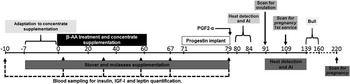
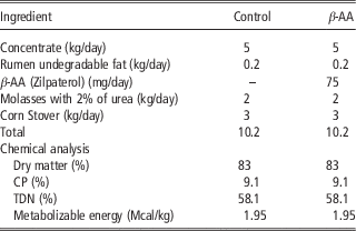
Diagrammatic representation of feeding, treatments and measurements are presented in Figure 1. During the study cattle were kept in native tropical pastures (Axonopus argentinus and Paspalum notatum) that if consumed at 3% of their BW would provide, in a conservative estimation, 98% of the daily protein and metabolizable energy requirements for a 60-day postpartum cow weighing 450 kg and producing 4.5 kg/day of milk (NRC, 1996). Seven days before the start of the experiment (i.e. 75.9±12 days postpartum), cows were weighed (401.80±9.18 kg), BCS recorded (3.10±0.22) and conditioned to handling procedures and to the feed supplement starting with 1 kg/day on day 1, and increasing it by 2 kg every 3 days until they consumed 5 kg/day. At the start of the study and for 60 days thereafter, cows were separated from their calves for 1 h each day beginning at 0800 h and individually fed a supplement (Table 1) that provided an additional 68% of their daily metabolizable energy requirements (NRC, 1996). After 60 days on study, the cows then received a supplement made of corn stover and molasses for an additional 20 days (that provided an additional 33% of their daily metabolizable energy requirements).
Experimental design. Cows were fed in pasture providing 98% of the daily protein and metabolizable energy requirements and additionally supplemented with 68% of metabolizable energy requirements for 60 days (Control group, n=15). Additionally cows in the β-adrenergic receptor agonist group (β-AA) received 0.15 mg/kg of BW of zilpaterol hydrochloride (Zilmax® MSD Animal Health, Santiago Tianguistenco, Edo. México; n=15). Oestrus was induced by a norgestomet implant treatment for 9 days (Crestar® MSD animal health). Cows in heat were inseminated in the next 60 days or served by a bull in the following 21-day period. The occurrence of ovulation and pregnancy was assessed by transrectal ultrasonography.

Daily allowances per dietary treatment and composition of feed ingredients

TDN=total digestible nutrient.
Cows were randomly assigned to one of two groups (n=15 per group) balanced by days postpartum and BCS. The control group received only the supplement while the β-AA group received the same amount of supplement plus zilpaterol hydrochloride (0.15 mg/kg of BW Zilmax® MSD Animal Health, Santiago Tianguistenco, Edo. México). To test the effect of the different body composition and its associated hormonal milieu, and yet avoid the acute effect of β-AA (Paredes et al., Reference Paredes, Salvetti, Diaz, Dallard, Ortega and Lara2011) and energy supplementation on follicular development (Gutiérrez et al., Reference Gutiérrez, Oldham, Bramley, Gong, Campbell and Webb1997) and LH secretion (Hess et al., Reference Hess, Lake, Scholljegerdes, Weston, Nayigihugu, Molle and Moss2005), β-AA treatment and concentrate supplementation were removed 12 days before the start of the oestrus induction treatment. Oestrus was induced by a norgestomet implant (3 mg) plus 5 mg of oestradiol valerate (Crestar® MSD animal health). After 8 days, the progestin implants were removed, cows were injected with PGF2-α (25 mg; Lutalyse; Pfizer, Cd. de México) and calves were temporarily weaned for 72 h. Cows were observed for signs of oestrus during 96 h and those standing to be mounted were inseminated ~10 h later. Heat detection was continued twice daily for an additional 60 days and cows were inseminated whilst in heat. All artificial inseminations were carried out using semen from a single bull. Thereafter, one bull was introduced for an additional 21 days for natural service of those cows returning to oestrus. The occurrence of ovulation and pregnancy to 1st service was assessed by real time B mode ultrasonography. Ovulation was considered to have occurred if a CL was present at the ultrasonographic examination 12 days after implant withdrawal (7 to 11 days after the first insemination). Pregnancy to 1st service was assessed by the direct observation of the embryo 30 days after progestin implant withdrawal (25 to 29 days after the first insemination). Percentage of pregnant cows was estimated 60 days after the end of the breeding period by transrectal ultrasonography.
All cows were bled once a week for the retrospective determination of luteal activity before the oestrus induction treatment. A cow was considered cycling if serum progesterone concentrations were above 1 ng/ml in two consecutive weekly samples (Zalesky et al., Reference Zalesky, Day, GArcia-Winder, Imakawa, Kittok, D’Occhio and Kinder1984; Berardinelli and Joshi, Reference Berardinelli and Joshi2005; Flores et al., Reference Flores, Looper, Kreider, Post and Rosenkrans2006).
Body composition and blood sampling
Cows were weighed and BCS recorded 10 days before the onset of the supplementation period and every 2 weeks throughout the experimental period. Back fat thickness and muscle depth were measured between the 12th and 13th thoracic vertebrae using an Aloka ultrasound with a 3.5 MHz linear probe (Brethour, Reference Brethour1992) and the back fat to muscle ratio was calculated. Calves were weighed at the beginning and at the end of the experiment. To evaluate the hormonal and metabolic changes associated to β-AA treatment, six cows per group were randomly selected and bled weekly from 1 week before the start of supplementation and during the entire experimental period. Blood samples were taken by venipuncture of the coccygeal vein into vaccutainer tubes containing either clotting gel or sodium citrate to obtain serum or plasma, respectively. Samples were kept in ice bath until centrifuged at 1000×g for 15 min. Serum and plasma were aliquoted in polypropylene tubes and stored at −20°C until assayed.
Hormones and metabolites assays
Serum concentrations of insulin (Siemens Healthcare, S.A. de C.V., Distrito Federal México; cat. number TKIN5), IGF-I (Diagnostic Laboratory Systems, Webster, Texas; cat. number 2800) and leptin (Linco XL-85K; Linco, St Louis, MO, USA) were determined in duplicate according to manufacturer recommendations and the intra-assay coefficient of variation (CV) were 1.54%, 9.72% and 8.23% for insulin, IGF-I and leptin, respectively. Insulin, IGF-I and leptin were measured on days −10, 3, 32, 59, 67 and 79 of the supplementation period. Glucose, urea and triglycerides were analysed in duplicate in all samples with commercial kits (BioSystems®; cat. numbers COD-11504, COD-11537 and COD-11592, respectively). Intra and inter-assays CVs were: 3.01% and 3.87% for glucose; 6.37% and 9.68% for urea and 17.12% and 17.23% for triglycerides. Non-esterified fatty acids were measured in all samples with the method described by Duncombe (Reference Duncombe1964) with slight modifications (Guzmán et al., Reference Guzmán, Gonzalez-Padilla, Garcés-Yepez, Rosete-Fernandez, Calderón-Robles, Murcia and Gutiérrez2012). Intra and interassays variation coefficients were 4.82% and 5.36%, respectively.
Statistical analyses
Values are presented as mean±standard error. All analyses were carried out using JMP-5 statistical software (SAS Institute Inc., Cary, NC, USA). The cow was used as the experimental unit for statistical analyses. BW, back fat, muscle depth and back-fat/muscle ratio were analysed by Student’s t-test. The BCS was analysed by Mann–Whitney U whereas differences in reproductive response were analysed by χ 2 tests. Hormone and metabolites concentrations were analysed using an ANOVA for repeated measurements including treatment, day and their interaction as fixed variables and the animal nested within treatment as a random variable. The initial metabolite and hormone concentrations were tested as covariables resulting non-significant (P>0.05) and excluded from the final analyses. The effect of treatment was analysed using animal within treatment as an error term.
Results
BW, BCS, back fat and muscle depth
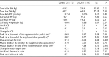
BW, BCS and back fat and muscle measurements are shown in Table 2. Daily BW gain was not different between control and β-AA group (P>0.05). Both groups increased BCS, although the increase in BCS was greater in β-AA treated cows (P<0.05). In addition, β-AA treated cows had less back fat and lower fat to muscle ratio (P<0.05) and a greater depth of the Longissimus dorsi than the control cows (P<0.05). Calf BW did not differ between treatment groups (P>0.05).
Effect of treatment with β-adrenergic receptor agonist (β-AA; 0.15 mg/kg BW) on BW, body condition score (BCS), back fat and muscle depth in lactating beef cows fed in pasture capable of providing 98% of the daily protein and metabolizable energy requirements and additionally supplemented with 68% of their metabolizable energy requirements for 60 days previous to an oestrus induction treatment

a Measurement was performed by ultrasonography between the 12th and 13th thoracic vertebrae.
Reproductive response
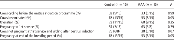
The reproductive responses to the oestrus induction treatment and after the breeding period are shown in Table 3. The number of cows with luteal activity before the oestrus induction treatment was similar between groups (P>0.05). In addition, the progesterone concentrations in the two consecutive weekly samples that determined luteal activity did not vary between groups (4.1±1.19 and 4.9±1.02 ng/ml for the control and β-AA groups, respectively). However, cows displaying oestrus and which were therefore inseminated after progestin treatment were lower in the β-AA (8/15) than in the control (13/15) group (P<0.05). Percentage of cows that ovulated and pregnancy to 1st service (P>0.05) did not differ between groups. Finally, the percentage of cycling cows after oestrus induction (P=0.07) and of pregnant cows at the end of the breeding period was lower (P<0.05) in β-AA than control cows. All cows that became pregnant in both groups did so within the 35 days that followed the synchronization protocol.
Effect of β-adrenergic receptor agonist (β-AA; 0.15 mg/kg BW) on reproductive response to an oestrus induction treatment in lactating beef cows fed in pasture capable of providing 98% of the daily protein and metabolizable energy requirements and additionally supplemented with 68% of metabolizable energy requirements

Hormones and metabolite concentrations
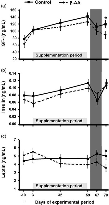
IGF-I concentrations increased (P<0.05) during the feed supplementation period in all animals, however when supplementation was withdrawn, IGF-I concentrations declined for both groups. Nineteen days after withdrawing the nutritional treatments, and coinciding with the end of the oestrus induction treatment, IGF-I concentrations were significantly lower (P<0.05) in β-AA treated cows (Figure 2a). Leptin concentrations (Figure 2c) in control cows did not change throughout the experimental period. In contrast, in the β-AA group leptin decreased (P<0.05) during the study and was significantly lower than in the control cows on day 78 (P<0.05). Insulin concentrations (Figure 2b) increased during the supplementation period and did not differ between groups (P>0.05).
Effect of β-adrenergic receptor agonist (β-AA; 0.15 mg/kg BW) on serum concentrations of IGF-I (a), insulin (b) and leptin (c) in lactating beef cows fed in pasture capable of providing 98% of the daily protein and metabolizable energy requirements and additionally supplemented with 68% of metabolizable energy requirements for 60 days. Grey bar: oestrus synchronization period. The asterisk* indicates differences (P<0.05) between β-AA and control group in the same day.

Discussion
In this study we successfully dissociated the existing positive relationship between BCS, body fat and leptin concentration in postpartum beef suckled cows in poor BCS. The use of β-AA increased BCS through muscle accretion, but not through fat accretion, and decreased leptin and IGF-I concentrations. These treatments resulted in a lower response to oestrus, lower percentage of cycling animals after progestin treatment and lower pregnancy at the end of the breeding period. Muscle mass influences BCS only at the lower range of the body condition scale (⩽4) (Apple et al., Reference Apple, Davis and Stephenson1999; BIF, 2010). However, the use of β-AA provoked a change in body composition by increasing muscle accretion (Winterholler et al., Reference Winterholler, Parson, Walker, Quinn, Drouillard and Johnson2008; Holland et al., Reference Holland, Krehbiel, Hilton, Streeter, Vanoverbeke, Shook, Step, Burciaga-Robles, Stein, Yates, Hutcheson, Nichols and Montgomery2010). Moreover, to avoid the direct effect of nutrient intake and/or β-AA treatment on follicular development and gonadotrophin secretion both treatment and feed supplementation were suspended prior to the initiation of the oestrus induction treatment. Hence the reproductive effects reported in this study are the result of body composition differing in their fat to muscle ratio and their associated hormonal milieu.
In a previous study in postpartum beef cows with low body fat, the increase in muscle to fat ratio, linked to β-AA treatment, reduced the response to oestrus induction treatment (Guzmán et al., Reference Guzmán, Gonzalez-Padilla, Garcés-Yepez, Rosete-Fernandez, Calderón-Robles, Murcia and Gutiérrez2012). This lower response was associated with a decrease in serum concentrations of IGF-I. However, serum concentrations of leptin were unaffected (Guzmán et al., Reference Guzmán, Gonzalez-Padilla, Garcés-Yepez, Rosete-Fernandez, Calderón-Robles, Murcia and Gutiérrez2012), perhaps because the cattle had very low BCS (3) and leptin concentrations were at the bottom of the physiological range (⩽3 ng/ml). Similarly, in the present experiment using beef cows that were in a better BCS, β-AA treatment also reduced the reproductive response to an oestrus induction treatment and, as before, serum concentrations of IGF-I were 25% lower than in control cows. Several investigations have reported that the reduction in serum concentrations of IGF-1 negatively affects reproductive function (Samadi et al., Reference Samadi, Phillips, Blache, Martin and D’Occhio2013). For example, in beef cows and heifers undergoing nutritional restriction, there is a reduction in serum concentrations of IGF-I and LH (Bossis et al., Reference Bossis, Wettemann, Welty, Vizcarra and Spicer2000) that delayed the first ovulation after re-feeding (Bossis et al., Reference Bossis, Wettemann, Welty, Vizcarra and Spicer2000). It is well recognized that IGF-I is a potent stimulator of GnRH secretion (Hess et al., Reference Hess, Lake, Scholljegerdes, Weston, Nayigihugu, Molle and Moss2005) and follicular development (Webb et al., Reference Webb, Nicholas, Gong, Campbell, Gutierrez, Garverick and Armstrong2003). Based on these observations we suggest these data provide evidence that IGF-I, plays a central role in the link between energy status and reproductive function in beef cows.
Leptin concentrations in the β-AA group were 28% lower than in the control cows and lower to the concentrations observed (5.1 ng/ml) previously for cows with good BCS (Strauch et al., Reference Strauch, Neuendorff, Brown, Wade, Lewis, Keisler and Randel2003). Changes in body fat, induced by β-AA treatment, caused a reduction in serum concentrations of leptin to levels observed in cows with low BCS in previous studies (León et al., Reference León, Hernández-Cerón, Keisler and Gutiérrez2004; Guzmán et al., Reference Guzmán, Gonzalez-Padilla, Garcés-Yepez, Rosete-Fernandez, Calderón-Robles, Murcia and Gutiérrez2012) and diminished the reproductive response. Leptin affects the reproductive function by regulating follicular steroid production (Kendall et al., Reference Kendall, Gutierrez, Scaramuzzi, Baird, Webb and Campbell2004) and GnRH-LH secretion (Zieba et al., Reference Zieba, Amstalden, Morton, Maciel, Keisler and Williams2004). In fasted beef heifers and cows, leptin injections into those animals increased secretions of LH (Zieba et al., Reference Zieba, Amstalden, Morton, Maciel, Keisler and Williams2004) and prevented the decline in LH-pulse frequency caused by fasting (Maciel et al., Reference Maciel, Zieba, Amstalden, Keisler, Neves and Williams2004). In cows with good BCS, there was a negative correlation (−0.27) between the length of the postpartum anoestrous period and leptin concentrations. Moreover, cows with shorter (30 to 37 days) postpartum anoestrus intervals had greater leptin concentrations (5.9 ng/ml) than cows with longer (78 to 132 days) anoestrous periods (Strauch et al., Reference Strauch, Neuendorff, Brown, Wade, Lewis, Keisler and Randel2003). In beef cows with low BCS (BCS⩽4: scale 1 to 9) the relationship between serum leptin concentrations (2.5–3.4 ng/ml) and reproductive function is not resolvable (Ciccioli et al., Reference Ciccioli, Wettemann, Spicer, Lents, White and Keisler2003; Konigsson et al., Reference Konigsson, Savoini, Govoni, Invernizzi, Prandi, Kindahl and Veronesi2008; Guzmán et al., Reference Guzmán, Gonzalez-Padilla, Garcés-Yepez, Rosete-Fernandez, Calderón-Robles, Murcia and Gutiérrez2012). Furthermore, similar to what was observed for the control cows in this study, in lean dairy cows (BCS⩽3: scale 1 to 5) the increase in BCS did not correlate with changes with leptin concentrations (Meikle et al., Reference Meikle, Kulcsar, Chilliard, Febel, Delavaud, Cavestany and Chilibroste2004). Together these data provide evidence that in cows with low body fat, due to low BCS or β-adrenergic receptor agonist treatment, serum concentrations of leptin are below a putative threshold of action and may not participate in the mechanisms which function to facilitate reproductive function.
In the present experiment, serum concentrations of insulin did not differ between control and β-AA treated cows. This is in sharp contrast with our previous report with similar lactating beef cows (BCS=3.1±1.4) where β-AA treatment reduced serum concentrations of insulin (Guzmán et al., Reference Guzmán, Gonzalez-Padilla, Garcés-Yepez, Rosete-Fernandez, Calderón-Robles, Murcia and Gutiérrez2012) and may be explained by the differences in BCS and energy concentrate supplementation between studies. Indeed, whilst the Guzmán et al. (Reference Guzmán, Gonzalez-Padilla, Garcés-Yepez, Rosete-Fernandez, Calderón-Robles, Murcia and Gutiérrez2012) cows were supplemented with 20% of their metabolizable energy requirements, in this experiment supplementation satisfied 68% of cow’s requirements. Hence, the high level of supplementation in the present study could have overridden the effect of the β-agonist on insulin concentrations. However, the better BCS of the cattle in this study also may have influenced the lack of effect, as neither zilpaterol nor ractopamin have been shown to affect serum concentrations of insulin in fed steers in good BCS (Winterholler et al., Reference Winterholler, Parson, Walker, Quinn, Drouillard and Johnson2008; Carroll et al., Reference Carroll, Burdick-Sanchez, Buntyn, Sieren, Jones and Schmidt2014). Nevertheless, similar to previous findings (Guzmán et al., Reference Guzmán, Gonzalez-Padilla, Garcés-Yepez, Rosete-Fernandez, Calderón-Robles, Murcia and Gutiérrez2012), insulin concentrations was not associated with the response to oestrus stimulation, suggesting that relative to IGF-I and leptin, insulin may have less of an effect on the control of reproduction in cattle.
Of interest, the changes in both IGF-I and leptin were only observed after β-AA treatment was withdrawn and supplementation reduced from 68% to 33% of the animal energy requirements. This strategy was chosen to overcome the stimulatory effects of nutritional supplementation on follicular development (Gutiérrez et al., Reference Gutiérrez, Oldham, Bramley, Gong, Campbell and Webb1997) and on GnRH-LH secretion (Wettemann et al., Reference Wettemann, Lents, Ciccioli, White and Rubio2003), and to avoid possible direct effects of β-AA on the ovaries (Paredes et al., Reference Paredes, Salvetti, Diaz, Dallard, Ortega and Lara2011). The circulating concentrations of IGF-I and leptin are related to the body condition of the animals and acutely modified by food consumption. Here however, we demonstrate that when the increase in body condition is not accompanied with a parallel increase in body fat, the endocrine milieu of the animal is changed and this impacts reproductive performance. For perspective, these associations and effects have also been observed in a similar animal model; the myostatin mutant model. More specifically, myostatin is the product of the MSTN gene, and is a cytokine that is produced and acts in the muscle to limit its growth and final size (Lee, Reference Lee2004). Several mutations exist that are reported and envisaged to disrupt the function of the myostatin protein in cattle (Grobet et al., Reference Grobet, Martin, Poncelet, Pirottin, Brouwers, Riquet, Schoeberlein, Dunner, Ménissier, Massabanda, Fries, Hanset and Georges1997 and Reference Grobet, Poncelet, Royo, Brouwers, Pirottin, Michaux, Ménissier, Zanotti, Dunner and Georges1998; McPherron et al., Reference McPherron, Lawler and Lee1997). Cattle affected by either of these mutations have 20% to 25% more muscle (Arthur, Reference Arthur1995; McPherron and Lee, Reference McPherron and Lee1997), a condition in cattle known as double muscling. In addition, myostatin is also expressed in the adipose tissue where it increases fatty acid oxidation (Choi et al., Reference Choi, Yablonka-Reuveni, Kaiyala, Ogimoto, Schwartz and Wisse2011; Zhang et al., Reference Zhang, McFarlane, Lokireddy, Masuda, Ge, Gluckman, Sharma and Kambadur2012). Hence, animals deficient in myostatin are very muscular and have low fat deposits, resulting in a high muscle to fat ratio (Clinquart et al., Reference Clinquart, Van Eenaeme, Mayombo, Gauthier and Istasse1995; Choi et al., Reference Choi, Yablonka-Reuveni, Kaiyala, Ogimoto, Schwartz and Wisse2011), similar to the animal model presented in this study. Historically, cattle carrying the mutation are known to have poor reproductive performance, including age at first calving and calving interval than the non-mutant cows from the same breed (Hanset et al., Reference Hanset, Michaux and Detal1989; Arthur, Reference Arthur1995). In addition, circulating concentrations of both IGF-I (Istasse et al., Reference Istasse, Van Eenaeme, Evrard, Gabriel, Baldwin, Maghuin-Rogister and Bienfait1990; Toscano-Pagano et al., Reference Toscano-Pagano, Lazzaroni, Pagano, Galeazzi and Biagini2001) and leptin (Daix et al., Reference de Medeiros Bastos, Brenner, Willke, Neves, de Oliveira, Bragança, Machado, Porciúncula and Gonçalves2008) are lower in double-muscle cattle compared with non-double-muscle cattle. Recently, the leucine allele in the MSTN F94L SNP, that causes muscle hypertrophy, was linked to a lower proportion of heifers cycling at 11, 12 and 13 months of age (Cushman et al., Reference Cushman, Tait, McNeel, Forbes, Amundson, Lents, Lindholm-Perry, Perry, Wood, Cupp, Smith, Freetly and Bennett2015). Thus, the β-AA treated cattle and the myostatin deficiency cattle are comparable in that both have a reduced fat to muscle ratio, and both have a decrease in serum concentrations of leptin and IGF-I. Similarly, both animal models share a delay in the initiation of ovarian cyclicity, and although the initiation of ovarian cyclicity is a similar endocrine process in puberty and postpartum, caution should be taken as postpartum cows have the burden of the prior parturition and lactation.
Beta-adrenergic receptor agonists cause redistribution of energy and improve daily weight gain and muscle accretion in steers (Winterholler et al., 2008). However, in a lactating cow, this energy redistribution could redirect nutrients towards milk production, changing the energy balance of the cows. Therefore, BW of the calves was measured to determine if such effect existed. Calves in both groups gained BW and no differences were observed between groups, suggesting that β-AA treatment did not affect milk production. Finally, in this study we used a β-AA to change the body composition of the cows by increasing lean muscle and decreasing body fat. Notwithstanding, β-AA treatment has been associated with increased uterine oestrogen sensitivity in calves (Paredes et al., Reference Paredes, Salvetti, Diaz, Dallard, Ortega and Lara2011), follicular cysts in pigs (Biolatti et al., Reference Biolatti, Castagnaro, Bollo, Appino and Re1994) and disruption of cyclicity in rats (Illera et al., Reference Illera, Silván, Martı́, Conley, Corbin, Blass, Lorenzo and Illera2003), thus to avoid the potential direct actions of the β-AA on reproductive function we withdrew the treatment 12 days before the start of the oestrus induction treatment. We considered this time long enough to eliminate direct effects of β-AA since the withdrawal period for zilpaterol in animals destined for human consumption is 3 days (Holland et al., Reference Holland, Krehbiel, Hilton, Streeter, Vanoverbeke, Shook, Step, Burciaga-Robles, Stein, Yates, Hutcheson, Nichols and Montgomery2010). In addition, evidence in finishing beef steers show that muscle accretion stopped 10 days after withdrawing zilpaterol (Holland et al., Reference Holland, Krehbiel, Hilton, Streeter, Vanoverbeke, Shook, Step, Burciaga-Robles, Stein, Yates, Hutcheson, Nichols and Montgomery2010). Hence, the direct effect of the β-AA on the variables measured is unlikely. Nonetheless, at the end of the synchronization protocol we corroborated the presence of differences in BCS, body composition and hormonal profiles. Holland et al. (Reference Holland, Krehbiel, Hilton, Streeter, Vanoverbeke, Shook, Step, Burciaga-Robles, Stein, Yates, Hutcheson, Nichols and Montgomery2010) had previously shown that in steers, the differences in body composition last for up to 24 days after the β-AA was withheld (Holland et al., Reference Holland, Krehbiel, Hilton, Streeter, Vanoverbeke, Shook, Step, Burciaga-Robles, Stein, Yates, Hutcheson, Nichols and Montgomery2010). In this study we are not certain how long the differences in body compositions persisted, however we are certain that the responses reported herein occurred when this difference was present.
In conclusion, our results provide evidence that the increase lean tissue in relation to body fat results in diminished serum concentrations of leptin and IGF-I. These hormonal changes compromise the response to oestrus induction resulting in reduced oestrus and cyclicity after treatment, and pregnancies at the end of the breeding period.
Acknowledgements
Financial support for this study was provided by Macroproyecto SDEI-PTID-7.31, UNAM. A. Guzmán thanks CONACYT for a Doctorate student grant. The authors thank Mrs Murcia, Mrs Rodriguez and Mrs Rojas for their help with assays.