Introduction
Total skin electron therapy (TSET) is a treatment that uses an electron beam to treat pathologies such as cutaneous T-cell skin lymphoma, Kaposi’s sarcoma, breast cancer and scleroderma.Reference Parida, Verma, Chander, Joshi and Rath1 The aim of TSET, performed typically using the Stanford dual six field technique, is to deliver a uniform dose of radiation to the entire skin, while minimising the dose delivered to the organs at risk. In this technique, in order for the whole surface of the body to be exposed uniformly, the first series of electron fields with the six symmetrical positions spaced at 60° intervals are given to the patient’s head and thorax and the second series with the same arrangement are given to the patient’s lower limbs.Reference Chen, Agostinelli, Wilson and Nath2–Reference Wood, Goaz and Jacobs6
Total body irradiation (TBI) delivered with megavoltage photon beams, in some countries, is the most common method used before bone marrow transplantation, which is used to treat many diseases such as leukemia, lymphoma, autoimmune diseases and congenital metabolic disorders.Reference Henig and Zuckerman7 Like TSET, the aim of this treatment is to deliver a uniform dose to the entire body and protect vital organs from unnecessary radiation.Reference Khan8
To check the homogeneity of dose delivery in both TBI and TSET, using a humanoid phantom, it is necessary to simulate geometry and composition of the human body. These phantoms are usually used for dosimetry studies to evaluate the therapeutic process and new methods of calculation. They have different structures and may consist of many layers of soft tissue, skeletal and lung equivalent material.Reference Goldstone9–Reference Goldstone11
The application of molded resin materials that match the attenuation of various anatomical tissues was first described by White et al. in 1977.Reference White, Martin and Darlison12 In this technique, the linear attenuation of body tissues was experimentally estimated. Then, material compositions with specific linear attenuation that could result in the same radiographic attenuation as the objective tissue were proposed.Reference White, Martin and Darlison12
Most dosimetry studies in diagnostic imaging and radiation therapy employ commercially available anthropomorphic phantoms such as RANDO phantoms (The Phantom Laboratory, Salem, NY, USA), ATOM phantoms (Computerized Imaging Reference System, Inc, Norfolk, VA, USA) and UF phantoms (a series of easily prepared low-cost phantom, University of Florida).Reference Winslow, Hyer, Fisher, Tien and Hintenlang10,Reference Behmadi, Gholamhosseinian, Mohammadi, Naseri, Momennezhad and Bayani13,Reference Falahati, Nedaie, Esfahani and Banaee14
The Alderson RANDO phantom is a valuable and useful tool in human body dosimetry measurements.Reference Santos, Gomez, da Silva, do Carmo Santana and Mourao15 This phantom consists of a human skeleton that is surrounded by a tissue equivalent plastic (with 0·985 g/cm3 mass density and 7·30 effective atomic number), which is shaped into the human body. The RANDO phantom also contains simulated lung tissue (with 0·32 g/cm3 mass density and 7·30 effective atomic numbers). The phantom is cut in cross sections with 2·5-cm (1 inch) standard thickness and is only produced in some special centres with high production costs that are not economically feasible in some clinic settings. Unfortunately, the clinical application of these phantoms is limited by their high cost.Reference Winslow, Hyer, Fisher, Tien and Hintenlang10,Reference Archer, Glaze, North and Bushong16 Furthermore, the RANDO phantoms are constructed with a natural human skeleton cast inside material that is radiologically equivalent to soft tissue and disintegrates over time.Reference DeWerd and Kissick17
Allahverdi et al.Reference Allahverdi, Geraily, Esfehani, Sharafi, Haddad and Shirazi18 studied TBI with cobalt-60 on the RANDO phantom; however, the authors did not consider the dosimetry for the lower limbs in the phantom and this is an important consideration when delivering TBI and TSET, since it is common for symptoms of skin cancers to first present on the lower limbs.
The aim of this study was to produce an anatomical model of adult male including lower limbs, to evaluate the dose distribution in 3D for dosimetry measurements of the whole body and the lower limbs, as an important site of the body in TBI and TSET. In addition, the authors aimed to offer a method for low-cost fabrication of an anthropomorphic phantom by using tissue substitute material as compared to a similar commercial phantom.
Materials and Methods
To design the anthropomorphic RANDO phantom, computed tomography (CT) scan images of the atomic energy organisation RANDO phantom were used. This RANDO phantom does not have lower limbs. Thus, to design and estimate phantom’s lower limbs dimensions, CT scan images of 20 healthy persons were averaged. GE scanner (64 slices light Speed, Scanner Siemens, USA) was used to obtain images in Digital Imaging and Communications in Medicine format under the radiation conditions of 120 kV and 120 mA. The designed RANDO phantom in this study has five different heterogeneities including bone, lung, muscles, soft tissue, air and three types of organs including the rectum, bladder and prostate.
Selection of Different Body Tissues Substitute Materials
Soft tissue substitute
According to the studies on mass density, linear attenuation coefficient and effective atomic numbers, Plexiglass (Polymethyl Methacrylate) that belongs to a group of polymers with glass-like appearance was used as soft tissue simulant.Reference Kucuk, Cakir and Isitman19 Another feature of Plexiglass is its high scatter-resistance when compared to glass. It is available in different colours, both opaque and translucent, with thickness ranging from 1 to 40 mm. The cutting of Plexiglass was performed by a computer numerical control machine using laser technology, which makes the slices smoother, cleaner and more delicate and as a result, increasing the quality of the work. Furthermore, mass density of the adipose tissue of different parts of the body (about 18200 g, according to ICRP89 for an adult maleReference Valentin20) was considered as soft tissue.
Bone tissue substitute
The bone tissue substitute material must mimic the mass density and effective atomic number of the true bone of the human body, and it should also be durable, in order to resist degradation over time. In this study, polytetrafluoroethylene (PTFE) with the chemical formula (C2F4)n was used according to ICRU44Reference Goldstone11 as bone tissue substitute. PTFE or Teflon is a rigid solid material that is white in colour.
Lung tissue substitute
Cork was used as lung equivalent material because of its spongy structure. It is a 100% natural material extracted from cork oak, with an elasticity and excellent flexibility suitable for lung tissue. The lungs weight for an adult male between 20 and 40 years old was considered as 1200 g, according to ICRP89.Reference Valentin20 Also, the air of the lung was simulated with an air cavity.
Organs substitute for rectum, bladder and prostate
Polyethylene (PE; C2H4)n was used to fabricate pelvic organs. PE or polyethene is one of the simplest and cheapest polymers. It is a semi-transparent, soft, flexible, waxy and inactive solid material obtained by ethylene polymerisation. The convenience of using this material in phantom structure, its durability and more stability over the time and ease of usage of films and dosimeters in the phantom have made it useful in phantom construction.
Phantom assembly
The designed male anthropomorphic RANDO phantom has four sections including head and neck, trunk, pelvis and lower limbs. To construct the head and neck section, a mould-like human head was made according to the actual molasses of the head. Its bony parts and intracranial soft-tissue substitute were simulated with white PTFE and colourless Plexiglass, respectively. Furthermore, due to the presence of the brain in the skull, which mainly consists of fat and protein, the Plexiglass (PMMA) was used to fill the intracranial space. The locations of the eyes, nose and mouth were also considered in the head section of the phantom and filled with soft tissue substitute material.
For the trunk section of the phantom, simulation of the spinal cord, ribs and lung was performed, similar to the anatomical condition. The pelvis section construction was similar to the trunk section. In the pelvic phantom construction, the shape of the obturator foramen is important, because it is oval in the male and triangular in female hips.
Then, two lower limbs including all body parts and soft tissue sections of the thighs, knees, legs and ankles were simulated with PTFE and PMMA, respectively. The steps of construction of the phantom sections are as follows:
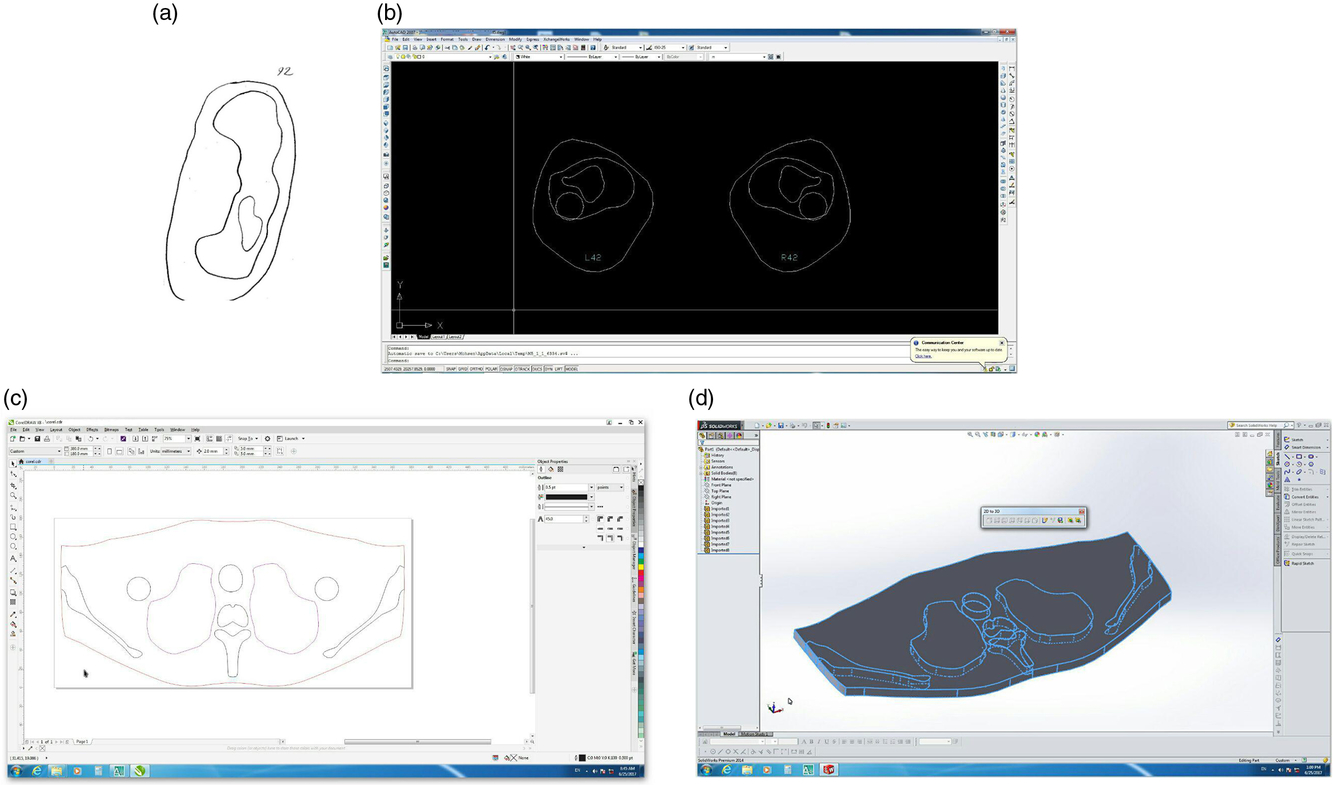
1. Drawing a simplified cross section of each part of the whole body in the calc sheets (Figure 1a).
2. Providing a scan from calc drawings to obtain the computerised file.
3. Importing information of each layer, including thickness and dimensions to the AutoCAD 2D and 3D tools (version 2014; Autodesk Inc., USA) to obtain a plotted map from scans (Figure 1b).
4. Providing 3D maps of drawings and putting them together in appropriate layers (Figure 1c).
5. Converting the file format from AutoCAD to Corel and Solid works for segment fabrication and assembly (Figure 1d).
The construction steps of the phantom sections: (a) simple cross-sectional drawing in calc sheets, (b) 2D image of body cross section in AutoCAD software, (c) assembly of segments in Corel software, and (d) assembly of segments in Solid works software.

Phantom validation
The constructed phantom (Figures 2a and 2b) must comply with the ICRU44 standards; thus, validation of the phantom should be carried out. Because this phantom is to be used for megavoltage beam measurements and the dominance of Compton scattering in this energy range, the electron density of tissue substitute material must match real tissue. Furthermore, the electron density is an important radiological characteristic of each substance and should be taken into consideration for phantom validation. Validation for our phantom was performed using two approaches as follows.
(a) Three-dimensional images of constructed RANDO phantom from different directions. (b) Better view of constructed RANDO phantom.

Conformity of the tissue’s CT numbers with the standard protocol
Because of different CT values of various human tissues, it is necessary to compare the CT numbers of different parts of the RANDO anthropomorphic phantom with the range of acceptable numbers of each tissue according to the standard protocol (ICRU44). To determine the CT number of different regions of the tissues in RANDO phantom, region of interest with dimensions of 1 cm2 at the centre of each tissue in the CT images of the phantom was selected and CT numbers were determined for each area.
Electron density measurement of tissue equivalent materials and comparison with standard values
The electron density is an important radiological characteristic of each substance and should be taken into consideration for phantom validation. Having the same electron density greatly guarantees the equivalent behaviour of materials against photon beams.
Linear attenuation coefficients
In general, two materials can be radiologically equivalent for megavoltage beams, if they have the same electron density. It is justified by the fact that the linear attenuation coefficient of the matter is directly proportional to its electron density. In this method, ρ e can be obtained from the following equation:
 $${\rho _e} = (\mu /{\mu _w}) \times {\rho _{e,w}}$$
(1)
where ρ e and ρ e,w are the electron density of tissue substitute and water, respectively; µ refers to the attenuation coefficient of the tissue substitute and µ w is the attenuation coefficient of water.
CT number (Hounsfield HU)
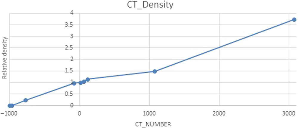
A different method for determining the electron density is using the CT number, relative electron density calibration curve of GE CT scanner (64 slices light Speed Scanner), by which our fabricated phantom was scanned (Figure 3).
Dependence of CT numbers on relative electron density for GE CT scanner.

Results
Selection of different body tissues substitutes materials
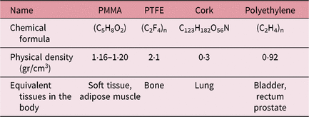
Our constructed anthropomorphic male RANDO phantom consists of 175 layers with 1-cm thickness, 175-cm height and 72-kg weight. The mass of the fabricated skeletal bone, bone tissue, skeletal muscle, brain, rectum, bladder and prostate phantom in this study is consistent with the reference values suggested by ICRP89 (Table 1). Also, bone tissues with 2710 cm3 volume and 17 m2 total surface areas, rectum with 11-cm length, and breasts with the total mass of 25 g absolutely complied with the defined standards for an adult male.
Description and physical properties of tissue substitutes used in this project

Conformity of the tissue’s CT numbers with the standard protocol
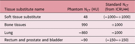
The results of the evaluation and comparison of CT numbers of tissue substitute materials and real human body tissue indicate absolute concordance of the range of CT numbers of different bone tissues and skeletal muscles, soft tissues, lung tissue and pelvic heterogeneities including rectum, bladder and prostate with defined standard values (Table 2).
Comparison between relative CT number of different tissue substitute material and standard data from ICRU44

Electron density measurement of tissue equivalent materials and comparison with standard values
Linear attenuation coefficients
Relative electron densities (reDLAC) were calculated from the linear attenuation coefficient for various body tissues and substitute material. Related data are presented in Table 3.
Relative electron densities calculated from linear attenuation coefficient (reDLAC) for body tissues and for material

CT number (Hounsfield HU)
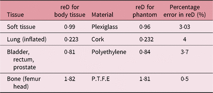
Relative electron density of tissue substitute materials and body tissue according to their CT number ranges and their percentage error were extracted using reD conversion curve (Figure 3).
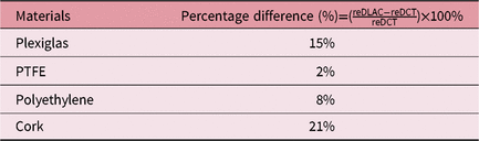
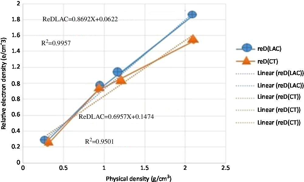
Results are shown in Table 4. To compare two validation methods, percentage error of CT calculated and linear attenuation coefficient calculated, relative electron densities of all tissue substitute materials were determined (Table 5). Furthermore, relative electron densities of tissue substitute materials were plotted as a function of the material’s mass density for these two methods. The correlation equations with the regression coefficients associated with each of them (R2) are located on the curve for both methods (Figure 4).
Relative electron density calculated from reD-CT number curve for tissue substitute materials and body tissue

Percentage error of relative electron density of CT number and linear attenuation correction method

reD for tissue equivalent material used in phantom construction, as a function of the material’s mass density for both linear attenuation coefficient and CT number methods.

Discussion
In this study, the RANDO anthropomorphic phantom was designed from cross-sectional CT images of the atomic energy organisation standard RANDO phantom as a reference phantom. For accurately measuring the dosimetry for techniques such as TSET and TBI, the design of the whole body phantom with two lower limbs is required.
Comparison between data in Table 2 showed that relative CT numbers of tissue substitute materials are within accepted ranges defined by ICRU44.
Percentage error data from Tables 3 and 4 showed that the PTFE for bone is the best choice of substitute material due to its minimum percentage error of 0·5%. Both methods (CT number and linear attenuation coefficients) illustrated that other tissue substitute materials have an equal deviation, up to 12% from body tissue which could encourage the researcher to find better materials in subsequent studies.
Higher correlation (R 2 = 0·9957) between the physical density of the tissue equivalent materials and the relative electron density obtained from the linear attenuation coefficient (reDLAC) in comparison with the corresponding regression coefficient between the physical density material and the relative electron density obtained through the CT number (reD-CT) with R 2 = 0·9501, was observed. This can be due to the beam-hardening effects, which are in good agreement with other studies. Therefore, the electron densities obtained from the linear attenuation coefficient (reDLAC) for the tissue equivalent material used in the phantom is a better option for drawing CT number to reD conversion curve for designing treatment planning software. According to the mentioned benefits, the constructed phantom in this study is recommended for TBI and TSET dosimetry.
Conclusion
This phantom has numerous advantages over the origin type of RANDO phantom. First, it was constructed using available facilities with lower costs. In RANDO phantom, the bony parts of the phantom, due to the use of human natural skeletons, disintegrate over time. Thus, the second advantage of this study is the use of high stability material (PTFE) as a bone substitute material which makes phantom more durable.
Another advantage of this phantom is the design and the construction of three intra pelvic organs (bladder, prostate and rectum) for further dosimetry studies and evaluation of the dose delivered to radiation sensitive organs in pelvis radiation therapy. Finally, the thickness of the phantom cross sections was considered as 1 cm as compared to 2·5 cm in the reference RANDO phantom; thus, better resolution for film and dosimeters placement in the phantom body for internal dose measurements was achieved. The phantom validation technique for our RANDO phantom confirmed its material selection validity.
Acknowledgements
This research was supported by Tehran University of Medical Sciences as a master thesis number of 94-03-30-29996.
Conflict of Interest
There is no conflict of interest.
Funding
This study was funded by grant number 2996.