Oropharyngeal candidiasis is an opportunistic infection caused primarily by Candida albicans (C. albicans), a ubiquitous fungal organism that is part of the normal oral microflora in up to 80% of healthy individuals (Ref. Reference Odds1). The transition from commensalism to infection in the oral mucosa is caused by changes in the local oral microenvironment, or by an inadequate host defence, which results in C. albicans overgrowth (Ref. Reference Scully, el Kabir and Samaranayake2). The prevalence of oropharyngeal candidiasis continues to be high because of an increasing population of immunocompromised patients, including individuals with diabetes, HIV, transplant recipients and patients with solid tumors or haematological malignancies receiving stem cell therapy (Refs Reference Rubin3, Reference Ohmit4, Reference Redding5, Reference Hermann6, Reference Penk and Pittrow7). In general, epidemiological studies suggest a key role of an intact immune system in protection against this infection, and indicate a specific role for T cell immunosuppression in susceptibility to infection (Refs Reference Rubin3, Reference Ohmit4, Reference Redding5, Reference Hermann6, Reference Penk and Pittrow7). Oral lesions are generally associated with local discomfort, altered taste sensation and dysphagia, resulting in poor nutrition (Ref. Reference Neville8). Moreover, oropharyngeal candidiasis is associated with esophageal candidiasis, a more invasive form of infection with the potential for systemic spread (Refs Reference Anaissie and Bodey9, Reference Richet10, Reference Samonis and Bafaloukos11). Immune defence against opportunist infection with C. albicans depends on proper host recognition, followed by activation of antifungal mechanisms in effector cells. This review discusses the current understanding of recognition of C. albicans by the host and activation of anti-Candida mechanisms in mucosal structural and immune cells. We also discuss how to apply this knowledge to the development of novel treatment strategies against this fungal organism.
Candida recognition by structural mucosal and innate immune cells
Activation of protective mechanisms in innate immune and nonimmune effector cells depends on appropriate pathogen recognition. Rapid recognition of pathogens is mediated by the expression of pattern-recognition receptors (PRRs) on host cells, which recognise conserved pathogen-associated molecular patterns (PAMPs) (Ref. Reference Mukhopadhyay12). These recognition receptors are expressed by numerous cells, including polymorphonuclear leukocytes (PMNLs), dendritic cells, monocytes, macrophages, B cells, T cells, endothelial and epithelial cells. Recognition of PAMPs by PRRs induces intracellular signalling pathways that modulate protective and inflammatory responses, such as activation of antimicrobial mechanisms, expression of costimulatory molecules, release of proinflammatory cytokines and modulation of acquired immunity.
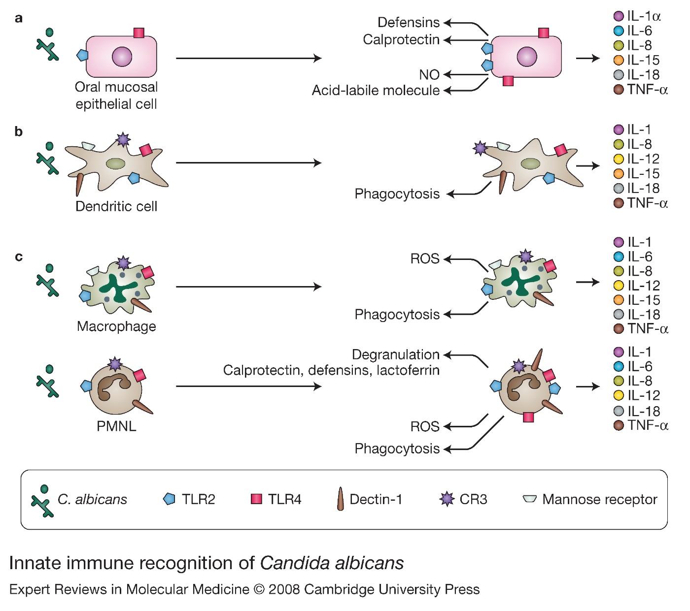
C. albicans possesses a variety of PAMPs, including chitin (a cellulose-like biopolymer consisting predominantly of N-acetyl-D-glucosamine), glucan (a β-linked branched-chain polymer of glucose), mannan (an α-linked polymer of mannose), mannoproteins and glycolipids. Differential recognition of these components on the C. albicans cell wall is mediated through numerous PRRs, such as Toll-like receptors (TLRs), mannose receptors, Dectin-1, complement receptor 3 (CR3) and mannose-binding lectins (Refs Reference Brown13, Reference Forsyth, Plow and Zhang14, Reference Underhill15) (Fig. 1).
Innate immune recognition of Candida albicans. PRRs are expressed in oral mucosal surveillance cells including: (a) structural oral epithelial cells, (b) dendritic cells and (c) professional phagocytes, such as macrophages and neutrophils. Recognition of C. albicans by PRRs induces intracellular signalling pathways that modulate protective, inflammatory and regulatory responses, including activation of antimicrobial mechanisms, and release of proinflammatory and immune-instructive cytokines. Abbreviations: CR3, complement receptor 3; IL-8, interleukin-8; NO, nitric oxide; PMNL, polymorphonuclear leukocyte; PRR, pattern-recognition receptor; ROS, reactive oxygen species; TLR, Toll-like receptor; TNF, tumour necrosis factor.

Toll-like receptors
TLRs constitute a family of PRRs that mediate microbial recognition and subsequent activation of phagocytosis, inflammatory responses and production of effector molecules, such as nitric oxide and reactive oxygen species (ROS) (Ref. Reference Takeda16). These receptors are type 1 transmembrane proteins, which are characterised by an extracellular domain containing 25 leucine-rich repeats and an intracellular Toll–IL-1 receptor (TIR) domain (Ref. Reference Janeway and Medzhitov17). Following ligand binding, the TLR-signalling pathway culminates in the migration of the transcription factor NF-κB into the nucleus, an event that in turn initiates the transcription of several inflammatory genes (Ref. Reference Roeder18). A role for TLRs in protection against C. albicans infection has been demonstrated by the increased susceptibility of myeloid differentiation factor 88 (Myd88)-knockout mice, which lack the MyD88 intracellular adaptor of the TLR TIR domain, to systemic candidiasis (Refs Reference Bellocchio19, Reference Gozalbo20, Reference Marr21). More recently, both TLR2 and TLR4 have been specifically implicated in the host defence against C. albicans (Ref. Reference Netea22). The first observation that indicated a possible protective role of TLR2 during C. albicans infection came from the increased susceptibility of TLR2-deficient mice to haematogenously disseminated candidiasis, an effect that was attributed to impaired production of tumour necrosis factor-α (TNF-α) and macrophage-inflammatory protein-2 (MIP-2) as well as decreased PMNL recruitment into infected organs (Ref. Reference Villamon23). However, other studies demonstrated reduced mortality and a lower fungal burden in TLR2-deficient mice (Refs Reference Bellocchio19, Reference Netea24). Greater resistance of these mice to C. albicans infection is associated with reduced production of T-helper 2 (Th2) cytokines, increased levels of interferon-γ (IFN-γ) and interleukin-12 (IL-12), and decreased numbers of CD4+ CD25+ regulatory T cells (Refs Reference Bellocchio19, Reference Netea24). Moreover, in the absence of TLR2 receptors, macrophages show increased chemotaxis into infected sites and augmented fungicidal activity (Ref. Reference Netea24).
TLR4 has also been implicated in host protection against C. albicans. TLR4-deficient mice have an increased susceptibility to primary disseminated, intragastric and secondary infection with C. albicans (Refs Reference Bellocchio19, Reference Netea22). The increased fungal load in the organs of TLR4-knockout mice is associated with impaired release of CXC chemokines, including keratinocyte-derived chemokine (KC) and MIP-2, which results in reduced recruitment of PMNLs to the sites of infection (Ref. Reference Netea22). Surprisingly, the local production of proinflammatory cytokines in response to C. albicans was minimally affected by the absence of TLR4 and the fungicidal activity of TLR4–/– PMNLs remained unaltered (Ref. Reference Netea22). However, this increased susceptibility of TLR4-deficient mice to Candida infection was not confirmed in additional studies using haematogenous models of disseminated candidiasis (Refs Reference Bellocchio19, Reference Murciano25).
The reason for the divergent data in TLR2- and TLR4-knockout mice in different studies is not understood. However, discrepant findings could be at least partially explained by the use of animals with distinct genetic backgrounds challenged with different fungal strains and examination of fungal burden in different organs. The latter may be an important factor, because fungal growth and morphogenesis may be differentially regulated in different tissue environments. In addition, a tissue-specific protective immunity has been shown to exist in Candida infections (Ref. Reference Fidel26). Thus, depending on the unique immune regulatory and effector mechanisms in each tissue or organ, TLR-mediated signalling may or may not play an important role in protection.
The participation of other TLRs in the host response to C. albicans has not been extensively investigated. Nevertheless, TLR9-knockout mice failed to show an increased susceptibility to haematogenously disseminated candidiasis (Ref. Reference Bellocchio19). In fact, TLR9-deficient mice had a lower fungal burden than control mice (Ref. Reference Bellocchio19). Lower levels of IL-12 and higher levels of IL-4 and IL-10 were detected in TLR9-knockout mice (Ref. Reference Bellocchio19), which raises the question of how a shift towards a Th2 response, which is generally considered nonprotective (Ref. Reference Romani27), did not result in increased susceptibility to candidiasis.
Most of the studies on Candida recognition by TLRs focus on the role of these receptors in systemic infection. Owing to the differences in protective immunity between systemic and mucosal Candida infections (Ref. Reference Fidel28), the role of each TLR in mucosal protection may not be extrapolated from systemic models of infection. However, constitutive expression of several TLRs in structural oral mucosal cells suggests a potential role in mucosal surveillance and protection (Refs Reference Uehara, Sugawara and Takada29, Reference Hayashi, Means and Luster30, Reference Cottalorda31, Reference Mahanonda and Pichyangkul32). Notably, the ability of epithelial cells to kill C. albicans is inhibited by anti-TLR2 and -TLR4 antibodies (Ref. Reference Pivarcsi33). In an in vitro model of the human oral mucosa where neutrophils were basally added, C. albicans induced the expression of TRL4 on oral epithelial cells. In this model, epithelial TLR4 expression requires the presence of PMNLs and correlates with protection against fungal invasion and cell injury, as demonstrated by the increased cell damage and fungal invasion in response to neutralising TLR4 antibodies, or partial TLR4 gene silencing using RNA interference (Ref. Reference Weindl34). By contrast, TLR2 failed to provide host protection in this infection model (Ref. Reference Weindl34). Although these in vitro experiments suggest a more active role of TLR4 in oral mucosal protection, they need to be confirmed in animal models of oropharyngeal candidiasis. In addition, the functional activation of neutrophils has not been directly assessed in these three-dimensional oral mucosal models (Ref. Reference Weindl34).
Mannose receptors
Mannose receptors have been implicated in the recognition of C. albicans highly branched N-linked mannosyl residues and stimulation of T-helper 1 (Th1) cytokine production (Ref. Reference Netea35). Macrophages are able to ingest and kill C. albicans through mannose receptors (Refs Reference Marodi, Forehand and Johnston36, Reference Marodi, Korchak and Johnston37), which are involved in TNF-α, IL-1β, IL-6 and granulocyte macrophage colony-stimulating factor (GM-CSF) responses (Refs Reference Netea35, Reference Yamamoto, Klein and Friedman38). Exploitation of mannose receptors on dendritic cells by C. albicans also leads to development of a Th1 response (Ref. Reference Romani39). Furthermore, a mannose-binding receptor is expressed on human keratinocytes and mediates anti-Candida activity, possibly via nitrous oxide production (Refs Reference Dobozy40, Reference Szolnoky41). The role of mannose receptors during infection has been demonstrated in a murine model of candidiasis in which intraperitoneal administration of C. albicans results in an increased fungal burden in mannose-receptor-knockout mice (Ref. Reference Lee42).
Dectin-1
Recently, a receptor named Dectin-1 has been shown to recognise C. albicans. Dectin-1 is a type II transmembrane receptor expressed on macrophages, monocytes, neutrophils, natural killer cells and T cells (Ref. Reference Brown43). Dectin-1 has been reported to be the major receptor for a variety of β-1,3-linked and β-1,6-linked glucans (Ref. Reference Brown and Gordon44), and the high level of expression of this receptor in the lungs and gastrointestinal tract suggests a role in mucosal pathogen recognition and host defence (Ref. Reference Brown43). Dectin-1 mediates macrophage recognition of C. albicans yeast cells, but not filaments. This appears to correlate with the functional accessibility of β-glucan in the cell wall of different C. albicans morphotypes. Whereas β-glucan is exposed at sites of budding growth and septation on yeast cells, C. albicans filaments display no functionally accessible β-glucan under certain experimental conditions (Ref. Reference Gantner, Simmons and Underhill45). Interestingly, we have found that β-glucan is accessible to anti-β-glucan antibody at the tips of hyphal organisms growing in contact with a three-dimensional oral mucosa culture system (Ref. Reference Dongari-Bagtzoglou46). It is possible that active cell-wall synthesis in organisms undergoing sessile growth on the surface of biological surfaces exposes β-glucan components, which in time might become masked by surface mannans or mannoproteins.
Expression of Dectin-1 in macrophages enhances the ability of these cells to bind to C. albicans and produce TNF-α (Ref. Reference Gow47). However, fungal recognition and killing are thought to occur through different mechanisms, because Dectin-1-knockout macrophages show no defect in their ability to kill C. albicans (Ref. Reference Brown43). Nevertheless, overexpression of Dectin-1 on macrophages enhances the release of arachidonic acid, COX2 expression and prostaglandin production in response to C. albicans, suggesting a role of Dectin-1-mediated signalling on phospholipase A2 (PLA2) activation and arachidonic acid release (Ref. Reference Suram48). Dectin-1 also mediates non-opsonic recognition and internalisation of C. albicans by PMNLs (Ref. Reference Kennedy49). Moreover, after fungal internalisation, Dectin-1 elicits fusion of cytoplasmic granules with phagosomes and production of ROS by PMNLs, which in turn enhances their ability to kill C. albicans (Ref. Reference Kennedy49).
Recent evidence also indicates that Dectin-1 cooperates with TLR2 in the induction of TNF-α and IL-12 responses, and NADPH activation in phagocytes (Refs Reference Rogers50, Reference Gantner51). During macrophage and dendritic cell recognition of particulate β-glucan, both Dectin-1 and TLR2 are recruited to phagosomes, where Dectin-1 binds to β-glucans and TLR2/CD14 recognises other components of the fungal cell wall (Ref. Reference Gantner51). Finally, Dectin-1 is also required for zymosan-mediated activation of NADPH oxidase – a response that is primed by TLR4 activation (Ref. Reference Gantner51).
Based on its ability to: (1) recognise fungal-cell-wall products; (2) facilitate fungal phagocytosis and killing; (3) induce NADPH oxidase activation; and (4) stimulate and regulate cytokine responses in cooperation with TLRs, Dectin-1 has recently been proposed to play a key role in fungal recognition and antifungal immunity (Ref. Reference Brown43). In agreement with this, Dectin-1-knockout mice showed lower survival rates than wild-type mice after systemic infection with C. albicans (Ref. Reference Taylor52). Moreover, in a peritoneal model of C. albicans infection, Dectin-1-knockout mice had fewer inflammatory leukocytes, such as PMNLs, monocytes and eosinophils, at the infected sites (Ref. Reference Taylor52). Reduced recruitment of inflammatory cells was associated with lower levels of cytokines (IL-6) and growth factors (G-CSF and GM-CSF) (Ref. Reference Taylor52). However, these results are in disagreement with other studies demonstrating that Dectin-1-knockout mice showed no difference in survival after intravenous infection with C. albicans compared with wild-type mice (Ref. Reference Saijo53). It is possible that the conflicting results observed in these studies are related to differences in the infecting strain, as well as the growth conditions prior to challenge, or the morphological state of Candida in the animals, which might affect the levels of β-glucan exposed in C. albicans upon entry into the bloodstream.
Other pattern-recognition receptors
Two other PRRs have recently been found to participate in the process of C. albicans recognition. The C-type lectin receptor DC-SIGN (dendritic-cell-specific, intercellular adhesion molecule-3-grabbing, non-integrin) was shown to mediate the uptake of C. albicans by dendritic cells (Ref. Reference Cambi54), whereas a DC-SIGN mouse homologue, termed SIGNR1 (SIGN-related 1) was identified as a macrophage receptor involved in yeast recognition (Ref. Reference Taylor55).
Candida recognition systems in the oral mucosa
It is clear that the recognition of C. albicans is a complex process involving multiple PRRs. Activation of different PRRs in response to C. albicans is likely to provide a broad range of possibilities for effective inflammatory responses. However, the relative contribution of specific candidal PAMPs and their respective receptors in the activation of effector cells, generation of cytokine responses and modulation of the adaptive immune response in the oral mucosa remain to be defined. There is evidence suggesting that the innate host recognition system has sufficient redundancy to ensure adequate microbial recognition and host response (Ref. Reference Mukhopadhyay12). This inherent redundancy complicates the accurate determination of the relative importance of each recognition receptor during infection. Upon ligand binding, TLR2 and TLR4 activate common intracellular pathways that rely on the recruitment of the adaptor protein MyD88 (Ref. Reference O'Neill56). This may explain why TLR2- and TLR4-deficient mice fail to show increased susceptibility to C. albicans infection in some studies (Refs Reference Bellocchio19, Reference Netea24), unlike MyD88-deficient mice (Ref. Reference Villamon57). It has also been suggested that the TLR2- and TLR4-signalling pathways might exert opposite effects in immune protection to systemic candidiasis, by differentially tipping the balance of pro- versus anti-inflammatory cytokines (Ref. Reference Netea58). However, most of the work so far in innate recognition systems, signalling pathways and effector mechanisms was carried out in models of disseminated, and not oral, candidiasis. Disseminated models offer minimal insight into the pathogenesis of oral infection. One of the reasons for this is that, at least in the initial stages of infection, the yeast form is more important in establishing disseminated infection (Ref. Reference Saville59). In contrast to haematogenous infection, the hyphal form of the organism rather than the yeast form may be more important in establishing tissue invasion in the oral mucosa – a process that distinguishes asymptomatic colonisation from infection in the alimentary tract (Ref. Reference Mathieson and Dutta60). It has been established that disparate cell wall components and recognition systems exist in these two morphogenetic strains (Refs Reference Romani39, Reference Netea61). For example, recognition and phagocytosis of unopsonised C. albicans yeasts and hyphae by dendritic cells occurs through different receptors (Ref. Reference Romani39). Other evidence also suggests that, although yeast cells stimulate both TLR2 and TLR4, hyphal cells are not recognised by TLR4 and induce a strong anti-inflammatory cytokine response mediated by TRL2 (Ref. Reference van der Graaf62). Based on these findings and the tissue-specific role of different fungal morphotypes in pathogenesis, the role of specific PRRs and their respective PAMPs in the pathogenesis of oral and disseminated infection is predicted to be different.
Immune and nonimmune antifungal effector cells in the oral mucosa
Multiple immune and nonimmune cells contribute directly or indirectly to protection against mucosal infection by recognising C. albicans, modulating protective and inflammatory responses or having direct antifungal effector functions. One of the consequences of C. albicans recognition by the host is induction of inflammatory and immune regulatory cytokines such as IL-1, TNF-α, IFNγ, IL-4, IL-5, IL-6, IL-8, IL-13, IL-17 and several chemokines (Refs Reference Lilly63, Reference Dongari-Bagtzoglou and Kashleva64, Reference Li, Kashleva and Dongari-Bagtzoglou65, Reference Li and Dongari-Bagtzoglou66, Reference Djeu67, Reference Djeu, Serbousek and Blanchard68) (Fig. 1). Antimicrobial effector functions of structural and immune cells are highly inducible via these signalling molecules. Direct candidacidal activity has been demonstrated for four cell types: PMNLs, monocytes/macrophages, non-MHC-restricted CD8+ T cells and epithelial cells (Refs Reference Marquis69, Reference Romani70, Reference Steele71, Reference Steele72) (Fig. 1).
Neutrophils
It is generally accepted that PMNLs are the most important effector cells in the host response against systemic, but not vaginal, infection with C. albicans (Refs Reference Djeu, Serbousek and Blanchard68, Reference Marquis69, Reference Fidel73). Although the role of PMNLs during oral candidiasis has not been fully elucidated, their protective function is suggested by an increased severity of oral infection in BALB/c and CBA/CaH mice following antibody-mediated depletion of PMNLs (Ref. Reference Farah74). The protective role of PMNLs during oropharyngeal candidiasis requires activation signals provided by CD4+ T cells, because PMNLs failed to limit chronic oral infection of C. albicans in transgenic mice expressing human immunodeficiency virus type 1 (Ref. Reference Marquis75). During infection, local production of multiple cytokines and chemokines induces the recruitment and activation of PMNLs from the vascular compartment to the oral mucosa. IL-8 is one of the key chemokines involved in the recruitment of PMNLs to sites of infection (Refs Reference Baggiolini, Walz and Kunkel76, Reference Casale and Carolan77). Oral epithelial cells produce high levels of IL-8 in response to C. albicans (Ref. Reference Dongari-Bagtzoglou, Villar and Kashleva78) (Fig. 1), which might explain the substantial migration of PMNLs into Candida-infected oral mucosal sites.
PMNLs ingest large numbers of Candida yeast cells (Ref. Reference Pereira and Hosking79) and can kill a large percentage of ingested organisms (Ref. Reference Richardson and Smith80). Much of the killing that takes place upon interaction with C. albicans is due to activation of respiratory burst and release of ROS (Ref. Reference Romani70). ROS are essential to the role of PMNLs in host defence, and animals with defects in the production of these bioactive molecules are highly susceptible to C. albicans infection (Ref. Reference Aratani81). IFN-γ, TNF-α, IL-8 and GM-CSF stimulate the production of ROS by PMNLs (Refs Reference Diamond, Lyman and Wysong82, Reference Le and Vilcek83, Reference Tan84, Reference Gadish85, Reference Schroder and Christophers86) and not surprisingly, they effectively augment the candidacidal activity of human PMNLs (Refs Reference Diamond, Lyman and Wysong82, Reference Djeu87) (Fig. 1). It is interesting to note that death of neutrophils at the site of infection, might also contribute to clearance of C. albicans, because dying PMNLs undergo degranulation and release several antimicrobial peptides, including calprotectin, defensins and lactoferrin (Refs Reference Steinbakk88, Reference Sohnle, Collins-Lech and Wiessner89) (Fig. 1).
Monocytes/macrophages
The role of macrophages in disseminated infection with C. albicans is well established, (Refs Reference Hurtrel and Lagrange90, Reference Jensen, Warner and Balish91, Reference Qian92); however, few studies have assessed their role in resistance to oropharyngeal infection. Macrophage inactivation significantly increases the severity of oral infection compared with that of controls (Ref. Reference Farah74). These mononuclear cells have been reported to ingest and kill 30–60% of unopsonised C. albicans yeast cells (Refs Reference Marodi, Korchak and Johnston37, Reference Vonk93, Reference Netea, Kullberg and Van der Meer94). Moreover, antibody opsonisation of C. albicans in either the yeast or hyphal form further increases the ability of human monocytes to ingest these organisms (Refs Reference Wellington, Bliss and Haidaris95, Reference Wellington, Dolan and Haidaris96) (Fig. 1). Notably, although antibody opsonisation enhances the ability of monocytes to damage C. albicans yeast cells, it has no effect on monocyte-mediated damage to C. albicans hyphal cells (Ref. Reference Wellington, Dolan and Haidaris96), suggesting that hyphal cells are either processed in a different pathway or are generally more resistant to monocyte-mediated damage.
CD8+ T cells
A direct protective role of CD8+ T cells against oropharyngeal candidiasis has also been suggested. CD8+ T cells exert growth inhibition against the hyphal form of C. albicans in vitro (Ref. Reference Beno, Stover and Mathews97). CD8+ T cells were required to limit chronic oral carriage of C. albicans in one animal model of infection (Ref. Reference Marquis69), but other studies failed to show a role of CD8+ T cells in oral protection (Ref. Reference Farah98). Accumulation of CD8+ T cells at the interface of the oral epithelium and the lamina propria further suggests a role for CD8+ T cells in protection against oropharyngeal candidiasis (Ref. Reference Leigh, McNulty and Fidel99). The exact mechanism of action of CD8+ T cells against C. albicans infection is currently unknown. Cytotoxic activity by CD8+ T cells is not effective, suggesting that its antifungal property is solely based on growth inhibition activity (Ref. Reference Beno, Stover and Mathews97).
Epithelial cells
Although structural mucosal cells were thought to merely act as a physical barrier against infection, recent data suggest that oral epithelial cells possess a direct antimicrobial function, which may contribute to host defence against C. albicans. Expression of inducible nitric oxide synthase by oral epithelial cells has been shown to exert bactericidal activity (Ref. Reference Deitch100), and might contribute to the host defence against C. albicans (Fig. 1). NO diffuses across cell membranes and is capable of reacting with DNA and a variety of proteins or lipids, causing cell damage. Although the mechanism of action of NO against C. albicans has not been specifically investigated, NO has been associated with candidacidal activity and resistance to mucosal candidiasis (Ref. Reference Jones-Carson101). However, oral keratinocytes might also control C. albicans infection via secretion of antimicrobial peptides, such as members of the β-defensin family of peptides and calprotectin (Refs Reference Dunsche102, Reference Dunsche103, Reference Harder104, Reference Liu105, Reference Mathews106, Reference Schroder and Harder107) (Fig. 1). β-defensin-2 and β-defensin-3 have been shown to possess the greatest candidacidal activity (Ref. Reference Feng108); they are upregulated in response to IFN-γ and TNF-α (Refs Reference Harder104, Reference Garcia109) and cause disruption of the cell membrane (Ref. Reference Feng108).
In vitro studies have shown that oral epithelial cells inhibit the growth of Candida species by 60–90%, through a mechanism that requires direct contact between epithelial cells and the fungal organism (Refs Reference Steele71, Reference Steele72, Reference Nomanbhoy110). Although the mechanism responsible for this inhibitory action has not been fully identified, it is generally accepted that it does not involve phagocytosis, oxidative killing or nonoxidative antimicrobial mechanisms via calprotectin or defensins (Ref. Reference Steele111). Enzymatic removal of carbohydrates from oral epithelial cells has been shown to eliminate this anti-Candida activity, suggesting that it occurs via a cell-associated carbohydrate moiety (Refs Reference Nomanbhoy110, Reference Steele111). However, more recent work showed that the antifungal activity is actually due to an acid-labile protein moiety (Ref. Reference Yano112) (Fig. 1). The involvement of high molecular weight hyaluronan in the anti-Candida activity of human oral epithelial cells has also been suggested, but not proven (Ref. Reference Sakai113).
Role of regulatory T cells in oral mucosal protection
T-cell-deficient mice have been used to demonstrate that CD4+ T cells play a key regulatory role in the immune response against oral candidal infection. Even though the number and function of neutrophils and monocytes in these mice were normal, they could not clear C. albicans oral mucosal infection (Refs Reference Farah98, Reference Cantorna and Balish114). Moreover, reconstitution of these immunodeficient mice with naive CD4+ T cells (but not CD8+ T cells) significantly decreased oral colonisation compared with that in controls. Similarly, depletion of CD4+ T cells in the oral mucosa of BALB/c mice has been shown to increase the severity of oral infection with C. albicans (Ref. Reference Farah98). Interestingly, high levels of IL-12 and IFN-γ are produced by lymphocytes derived from the lymph nodes of resistant mice following C. albicans infection, suggesting that CD4+ T cells may augment phagocytic cell function via secretion of Th1 cytokines (Ref. Reference Farah98). However, there appears to be no protective role of IFN-γ against Candida in the oral mucosa (Ref. Reference Farah and Ashman115).
More recently, the role of CD4+ CD25+ regulatory T (Treg) cells in host protection against C. albicans has also been investigated in animal models of candidiasis. Two different types of Treg cells participate in the immune response against C. albicans – naturally occurring Treg cells and inducible Treg cells (Ref. Reference De Luca116). Naturally occurring Treg cells migrate to inflamed sites where they modulate the effector function of PMNLs and limit early inflammatory responses. Although migration of these Treg cells into infected sites requires activation of the TRIF-signalling pathway, their anti-inflammatory activity is suppressed by the MyD88-signalling pathway (Ref. Reference De Luca116). Later, inducible populations of Treg cells expand in both disseminated and gastrointestinal infection with C. albicans (Ref. Reference De Luca116). The induction of these cells during C. albicans infection is determined by the level of expression of costimulatory molecules, such as CD28 and CD86, and by signals provided by TLR2- and TRIF-signalling pathways (Refs Reference Netea24, Reference De Luca116, Reference Montagnoli117, Reference Sutmuller118). During infection, inducible Treg cells producing IL-4, IL-10 and transforming growth factor-beta (TGF-β) restrict inflammatory Th1 and Th17 responses, which on one hand limits the ability of the immune system to control fungal growth (Refs Reference Netea24, Reference Montagnoli117, Reference Zelante119), but on the other hand increases host resistance to reinfection (Ref. Reference Montagnoli117).
Reports from systemic infection models have suggested that memory CD4+ T cells producing IL-17 (Th17 cells) are generated as part of the immune response to C. albicans (Ref. Reference LeibundGut-Landmann120). Generation of Th17 cells is triggered by exposure of newly primed CD4+ T cells to IFN-γ, TGF-β, IL-6 and IL-23 (Refs Reference Veldhoen121, Reference Mangan122, Reference Bettelli123). Exposure of CD4+ T cells to these cytokines induces the expression of transcription factor retinoic acid receptor (RAR)-related orphan receptor gamma (RORγ), which controls the expression of IL-23 receptors on CD4+ T cells (Ref. Reference Ivanov124). Increased expression of IL-23 receptors on CD4+ T cells in turn renders these cells susceptible to IL-23 – a cytokine that promotes proliferation and differentiation of T-helper cells into Th17 cells (Refs Reference Aggarwal125, Reference Langrish126). C. albicans hyphal cells stimulate the production of IL-23 by monocyte-derived dendritic cells (Ref. Reference Acosta-Rodriguez127). Activation of Dectin-1 signalling through the Syk-CARD9 pathway induces maturation of dendritic cells and secretion of IL-6, IL-23, and TNF-α by these cells, which in turn, induces differentiation of naive T cells into Th17 cells (Ref. Reference LeibundGut-Landmann120). This Dectin-1–Syk–CARD9 signalling pathway has been shown to be required for the development of a potent Th17 response during haematogenouly disseminated candidiasis (Ref. Reference LeibundGut-Landmann120).
Production of IL-17 by Th17 cells in response to C. albicans (Ref. Reference LeibundGut-Landmann120) can potentially induce the recruitment and activation of neutrophils, and production of proinflammatory cytokines by epithelial cells, endothelial cells and macrophages (Ref. Reference Weaver128). In fact, defective mobilisation and influx of neutrophils into infected sites, increased fungal burden and impaired survival have all been shown in IL-17-receptor-knockout mice after systemic challenge with C. albicans (Ref. Reference Huang129). However, the increased susceptibility of IL-17-deficient mice to candidal infection was not confirmed in subsequent studies using animal models of systemic and gastrointestinal infections. By contrast, IL-17-knockout mice were more resistant to candidal infection, and the increased resistance was associated with a greater number of IFN-γ-producing Th1 cells (Ref. Reference Zelante119). These recent data suggest that a crossregulation exists between IFNγ/Th1 and IL-23/Th17 pathways. Th17 responses have not been characterised in oral models of infection; however, it is speculated that Th17 cells, which express chemokine receptors involved in mucosal homing (Ref. Reference Acosta-Rodriguez127), may play a role in the immune response to oropharyngeal candidiasis.
Cytokines as adjuncts to traditional antifungal therapies in the oral mucosa
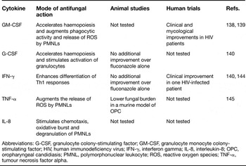
Most cases of oropharyngeal candidiasis are treated with antifungals, such as azoles or nystatin; however, prolonged use of antifungals is associated with emergence of resistant strains (Ref. Reference Rogers130). Moreover, severely immunocompromised patients often relapse after antifungal treatment (Ref. Reference Rogers130), indicating the necessity for more effective long-term therapeutic approaches. Growth factors and cytokines are signalling molecules that regulate the differentiation, proliferation and function of immune cells. In this context, presence of these bioactive molecules in the infected tissues dictates the outcome of the host defence against microbial infection. Increased levels of GM-CSF, IFN-γ, TNF-α IL-8, IL-18 and IL-1α have been identified in the oral mucosa and saliva of subjects with oral candidiasis and in experimental models of oral candidiasis (Refs Reference Lilly63, Reference Schaller131, Reference Tardif132). Since antimicrobial effector functions of immune cells are highly inducible by these signalling molecules, these molecules are expected to play a central role in the protection against C. albicans. Few cytokine-based therapies have been investigated in both murine models and human cases of oropharyngeal candidiasis (Table 1).
Cytokines with potential therapeutic applications in oropharyngeal candidiasis

Abbreviations: G-CSF, granulocyte colony-stimulating factor; GM-CSF, granulocyte monocyte colony-stimulating factor; HIV, human immunodeficiency virus; IFN-γ, interferon gamma; IL-8, interleukin-8; OPC, oropharyngeal candidiasis; PMNL, polymorphonuclear leukocyte; ROS, reactive oxygen species; TNF-α, tumour necrosis factor alpha.
GM-CSF has been shown to accelerate haemopoiesis in the early phases of differentiation of myeloid cells, resulting in elevated production of monocytes, neutrophils and eosinophils. In addition to its ability to increase cell numbers, GM-CSF elicits the expression of chemotaxis receptors in PMNLs, stimulates chemotaxis, phagocytic activity and the release of ROS by PMNLs (Refs Reference Gadish85, Reference Ziege133), and prolongs their survival by delaying apoptosis (Ref. Reference Richardson, Brownlie and Shankland134). GM-CSF also stimulates TLR2 expression in PMNLs (Ref. Reference Kurt-Jones135) and Dectin-1 expression in macrophages (Ref. Reference Willment136), which can potentially contribute to the process of pathogen recognition. Furthermore, GM-CSF may increase antifungal resistance by inducing the expression of chitotriosidase by professional phagocytes – an enzyme that attacks chitin-containing fungal cell walls (Ref. Reference van Eijk137).
Owing to its potential ability to promote granulocyte and monocyte activation and influx into infected sites (Ref. Reference Richardson, Brownlie and Shankland134), the therapeutic use of GM-CSF has been investigated for oropharyngeal candidiasis. In one human pilot study, GM-CSF, in association with either fluconazole or amphotericin B, resulted in modest clinical and mycological improvements in patients with acquired immunodeficiency syndrome (AIDS) with oropharyngeal candidiasis that was resistant to antifungals (Refs Reference Vazquez, Gupta and Villanueva138, Reference Vazquez, Hidalgo and De Bono139). Although adjunctive GM-CSF therapy somewhat promoted resolution of oropharyngeal and esophageal lesions, patients relapsed within 2 weeks after discontinuation of GM-CSF (Refs Reference Vazquez, Gupta and Villanueva138, Reference Vazquez, Hidalgo and De Bono139), suggesting that an intact immune response is important for sustained fungal clearance and that fungal eradication in patients with advanced AIDS might be a difficult task even when those patients are treated with adjunctive immunoregulatory agents. Moreover, G-CSF in combination with fluconazole showed no additional benefit over fluconazole monotherapy in reducing fungal burden in a murine model of chronic orogastrointestinal candidiasis (Ref. Reference Clemons and Stevens140).
IFN-γ acts as a major regulator of the Th1/Th2 response, enhancing differentiation of Th1-cell-mediated immunity, which is considered to be protective against C. albicans infection (Ref. Reference Romani70). IFN-γ has been shown to stimulate the activation of several host effector cells, including macrophages, monocytes, natural killer cells and neutrophils (Refs Reference Gallin141, Reference Murray142, Reference Torrico143). Moreover, it induces the expression of defensin-3, TLR-2 and TLR-4 in epithelial cells (Refs Reference Uehara, Sugawara and Takada29, Reference Garcia109). As a key activator of Th1-cell-mediated immunity (Ref. Reference Romani70), IFN-γ represents a promising treatment for Candida infection. Subcutaneous administration of IFN-γ resulted in a drastic improvement of clinical signs and symptoms in a HIV-infected patient with azole-resistant oropharyngeal candidiasis (Ref. Reference Bodasing144). However, this cytokine failed to improve the efficacy of fluconazole in the treatment of oropharyngeal candidiasis in severe combined immunodeficiency (SCID) mice (Ref. Reference Clemons and Stevens140).
Finally, reduced levels of C. albicans were found on the tongues of infected mice treated by oral administration of TNF-α, supporting a protective role of TNF-α during oral candidiasis (Ref. Reference Ohta145). TNF-α is a proinflammatory cytokine required for the development of effective innate and adaptive immunity to fungal infection. First, TNF-α plays a key role in the process of transmigration of neutrophils into infected sites by inducing the expression of leukocyte and endothelial cell-adhesion molecules (Ref. Reference Gamble146). Second, this cytokine has been shown to increase the microbicidal activity of neutrophils via increased secretion of ROS (Refs Reference Diamond, Lyman and Wysong82, Reference Le and Vilcek83, Reference Tan84). Third, TNF-α induces secondary production of other cytokines, such as IFN-γ, IL-1, IL-6 and IL-12 (Ref. Reference Netea, Kullberg and Van der Meer94), which further amplify the protective immune response to C. albicans infection. Although data from in vitro and animal studies suggest that TNF-α plays a central role in host protection against C. albicans infection, the effects of TNF-α treatment on human cases of oropharyngeal candidiasis are not known.
Future approaches for antifungal discovery
Treatment of oropharyngeal candidiasis has relied strongly on the use of antimicrobial drugs. However, unsatisfactory results with drug monotherapy and emergence of resistant strains in extremely immunocompromised patients have prompted investigations on the use of adjunctive immunotherapies. The major promise of new adjunctive immunoregulatory therapies lies in their potential to generate and/or augment an appropriate host immune response that is required for control of the microorganisms. This would be particularly helpful in immunocompromised patients who suffer from persistent or recurrent episodes of oropharyngeal candidiasis.
A small spectrum of immunomodulators has been tested in animal models of oropharyngeal candidiasis. Clinical experience with immunoregulatory cytokines is limited, and controlled multicentre clinical trials will be necessary to validate the initial results from small studies or case reports. For example, although GM-CSF was shown to temporarily improve the immune response and positively influenced resolution of oropharyngeal candidiasis in a very small group of HIV-infected patients (Refs Reference Vazquez, Gupta and Villanueva138, Reference Vazquez, Hidalgo and De Bono139), larger-scale human clinical trials are still required to establish its therapeutic potential. Moreover, the role of other cytokines in the treatment or prevention of oropharyngeal candidiasis still needs to be assessed. Owing to its positive effects on the migration and activation of PMNLs (Refs Reference Baggiolini, Walz and Kunkel76, Reference Casale and Carolan77, Reference Schroder and Christophers86), IL-8 might represent a potential candidate cytokine for the treatment of chronic oropharyngeal candidiasis. Finally, because of the high potential for toxicity, data on the safety of the clinical use of cytokines administered locally or systemically, need to be fully disclosed before large-scale incorporation in therapeutic regimens. One of the advantages of treating fungal infection in the oral mucosa is the accessibility of this tissue to drug delivery, which allows local as opposed to systemic therapeutic interventions. Thus, as long as local delivery is accompanied by adequate substantivity, locally applied cytokines may find a therapeutic application in oropharyngeal candidiasis. Nevertheless, the design of an appropriate delivery system that keeps the immunomodulator in place for a sufficient period will be as important as the choice of the immunomodulator itself, for clinical success.
The identification of new PRR-signalling agonists that can amplify a protective inflammatory response to invasive fungal infection may also provide novel therapeutic targets for the treatment of oropharyngeal candidiasis in the immunocompromised host. For example, oral delivery and gastrointestinal absorption of soluble glucans has been shown to modulate the expression of PRRs, such as Dectin-1 and TLR2, and to increase systemic levels of IL-12, which in turn promotes protection against C. albicans infection (Ref. Reference Rice147).
To date, most TLR-signalling-based therapeutics that have been developed are low molecular mass inhibitors that suppress inflammatory cytokines in chronic inflammatory disorders, agonists used as adjuvants in vaccine development or agonists used as adjuvants in cancer chemotherapy (Refs Reference Verstak, Hertzog and Mansell148, Reference Garay149). The design of novel pathogen (ligand)-based or adaptor-molecule-based mimetics that can trigger the TLR-mediated protective immune response to pathogens is still in its infancy. TLR2 and TLR4 agonists show promise as molecules for treatment of fungal infection because they can trigger transitory increases in pro- and anti-inflammatory cytokines, as well as activating an array of antifungal functions of neutrophils and macrophages. The kinetics and array of cytokines triggered by TLR2 agonists versus TLR4 agonists differ; thus, the outcome of an inflammatory response to infection can be selectively manipulated using one or a combination of such agonists. For example, it is known that the transcription of several cytokine genes remains active for extended periods in cells stimulated via TLR2, but rapidly declines in cells stimulated by TLR4 (Ref. Reference Re and Strominger150). In addition, the release of the anti-inflammatory cytokine IL-10 is preferentially activated via TLR2 signalling (Ref. Reference Netea24). Therefore, by administering a TLR2-signalling agonist during the later stages of infection, it may be possible to limit host damage from a proinflammatory cytokine response triggered by a TLR4 agonist.
Several chemically pure TLR agonists that have been tested in cancer immunotherapy (Ref. Reference Garay149) could have potential applications as adjuvants in the treatment of drug-resistant or recurrent fungal infections. For example, the muramyl dipeptide-Lys18 (L18), which is a TLR2 agonist, restores PMNL function in immunosuppressed hosts (Ref. Reference Namba151), and given the importance of this cell type in oral infection they could provide an attractive therapeutic target. Another TLR agonist with potential application in fungal infections is a bacterial lipid-A derivative (OM-174), which stimulates both TLR2- and TLR4-mediated pathways. In addition to being well tolerated in human clinical trials, this TLR2/4 agonist triggers a biphasic cytokine response, with an early proinflammatory response followed by a delayed IL-10 response, which limits inflammatory toxicity and host damage (Ref. Reference Garay149). Other properties of this molecule, which may enhance its potential as an antifungal, are the ability to induce iNOS expression and the activation of professional phagocytic cells (Ref. Reference Onier152). It is expected that the field of immunoenhancement strategies will grow in this direction in future years, and might revolutionise the therapy of recurrent oropharyngeal candidiasis or other deep-seated fungal infections in immunocompromised hosts.
In addition to much-needed progress in investigations focusing on the therapeutic use of cytokines and PRR agonists in the treatment of candidiasis, new strategies for the development of effective vaccines in high-risk populations are also needed. Vaccines developed so far are based on C. albicans adhesins Als1p and Als3p. Experiments with Als1p and Als3p vaccines were first conducted in animal models of disseminated candidiasis (Refs Reference Spellberg153, Reference Spellberg154, Reference Ibrahim155, Reference Ibrahim156). Both Als1p and Als3p vaccines caused a marked improvement in the fungal burden and survival of both immunocompromised and immunocompetent mice infected with C. albicans (Refs Reference Spellberg153, Reference Spellberg154, Reference Ibrahim155, Reference Ibrahim156). Positive results in the systemic models of infection prompted the evaluation of the efficacy of Als1p and Als3p vaccines in murine models of oropharyngeal candidiasis. These vaccination strategies have shown promising results in mucosal infection, because they reduce both the tongue fungal burden and the severity of lesions (Refs Reference Spellberg154, Reference Ibrahim156). Although these vaccines increase Candida resistance in animal models of infection, their clinical success remains to be established in human trials.
Acknowledgements
We thank the reviewers for their constructive criticism, which has resulted in a stronger publication. This work was supported by NIH/NIDCR grants RO1DE13986 and R21DE16466 to ADB.