The clubbed tunicate, Styela clava, is a solitary stolidobranch ascidian native to the north-west Pacific Ocean. It was originally described by Herdman (Reference Herdman1881) from specimens dredged from Osaka Bay off Kobe, Japan (34°35′N 135°10′E) from 8 fathoms and 50 fathoms. This species is a major invasion menace to all temperate marine ecosystems and has successfully invaded both coasts of North America, Europe, and Australasia (Carlisle, Reference Carlisle1954; Abbott & Johnson, Reference Abbott and Johnson1972; Clarke & Therriault, Reference Clarke and Therriault2007; Davis & Davis, Reference Davis and Davis2007) posing a threat to shellfish aquaculture by smothering target species and fouling gear (LeBlanc et al., Reference LeBlanc, Davidson, Tremblay, McNiven and Landry2007).
Styela clava occurs mainly in low intertidal to shallow subtidal zones and is common on rocks, floats, and pilings in protected waters. It is a sessile suspension feeder and normally has two anteriorly directed siphons: one oral (incurrent) and one atrial (excurrent). This note describes an abnormal specimen of S. clava with a single oral siphon and two atrial siphons.
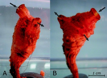
A normal S. clava is pictured in Figure 1A. The abnormal specimen (Figures 1B & 2) was discovered on a Pacific oyster culture tray in Deep Bay, Vancouver Island, British Columbia, Canada (49°27.60′N 124°43.80′W) in November 2007. It had three functional siphons and measured 4.5 cm in total length. The specimen was brought to the laboratory and held in a 20-l tank in a static seawater system at 11°C. In order to determine which siphon(s) was incurrent, we introduced Red Food ColourTM dye (McCormick Canada, London, Canada) to the aquarium and monitored the water currents. The specimen showed no adverse reaction to the dye, although it briefly retracted the siphons in response to the dye being pipetted into the tank. Water currents created were not very pronounced; S. clava probably exhibits the same feeding mechanism as S. montereyensis, i.e. mainly utilizes ambient water flow rather than a stigmata-generated one (Young & Braithwaite, Reference Young and Braithwaite1980). Visible currents indicated that our specimen had one oral and two atrial siphons.
Styela clava: (A) normal specimen with two siphons; (B) abnormal specimen with three siphons. Arrows show the direction of water flow.

Abnormal specimen of Styela clava with siphons closed. Arrows show the direction of water flow.

The specimen was then dissected in order to examine its internal structure. It was first anaesthetized in menthol in seawater for two hours to avoid siphon retraction and then preserved in 10% seawater formalin. After the tissues had hardened for 48 hours, the tunic was opened by a longitudinal incision extending from the base of the stalk to the oral siphon. The body was dissected by cutting from the oral siphon, along the ventral side, to the posterior end.
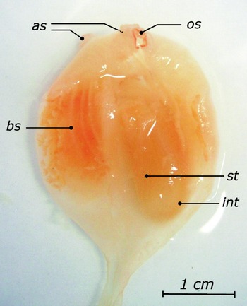
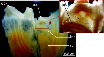
The dissection showed that the specimen had a complete set of properly positioned internal organs (Figure 3). The abnormality was observed only in the structure of the digestive tract. The intestine ran towards both atrial siphons and branched at the very end (Figure 4). Thus, it is probable that the animal was excreting waste products through both of its atrial siphons.
Styela clava dissected along the ventral side: as, atrial siphons; bs, branchial sac; int, intestine; os, oral siphon; st, stomach.

Styela clava dissected and opened along the ventral side, with a close-up of the intestine. Abbreviations as in Figure 3.

The three-siphon abnormality described is especially interesting since S. clava belongs to the order Stolidobranchia. Individuals from the order Phlebobranchia (e.g. Ciona, Ascidia, Phallusia, Ascidiella, Corella, Diazona spp.) go through a stage in their early development when they have three siphons, one oral and two peribranchial. Later, the pair of peribranchial siphons fuse to become a single mid-dorsal atrial siphon (Berrill, Reference Berrill1947; Niermann-Kerkenberg & Hofmann, Reference Niermann-Kerkenberg and Hofmann1989). Sometimes this fusion does not occur, and occasional adult Ciona, Ascidiella, Phallusia and other phlebobranchs have been noted with two atrial siphons (Thompson, Reference Thompson1933; G. Lambert, personal communication, 2008). However, stolidobranchs normally have only a single atrial siphon from metamorphosis, and thus the described specimen of S. clava is unlikely to represent a case of neotenous development. The animal may have developed an additional siphon during metamorphosis. Alternatively, siphon duplication could possibly be a result of a regeneration process due to an injury. George (Reference George1937) describes the formation of a new siphon in Styela plicata following the ‘closure’ (placement of a tight ligature) of one of the siphons; however, in George's experiments the tied-off original siphon did not function and turned into a ‘mass’ attached to the side of a new siphon. The abnormality described in this paper is likely the first case of a stolidobranch tunicate with two functional atrial siphons. This finding may be significant to morphologists, embryologists, and evolutionary biologists.
In December 2007, 1114 S. clava specimens were collected from the same location and examined. None of them had the abnormality described.
ACKNOWLEDGEMENTS
We are grateful to Gretchen Lambert (University of Washington Friday Harbor Laboratories) for providing invaluable advice and training in ascidian dissection techniques. We also thank John Blackburn (Fisheries and Oceans Canada) and David Cassis (University of British Columbia) for collecting the samples and Ted Sweeten (Fisheries and Oceans Canada) for assistance in taking the microphotographs. Support for this work was provided by funds from the Aquatic Invasive Species programme of Fisheries and Oceans Canada. A. Epelbaum was funded through the Visiting Fellowship programme of the Natural Sciences and Engineering Research Council of Canada.