Non-technical Summary
This study revises the fossil history of the sea urchin genus Arbacia in the Caribbean and southeastern United States. By examining > 400 fossil collections, the authors clarify which species are truly distinct, show how different species succeeded one another through time, and describe a new species from Venezuela, the Caribbean, and southern Florida. The results also suggest that western Atlantic Arbacia followed two different evolutionary histories, one of them more closely related to Pacific forms, making this genus a useful model for understanding the evolution of sea urchins in the region.
Introduction
The echinoid genus Arbacia Gray, Reference Gray1835 (Arbacioida, Echinoidea) is a well-studied taxonomic group often employed in analyses of toxicology and ecophysiology (e.g., Vazzana et al., Reference Vazzana, Mauro, Ceraulo, Dioguardi and Papale2020; Burns et al., Reference Burns, Mitchelmore, Williams, Heyes, Gonsior and Davies2025). With six extant shallow-water species inhabiting tropical and temperate seas across the eastern Pacific, western and eastern Atlantic, and the Mediterranean Sea, Arbacia has also been a focal point of phylogenetic and biogeographic studies (Metz et al., Reference Metz, Gómez–Gutiérrez and Vacquier1998; Lessios et al., Reference Lessios, Lockhart, Collin, Sotil and Sanchez–Jerez2012; Wangensteen, Reference Wangensteen2013; Courville et al., Reference Courville, Poulin, Saucède, Mooi, Lessios, Martinez Salinas and Diaz2023c, Reference Courville, Mongiardino Koch, Jossart, Moreau and Mooi2025). A recent revision of the phylogeny of Arbacia supports an initial divergence between the modern Atlantic and Pacific species (Courville et al., Reference Courville, Mongiardino Koch, Jossart, Moreau and Mooi2025). This divergence likely occurred ~ 9 Ma (95% HPD: 3.86–19 Ma) and underscores the pivotal roles of paleogeography and paleoceanography in our understanding of the evolutionary history of the genus, including the emergence of the Isthmus of Panama and the acceleration of the Gulf Stream and North Atlantic Current (Courville et al., Reference Courville, Mongiardino Koch, Jossart, Moreau and Mooi2025).
Although recent work has provided a clearer understanding of the origins of diversity and distribution of extant species, the paleontological diversity of the fossil taxa remains significantly underestimated and explored, particularly from a paleobiogeographic perspective. This is not uncommon among regular echinoids like arbacioids, the poor fossil record of which has long been cited (Kier, Reference Kier1977).
Descriptions of three new fossil species of Arbacia from northern Chile (Courville et al., Reference Courville, González, Mourgues, Poulin and Saucède2023b) and one from Baja California, Mexico (Courville et al., Reference Courville, Mooi, Poulin and Saucède2023a), indicate that the genus was already diverse and widely distributed in the Pacific Ocean by the Late Miocene, well before the final closure of the Isthmus of Panama ~ 3 Ma (O’Dea et al., Reference O’Dea, Lessios, Coates, Eytan and Restreo-Moreno2016).
On the Atlantic side, fossils of Arbacia are particularly abundant and widespread in upper Neogene deposits of the eastern coast of North America (e.g., Conrad, Reference Conrad1843a; H.L. Clark, Reference Clark1923; Cooke, Reference Cooke1941, Reference Cooke1959, Reference Cooke1961; Kier, Reference Kier1963, Reference Kier1972, Reference Kier1983, Reference Kier1997; Ciampaglio et al., Reference Ciampaglio, Osborn and Weaver2009; Ciampaglio and Osborn, Reference Ciampaglio and Osborn2011; Osborn et al., Reference Osborn, Mooi and Ciampaglio2016, Reference Osborn, Portell and Mooi2020; Osborn and Ciampaglio, Reference Osborn and Ciampaglio2019). The genus is here considered to include four fossil species in the eastern USA: the Middle to Late Pliocene Arbacia improcera (Conrad, Reference Conrad1843a) synonymizing Arbacia crenulata Kier, Reference Kier1963 and Arbacia sloani (W.B. Clark in W.B. Clark and Twitchell, Reference Clark and Twitchell1915); the Late Pliocene and Early Pleistocene Arbacia waccamaw Cooke, Reference Cooke1941; the earliest Pleistocene Arbacia rivuli Cooke, Reference Cooke1941; and the Late Pleistocene to extant Arbacia punctulata (Lamarck, Reference Lamarck1816).
A comprehensive review of > 400 Arbacia specimen lots from international institutions has revealed that a re-evaluation of the fossil record of the genus is long overdue. This material includes exceptionally well-preserved specimens with previously undocumented preservation of spination and microscopic anatomy of the test, providing an opportunity to study intraspecific variation of most species. This is crucial in efforts toward evaluating taxonomic validity and in turn, assessing more accurately the species-level biodiversity of fossil Arbacia from the region. Through this work, we provide an updated taxonomy and document known distributions, including extensive range expansions.
We also describe new fossil material of Arbacia from the southeastern USA, West Indies, and Venezuela, increasing the fossil diversity of the genus. One new Pliocene species is particularly noteworthy because it occurs across a wide geographic range from Venezuela to the Lesser Antilles and southern Florida. This and other findings alter former views of the paleobiogeography and fossil diversity of the genus, especially regarding relationships between Pacific and Atlantic forms. The rich fossil record of Arbacia offers a unique and almost unprecedented opportunity for a thorough paleobiogeographic survey of a regular echinoid, a relatively diverse group of urchins with an incomplete fossil record not usually supporting such studies.
Review of paleontological and geological setting
Stratigraphic formations in the southeastern USA
Neogene deposits of the eastern coast of North America are known for diverse fossil echinoid faunas that peaked in diversity during the Late Pliocene (Osborn et al., Reference Osborn, Portell and Mooi2020). After a sea-level regression in the Early Pliocene (Osborn et al., Reference Osborn, Portell and Mooi2020), the Late Pliocene of the eastern coast of North America was marked by a significant marine transgression recorded by extended synchronous deposits. The beginning of the Late Pliocene is associated with a return of warm tropical to subtropical climates (Mid-Pliocene Warming Event), with average temperatures estimated to be 3.5°C higher than today (Raymo et al., Reference Raymo, Grant, Horowitz and Rau1996; Petuch, Reference Petuch2004). These elevated temperatures and the rise in sea level likely contributed to the diversification of echinoids. Piacenzian deposits of the region contain a very diverse echinoid fauna, exceeded only by the Eocene echinoid fauna in the region (Osborn et al., Reference Osborn, Mooi and Ciampaglio2016).
Pliocene deposits in the southeastern USA
During the period of expansion of warmer seas in the Middle to Late Pliocene, Arbacia diversified throughout the eastern USA. The oldest Arbacia in the region is A. improcera, from the Yorktown Formation that crops out in Virginia and North Carolina (Conrad, Reference Conrad1843a). Arbacia improcera was described from the Yorktown Formation north of Smithfield, Isle of Wight County, Virginia. Kier (Reference Kier1972) called this locality Mogarts Beach while discussing this and other sites’ echinoid faunas in the Yorktown Formation of Virginia. Kier (Reference Kier1972) followed Gibson (Reference Gibson1967) and Hazel (Reference Hazel1971) in attributing these echinoid-rich strata to the Late Miocene. Ward and Blackwelder (Reference Ward and Blackwelder1980) reassigned the lower portion of the Formation to the Upper Miocene Eastover Formation. Based on molluscan faunas, they assigned the Yorktown Formation to the Early Pliocene and described four members: Sunken Meadow, Rushmere, Mogarts Beach, and Moore House, in stratigraphic order. M.R. Campbell (Reference Campbell1995) advised caution about these designations because the Rushmere, Mogarts Beach, and Moore House Members are not pervasive, single-lithology units. Mogarts Beach (now covered by erosion control efforts) is likely the type locality of A. improcera and was referred to the Late Pliocene (Dowsett et al., Reference Dowsett, Robinson, Foley and Herbert2021).
Arbacia improcera (see Kier, Reference Kier1983) and Arbacia cf. A. sloani (see Weaver et al., Reference Weaver, Richard and Chandler2006) were reported from the Yorktown Formation in the PCS Phosphate (Lee Creek) Mine near Aurora in Beaufort County, North Carolina. Specimens were attributed to the Lower Pliocene Sunken Meadow Member (Dowsett et al., Reference Dowsett, Robinson, Foley and Herbert2021), but evidence is insufficient to support this without access to in situ exposures in the quarry’s high walls. Yorktown Formation material was usually float amassed as overburden while quarrying continued in the underlying phosphate-rich strata of the Miocene Pungo River Formation. The type specimen was more likely collected from mixed spoil, and not likely to represent the only known Early Pliocene occurrence of this taxon.
Arbacia improcera occurs in other localities in Virginia (Kier, Reference Kier1972) and south of the type locality to Florida. It is reported in the Upper Pliocene lower Goose Creek Limestone of South Carolina (M.R. Campbell and Campbell, Reference Campbell and Campbell1995; Ciampaglio et al., Reference Ciampaglio, Osborn and Weaver2009), the Upper Pliocene Intracoastal Formation (Ciampaglio and Osborn, Reference Ciampaglio and Osborn2011; Osborn et al., Reference Osborn, Portell and Mooi2020), and the Tamiami Formation of southern Florida (Kier, Reference Kier1963; Osborn et al., Reference Osborn, Portell and Mooi2020). Along with Echinocardium orthonotum (Conrad, Reference Conrad1843b), it is the most widespread Late Pliocene echinoid in the eastern USA.
The most diverse Pliocene echinoid fauna of the Atlantic coast is found within the Goose Creek Limestone (GCLS) in eastern South Carolina (Osborn and Ciampaglio, Reference Osborn and Ciampaglio2019). Tuomey (Reference Tuomey1848) originally described outcrops eventually referred to as Goose Creek Limestone in and around Charleston, South Carolina (M.R. Campbell and Campbell, Reference Campbell and Campbell1995; Tuomey, Reference Tuomey1848). Although Sloan (Reference Sloan1908) coined the name Goose Creek phase for a section of strata along Goose Creek, north of Charleston, it was poorly defined until Weems et al. (Reference Weems, Lemon, McCartan, Bybell and and Sanders1982) formalized the Goose Creek Limestone for Charleston’s Pliocene limestones.
M.R. Campbell and Campbell (Reference Campbell and Campbell1995) subdivided the Goose Creek Limestone Formation into informal lower Goose Creek Limestone (LGCLS) and upper Goose Creek Limestone (UGCLS) members, separated by an unconformity. These included strata formerly referred to as the Duplin Marl in the LGCLS and the Bear Bluff Formation in the UGCLS. These are lithologically identical but differ in the composition of the molluscan fauna (M.R. Campbell and Campbell, Reference Campbell and Campbell1995). Although the LGCLS contains echinoids—Encope macrophora (Ravenel, Reference Ravenel1842) and A. improcera—a much more diverse echinoid fauna is found in the UGCLS (M.R. Campbell and Campbell, Reference Campbell and Campbell1995; Ciampaglio et al., Reference Ciampaglio, Osborn and Weaver2009; Ciampaglio and Osborn, Reference Ciampaglio and Osborn2011; Osborn and Ciampaglio, Reference Osborn and Ciampaglio2019), which contains A. waccamaw and 13 additional species. The presence of E. macrophora and A. improcera in the LGCLS indicates that it is at least partially correlated with the Tamiami Formation of southern Florida (Ciampaglio and Osborn, Reference Ciampaglio and Osborn2011). However, the Tamiami Formation also shares many species with the UGCLS, including Lytechinus variegatus (Leske, Reference Leske1778); Mellita caroliniana (Ravenel, Reference Ravenel1842); Echinocardium orthonotum; Plagiobrissus sarae (Ciampaglio, Osborn, and Weaver, Reference Ciampaglio, Osborn and Weaver2009); Brissus glenni Cooke, Reference Cooke1959; and Agassizia porifera (Ravenel, Reference Ravenel1848) (Ciampaglio and Osborn, Reference Ciampaglio and Osborn2011; Osborn et al., Reference Osborn, Portell and Mooi2020). Therefore, the Tamiami Formation could be at least partially correlated with both the upper and lower portions of the Goose Creek Limestone (Ciampaglio and Osborn, Reference Ciampaglio and Osborn2011).
As the dominant Pliocene unit of southern Florida, the Tamiami Formation is underlain by Early Pliocene strata poorly represented in Florida due to the regression event mentioned above. The Early Pliocene is perhaps best represented in the upper portion of the Peace River Formation, but echinoids have not yet been documented there (Osborn et al., Reference Osborn, Portell and Mooi2020).
W.B. Clark and Twitchell (Reference Clark and Twitchell1915) and Mansfield (Reference Mansfield1932) described the first Pliocene echinoids of Florida, but additional collecting (enabled by significant quarrying activity fueled by the surge in population of the state throughout the twentieth century) expanded these records from three to 20 species (Osborn et al., Reference Osborn, Portell and Mooi2020). Strontium isotope dating provided an age of 2.79 to 4.70 Ma (Missimer, Reference Missimer, Zullo, Harris, Scott and Portell1993) and radiometric dating (U-Pb) produced an age of 2.99 ± 0.11 Ma (Klaus et al., Reference Klaus, Meeder, McNeill, Woodhead and Swart2017) for the Tamiami Formation, but these ages are still debated. Biostratigraphy, lithostratigraphy, and radiochronology provided age estimates from the Early to the Late Pliocene (Kier, Reference Kier1963; Missimer, Reference Missimer, Scott and Allmon1992; Waller, Reference Waller2018; Osborn et al., Reference Osborn, Portell and Mooi2020). Osborn et al. (Reference Osborn, Portell and Mooi2020) considered the Tamiami Formation as Late Pliocene, noting that with 15 species, it contains the richest echinoid fauna in the Neogene of Florida (Osborn et al., Reference Osborn, Portell and Mooi2020).
The Late Pliocene is represented in Florida by three additional units: the Intracoastal and Jackson Bluff formations, and the lower portion of the Nashua Formation. The Intracoastal Formation is an echinoid-rich unit (Scott, Reference Scott2001). Restricted to the Apalachicola Embayment of the south-central Florida panhandle (Huddleston, Reference Huddleston1976), it shares at least five echinoid species with the Tamiami Formation, including Arbacia improcera (see Osborn et al., Reference Osborn, Portell and Mooi2020).
The Upper Pliocene Jackson Bluff Formation is restricted to Leon, Liberty, and Wakulla counties of the Florida panhandle (Vernon and Puri, Reference Vernon and Puri1964). Although it has a rich and diverse molluscan assemblage, only three echinoid taxa are known, including A. improcera (see Osborn et al., Reference Osborn, Portell and Mooi2020).
The still poorly understood echinoids of the Nashua Formation were first documented by Oyen and Portell (Reference Oyen and Portell2001) and later summarized by Osborn et al. (Reference Osborn, Portell and Mooi2020). The unit likely contains both Late Pliocene and Early Pleistocene taxa, but the known specimens of Arbacia are unidentifiable to species (Osborn et al., Reference Osborn, Portell and Mooi2020).
Although only two species of Arbacia (A. improcera and A. percaribbeana new species) are known from the Pliocene of the region, the overlying Pleistocene contains three: A. waccamaw, A. rivuli, and A. punctulata. The latter are most common in two areas: southern Florida and the coastal plain of the Carolinas (Osborn and Ciampaglio, Reference Osborn and Ciampaglio2019; Osborn et al., Reference Osborn, Portell and Mooi2020).
Oyen and Portell (Reference Oyen and Portell2001) noted a significant reduction in echinoid taxa in Florida from the Pliocene (20) to the Pleistocene (6), likely due to sampling and preservation biases, shorter duration of the Pleistocene, and other factors. This observed drop in diversity was challenged by stratigraphic revision at the base of the Pleistocene (Ogg et al., Reference Ogg, Ogg and Gradstein2008; Osborn et al., Reference Osborn, Portell and Mooi2020), resulting in 16 species of echinoid in the Pleistocene of Florida and 20 for the Pliocene. The addition of A. percaribbeana n. sp. increases the latter total to 21.
Pleistocene deposits in the southeastern USA
Southern Florida contains the richest Pleistocene echinoid faunas in North America. The Early Pleistocene of Florida is best represented in two units: the Caloosahatchee Formation, previously recognized as Upper Pliocene but reassigned to the lowermost Pleistocene (Ogg et al., Reference Ogg, Ogg and Gradstein2008), and the upper portion of the Nashua Formation, which contains Arbacia sp. The Caloosahatchee Formation is famous for its tropical to subtropical molluscan assemblage (Allmon et al., Reference Allmon, Emslie, Jones and Morgan1996) and the most diverse Pleistocene echinoid fauna of the region. We provide the first record of A. waccamaw from the formation, increasing the known number of Pleistocene echinoids in Florida to 17. This more complete picture of Pleistocene echinoid diversity is closer to that of the modern Floridian coastal fauna, with nine extant species (Osborn et al., Reference Osborn, Portell and Mooi2020).
The Middle Pleistocene of southern Florida is best illustrated by the Bermont Formation, which contains six echinoid species (Osborn et al., Reference Osborn, Portell and Mooi2020), but lacks known occurrences of Arbacia. Similar to the molluscan fauna of the unit, the echinoid fauna of the Bermont is decidedly modern. Only the cassiduloid, Rhyncholampas meansi Osborn, Portell, and Mooi, Reference Osborn, Portell and Mooi2020, is not represented in the Recent fauna of the region.
Well-exposed in eastern and southern Florida, the coastal Anastasia Formation contains A. punctulata (Herrera et al., Reference Herrera, Portell and Means2006; Osborn et al., Reference Osborn, Portell and Mooi2020). Murphy (Reference Murphy1973) used radiocarbon dating to suggest an age between 8.5–9.0 Ka for the type area on Anastasia Island, implying that at least some of the unit extends into the Holocene. Although no extinct molluscan taxa have been reported from the unit (Johnson, Reference Johnson1993), the cassiduloid R. meansi did not survive beyond the Pleistocene. It is likely that deposition of the formation continues today (Portell et al., Reference Portell, Turner and Beerensson2003).
The Early Pleistocene is represented in northeastern South Carolina and southeastern North Carolina by the Waccamaw Formation, which overlies the Upper Pliocene Goose Creek Limestone and outcrops as a narrow band stretching from Wilmington, North Carolina to Myrtle Beach, South Carolina (L. Campbell et al., Reference Campbell, Campbell, Colquhoun, Ernissee and Abbott1975). All occurrences are restricted to within 100 ft (30.5 m) of present mean sea level and no more than 75 mi (120.7 km) inland from the Atlantic (DuBar and Furbunch, Reference DuBar and Furbunch1965). The molluscan fauna of the Waccamaw Formation shares many similarities with the correlative Caloosahatchee Formation of southern Florida (DuBar, Reference DuBar1971; L. Campbell et al., Reference Campbell, Campbell, Colquhoun, Ernissee and Abbott1975; DuBar and DuBar, Reference DuBar and Dubar1980), but the echinoids are distinct. Both units contain 10 species (Osborn and Ciampaglio, Reference Osborn and Ciampaglio2019; Osborn et al., Reference Osborn, Portell and Mooi2020), but only A. waccamaw, L. variegatus, and A. porifera occur in both. Lytechinus variegatus is comparatively rare within the Waccamaw, and A. waccamaw is equally rare in the Caloosahatchee Formation.
There is a significant lithological distinction between the Upper Pliocene sandy gray limestone of the uppermost Goose Creek Limestone, which is capped with an indurated bed, and the overlying shelly marls of the Lower Pleistocene Waccamaw Formation in Horry County, South Carolina, but there are few changes in the echinoid fauna across the transition. Mellita caroliniana, A. waccamaw, and Rhyncholampas sabistonensis (Kellum, Reference Kellum1931) are abundant in both units on either side of the indurated bed separating them in Horry County (Osborn and Ciampaglio, Reference Osborn and Ciampaglio2019). The uppermost Goose Creek Limestone and Lower Waccamaw Formation also share A. porifera, L. variegatus, and B. glenni. Although A. waccamaw occurs in both the Upper Pliocene and Lower Pleistocene strata of the Carolinas, A. rivuli appears to be restricted to a very narrow stratigraphic horizon in the lowermost Waccamaw Formation in its type area in Horry County, South Carolina (Osborn and Ciampaglio, Reference Osborn and Ciampaglio2019).
The Waccamaw Formation is overlain by the Middle to Upper Pleistocene Canepatch Formation (DuBar, Reference DuBar1971), erected for a complex of sand, clay, silt, and peat deposits exposed along the Intracoastal Waterway in Horry County, South Carolina, and southeastern North Carolina. Bybell (Reference Bybell1990) stated that the Canepatch was deposited 450 Kya, and Lyons (Reference Lyons1991) asserted its correlation with the Upper Pleistocene Fort Thompson Formation of southern Florida. The Canepatch Formation contains abundant Mellita isometra Harold and Telford, Reference Harold and M. Telford1990 and Encope michelini Agassiz, Reference Agassiz1841, as well as rare occurrences of A. punctulata, which is usually represented by loose spines and test fragments.
Finally, Arbacia is found in the Upper Pleistocene Wando Formation at Simmons’ Bluff, which Holmes (Reference Holmes1849) described as the most extensive exposure of post-Pliocene strata in the Charleston, South Carolina area. The bluff, now covered, is on Yonges Island, 25 mi (40.2 km) southwest of Charleston on Wadlamaw Sound. Anapesus carolinus Holmes, Reference Holmes1860, described from the unit, was synonymized by Mortensen (Reference Mortensen1935) with A. punctulata, which occurs in the shell beds of Simmons’ Bluff. The Wando Formation includes strata previously attributed to the Pamlico Formation (Cooke, Reference Cooke1937) and Princess Anne Terrace (McCartan et al., Reference McCartan, Weems, Lemon, Sohl and Wright1980); McCartan (Reference McCartan1990) provided a Late Pleistocene age of 70–130 Ka for the unit.
Stratigraphic formations in the West Indies
The occurrence of Arbacia in the West Indies is poorly known. Donovan and Paul (Reference Donovan, Paul, Emson, Smith and Campbell1996, Reference Donovan and Paul1998) mentioned Arbacia sp. indet. from the Pliocene Bowden Formation in Jamaica (Donovan and Paul, Reference Donovan and Paul1998, pl. 2, figs. 2, 8, pl. 3, figs. 1–4, 6), and it was again referred to by Donovan (Reference Donovan2001), Donovan et al. (Reference Donovan, Portell and Veltkamp2005), Donovan and Portell (Reference Donovan and Portell2013), and Donovan et al. (Reference Donovan, Pickerill and Keighley2014). The material illustrated by Donovan and Paul (Reference Donovan and Paul1998) can be referred to as Lytechinus cf. L. variegatus. The specimen (NHMUK PI EE 5215-2) illustrated by Donovan et al. (Reference Donovan, Pickerill and Keighley2014) is also not an Arbacia, as evidenced by the spine structure and tubercle position. Other UF material from Jamaica does not belong to the genus Arbacia (UF 67430 and 108340 are too small to identify).
Donovan (Reference Donovan2003) mentioned an arbaciid from the Lower Pleistocene Manchioneal Formation in Jamaica. Donovan and Embden (Reference Donovan and Embden1996, fig. 2.9) depicted an internal mold (NHMUK PI EE 5102) with poor preservation. Additional material (NHMUK PI EE 5103, 5104) was also mentioned, but allocations remain uncertain (Donovan and Embden, Reference Donovan and Embden1996). Donovan et al. (Reference Donovan, Jones and Harper2016) also noted an ‘Arbacia? sp.’ from the Middle Miocene Cayman Formation of the Cayman Islands. The specimen (UA PI669) is a poorly preserved, small internal mold (< 15 mm) of uncertain affinities. Spines (e.g., UF 134468, 134901) from the Late Pleistocene Jaimanitas Formation of Cuba, can be attributed to Arbacia, because they are indistinguishable from those of A. punctulata, found rarely around Cuba.
Stratigraphic formations in Venezuela
The northwestern region of Venezuela (Falcón State) has fossil-rich upper Neogene deposits. These are extensively studied due to large, mostly terrestrial vertebrates, including crocodiles, xenarthrans, and testudines (Scheyer et al., Reference Scheyer, Aguilera, Delfino, Fortier, Carlini, Sánchez, Carrillo-Briceño, Quiroz and Sánchez-Villagra2013; Carrillo-Briceño et al., Reference Carrillo-Briceño, Sánchez, Scheyer, Carrillo and Delfino2021). The San Gregorio Formation, which is the youngest unit of the Urumaco stratigraphic section (Scheyer et al., Reference Scheyer, Aguilera, Delfino, Fortier, Carlini, Sánchez, Carrillo-Briceño, Quiroz and Sánchez-Villagra2013), is well-exposed in northwestern Venezuela near the town of Urumaco, where it crops out for 570 m (Carrillo-Briceño et al., Reference Carrillo-Briceño, Sánchez, Scheyer, Carrillo and Delfino2021).
The San Gregorio Formation was first subdivided into the Río Seco conglomerate, corresponding to the upper part of the formation, and a lower unnamed portion composed of uniform siltstones. Stainforth (Reference Stainforth1962) later subdivided the latter portion into two units based on fossil beds in its upper layers, the Vergel and Cocuiza members. The former covers the lowermost part of the San Gregorio Formation, spanning 350 m at the type section (Stainforth, Reference Stainforth1962), and is conformably overlain by the Cocuiza Member (Vucetich et al., Reference Vucetich, Carlini, Aguilera and Sánchez-Villagra2010). The Río Seco Member forms the uppermost part of the San Gregorio. Unlike others of the formation, the Cocuiza Member is composed of coastal marine sediments deposited under conditions of low to moderate energy (Mihaljevič et al., Reference Mihaljević, Klug, Aguilera, Lüthi and Sánchez–Villagra2010; Carrillo-Briceño et al., Reference Carrillo-Briceño, Sánchez, Scheyer, Carrillo and Delfino2021).
Age of the San Gregorio Formation was originally believed to be Early Pliocene (Rey, Reference Rey1990; Hambalek et al., Reference Hambalek, Rull, De Digiacomo and Díaz de Gamero1994). Carrillo et al. (Reference Carrillo, Amson, Jaramillo, Sánchez, Quiroz, Cuartas, Rincón and Sánchez-Villagra2018) proposed an age of ~ 1.8 Ma (Early Pleistocene) for the base of the formation, based on foraminiferan and nannoplankton assemblages, and magnetostratigraphy. The foraminiferans are poorly preserved and largely reworked specimens, unlike the nannoplankton assemblage that was dated to the early Pleistocene. 87Sr/86Sr geochronological analyses indicated an age ranging 1.59 ± 0.05 to 1.38 ± 0.06 Ma (Carrillo-Briceño et al., Reference Carrillo-Briceño, Sánchez, Scheyer, Carrillo and Delfino2021).
Specimens of A. percaribbeana n. sp. include UCMP 38200 (Linder et al., Reference Linder, Durham and Orr1988), collected by J.O. Nomland and L.W. Henry in 1931 from the San Gregorio Formation, Falcón State, Venezuela (UCMP locality S-18), ~ 2 km north of the Rio Seco Road of the Creole Petroleum Company. This is close to locality SRS-A of Carrillo-Briceño et al. (Reference Carrillo-Briceño, Sánchez, Scheyer, Carrillo and Delfino2021), at which the pumping station of the Creole Petroleum Company is mentioned, ~ 2 km south of the locality (Fig. 1). This locality provided an additional 18 specimens (UCMP 134665).
Distribution of study material of Arbacia percaribbeana n. sp. in Florida, Venezuela, and the Lesser Antilles: (1) type locality in Florida, USA (yellow star); (2) known distribution in Venezuela and the Lesser Antilles (blue stars).

Figure 1. Long description
A two-panel map set.
Panel 1: A large-scale map of the Americas. A yellow star marks the type locality in Florida, USA. A red bounding box highlights the northern region of South America, specifically Venezuela. The Atlantic Ocean is labeled to the Northeast, the Pacific Ocean to the Southwest, and South America at the center.
Panel 2: A detailed inset of the region within the red box. The map shows Venezuela in grey, bordered by Colombia to the West and Brazil to the South. Specific locations are marked with blue stars and labels:
* In the Northwest, a blue star is located in the Falcon State, which is highlighted in dark red.
* North of Falcon State, a blue star marks Curaçao.
* Moving East along the coast, a blue star marks Cubagua.
* In the Northeast, the island of Trinidad is highlighted in dark red with a blue star.
* North of Trinidad, a blue star marks Tobago.
A scale bar indicating 100 km and a North arrow are located at the bottom left of the panel.
Most specimens of the new species were collected in proximity to Urumaco (Falcón State, Venezuela), but without specific locality information. USNM PAL 805807 was collected near Rio Codore, below Urumaco; USNM PAL 805802 originated from eastern Falcón State, NW Venezuela; and USNM PAL 805803 was collected in Falcón State, Venezuela. Another specimen (USNM PAL 805805) was collected along Rio Seco, northeast of Urumaco, potentially corresponding to the locality SRS-A/B of Carrillo-Briceño et al. (Reference Carrillo-Briceño, Sánchez, Scheyer, Carrillo and Delfino2021), which is recognized as part of the Cocuiza Member containing a marine fauna.
A specimen identified as A. punctulata (see Mihaljevič et al., Reference Mihaljević, Klug, Aguilera, Lüthi and Sánchez–Villagra2010) is here referred to A. percaribbeana n. sp. It was collected in the locality of Cocuiza West (11°18’38.4"N; 70°14’26.2"W) in the Cocuiza Member of the San Gregorio Formation (Mihaljevič et al., Reference Mihaljević, Klug, Aguilera, Lüthi and Sánchez–Villagra2010), corresponding to locality NCH-B (Northward Chiguaje Hill) of Carrillo-Briceño et al. (Reference Carrillo-Briceño, Sánchez, Scheyer, Carrillo and Delfino2021), where the typical Cocuiza Member crops out (Carrillo-Briceño et al., Reference Carrillo-Briceño, Sánchez, Scheyer, Carrillo and Delfino2021).
USNM PAL 805806 comes from Cubagua Island (Venezuela) at which Neogene deposits are well-known. This is the only specimen identified from this locality. The Cubagua Group ranges from Late Miocene to Early Pleistocene (lower Gelasian; Landau et al., Reference Landau, Vermeij and da Silva2008).
USNM PAL 805804 comes from the Middle Pliocene (Piacenzian) Springvale Formation of Trinidad (Gazalie et al., Reference Gazalie, Lakhan and Archie2016), from Savonetta near Gran Couva, Savaneta River area, 1,700 ft (518 m) southwest of Philippine Estate, Bicchiu Castle Estate.
Donovan (Reference Donovan2001) identified Arbacia (NHMUK E 82828-9; 82880) from the coastal exposure at Rockly Bay Tobago, west of Scarborough, in the Balanus Bed (Bed 2) of the Rockly Bay Formation that Lewis and Donovan (Reference Lewis and Donovan1991) assigned to Arbacia improcera. The present work provides the first figures of these specimens, showing closer affinities with Arbacia percaribbeana n. sp. Although the formation is poorly documented, recent work suggests that it is Middle Pliocene (Donovan, Reference Donovan1989; Weber et al., Reference Weber, Arkle, Giorgis and Hippolyte2014; Hippolyte and Mann, Reference Hippolyte and Mann2021).
Fossils identified as Arbacia (UF 230081) from the Pliocene Seroe Domi Formation, Savarese Unit D of Curaçao are attributed to Arbacia percaribbeana n. sp. A previously collected specimen from the Seroe Domi Formation of Curaçao, identified as Arbacia waccamaw and illustrated by de Buisonjé (Reference de Buisonjé1974), is also referred to A. percaribbeana n. sp.
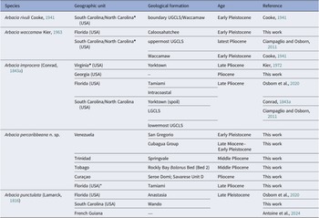
Material identified as Arbacia in earlier work but now assigned to A. percaribbeana n. sp. is listed in Table 1.
Reassignment of previously identified material to Arbacia percaribbeana n. sp.

Table 1. Long description
The table consists of eight rows of data under four headers.
Row 1: Arbacia sp. indet.; U C M P 38200, Linder and Durham 1988; San Gregorio Formation, Falcón State, Venezuela, UCMP locality S-18; Collected from the same fossil zone as additional material with consistent morphology.
Row 2: A. punctulata; Specimen figured by Mihaljevič et al. 2010; Cocuiza West, San Gregorio Formation, Venezuela; Locality and morphology consistent with the new species.
Row 3: Arbacia sp. indet.; U S N M P A L 805802, 805803, 805805, 805807; Falcón State and Urumaco region, Venezuela; Material from the same regional Pliocene-Pleistocene assemblage with matching morphology.
Row 4: Arbacia sp. indet.; U S N M P A L 805806; Cubagua Island, Venezuela; Consistent morphology.
Row 5: A. punctulata; U S N M P A L 805804; Springvale Formation, Trinidad; Morphology and age fit the new species.
Row 6: A. improcera; N H M U K E 82828, 82829, 82880; Rockly Bay Formation, Tobago, Balanus Bed; Re-examination shows closer affinity with the new species.
Row 7: Arbacia sp. indet. or A. waccamaw; U F 230081, de Buisonjé 1974 material; Seroe Domi Formation, Curaçao, Savarese Unit D; Specimens attributable based on morphology.
Row 8: Arbacia improcera; U F 289212, Osborn et al. 2020; Tamiami Formation; Specimens attributable based on morphology.
Materials and methods
Material studied
Recently, fossils were collected from a weakly indurated series of pale to dark gray sandy calcarenite of the Tamiami Formation in the Quality Materials Quarry in Charlotte County, Florida. These revealed a highly diverse, well-preserved community of at least 12 species, including numerous specimens of A. improcera retaining periproctal plates and spines (Osborn et al., Reference Osborn, Portell and Mooi2020). This same stratum has provided specimens referred to A. percaribbeana n. sp., which occurs with Arbacia improcera (previously identified as A. crenulata) at this locality.
The remaining material studied consists of 402 lots of Arbacia representing 419 complete or partial echinoid tests and hundreds of spines. These were collected over the past two centuries along the eastern coast of the USA (Virginia, the Carolinas, Florida, and Georgia), in the West Indies, and Venezuela, and are now reposited in the institutions listed below.
Taxonomy
The taxonomic revision and the description of the new species, Arbacia percaribbeana n. sp., are based on concurrent studies of the fossil species of Arbacia that support assignments using proposed diagnostic features of the genus provided by previous taxonomic studies (Smith et al., Reference Smith, Pisani, Mackenzie-Dodds, Stockley, Webster and Littlewood2006; Kroh and Smith, Reference Kroh and Smith2010; Courville et al., Reference Courville, Mooi, Poulin and Saucède2023a, Reference Courville, González, Mourgues, Poulin and Saucèdeb, Reference Courville, Mongiardino Koch, Jossart, Moreau and Mooi2025; Moreno-Mendoza et al., Reference Moreno-Mendoza, Courville, Angeles-Gonzalez, Duarte and Carulla2024). Systematic nomenclature and terminology for morphological descriptions largely follow Kroh (Reference Kroh2020). As for the extant species of Arbacia, important ontogenetic changes make it difficult to determine extremely small specimens to species level. The species descriptions below are based on the largest specimens, for which morphological differences among species are most strongly developed. The system of numbering ambulacra and interambulacra follows that of Lovén (Reference Lovén1874).
Anatomical abbreviations
Test diameter (TD); test height (TH); peristome diameter (PD).
Repositories and institutional abbreviations
The Academy of Natural Sciences of Drexel University, Philadelphia, Pennsylvania, USA (ANSP); Muséum national d’Histoire naturelle, Paris, France (MNHN); North Carolina Museum of Natural Sciences, Raleigh, USA (NCMNS); The Natural History Museum, London, UK (NHMUK); the Department of Earth and Atmospheric Sciences, University of Alberta, Edmonton, Canada (UA); University of California Museum of Paleontology, Berkeley, USA (UCMP); Invertebrate paleontology collections of the University of Florida, Florida Museum of Natural History, Gainesville, USA (UF); National Museum of Natural History [United States National Museum], Smithsonian Institution, Washington DC, USA (USNM) (paleontological collections, USNM PAL).
Systematic paleontology
Phylum Echinodermata Klein, Reference Klein1778
Class Echinoidea Schumacher, Reference Schumacher1817
Order Arbacioida Gregory, Reference Gregory, Bather, Gregory and Goodrich1900
Family Arbaciidae Gray, Reference Gray1855
Diagnosis
Mortensen (Reference Mortensen1935) provided an excellent description, but not a diagnosis in the sense that we use it elsewhere in the present work, so we have adapted it by excluding many features that are descriptive but not unique the Arbaciidae.
Arbacioids with compound plates in ‘arbacioid’ configuration, i.e., usually trigeminate, but rarely simple or polyporous. Tubercles imperforate, smooth, with ambulacral primary tubercles generally not smaller than those at interambulacral ambitus; the latter can be numerous, arranged in both horizontal and vertical series. Aboral side of test can be naked, lacking tubercles (and therefore, spines). Test usually carrying several epistromal papillae not bearing spines. Primordial interambulacral plate persistent throughout ontogeny, often conspicuously elongated, bearing a primary tubercle. Apical system usually dicyclic, but oculars partly insert in some taxa. Ocular pores usually double. Periproct covered by four or five large, triangular valves. Peristome often very large, variably covered with nonambulacral scales, sometimes naked. Buccal notches usually distinct. Primary spines with more or less developed cortical layer, which can be lacking on aboral spines; spines usually smooth, not thorny. Secondary spines little developed, can be totally lacking; when present, simple or club-shaped, not flattened, or scale-like. Pedicellariae tridentate, triphyllous and ophicephalous; stalk consisting of numerous filaments, not connected except distally. Sphaeridia usually in deep pits in ambulacral midline. Spicules in soft tissues often present as straight rods, widened and with a few central holes.
Genus Arbacia Gray, Reference Gray1835
Type species
Arbacia lixula Linnaeus, Reference Linnaeus1758. Distribution and occurrences provided by Courville et al. (Reference Courville, Poulin, Saucède, Mooi, Lessios, Martinez Salinas and Diaz2023c).
Diagnosis
Arbaciidae with ocular plates bearing small tubercle, usually resorbed during ontogeny; periproct slightly elongated in direction 1–IV and covered with four supra-anal plates; ocular pores double, not visible externally; peristome large (> 30% TD), ambulacra protruding to form ‘ambulacral lips’; buccal notches shallow, wide; tags narrow, more or less elongated; oral primary spines with cortex-layer ‘shoe’ or ‘cap’; ambital and upper spines without any cortex-layer; sphaeridia single, in small pit close to peristomial edge; epistroma conspicuously developed, can be of characteristic design, forming randomly distributed punctation, or arranged in horizontal or vertical series (Courville et al., Reference Courville, Poulin, Saucède, Mooi, Lessios, Martinez Salinas and Diaz2023c).
Arbacia punctulata (Lamarck, Reference Lamarck1816)
Fossil Arbacia punctulata (Lamarck, Reference Lamarck1816), Upper Pleistocene Anastasia Formation, Florida, USA: (1–3) UF 330485: (1) aboral view; (2) interambulacrum in lateral view; (3) ambulacrum in lateral view; (4) UF 324794, aboral view; (5, 6) UF 112190: (5) aboral view; (6) oral view. Scale bars = 10 mm.

Figure 2. Long description
A multi-panel photo containing six views of fossilized sea urchins.
* Panel 1 at the top-left shows an aboral view of specimen U F 330485, featuring a circular central opening and five radiating ambulacral segments separated by wider interambulacral areas covered in rounded tubercles.
* Panel 2 at the top-center shows a lateral view of the same specimen, focusing on the interambulacrum which displays large, prominent tubercles arranged in vertical rows.
* Panel 3 at the top-right shows a lateral view of the ambulacrum, characterized by smaller, more densely packed pore pairs and tubercles.
* Panel 4 at the bottom-left shows an aboral view of specimen U F 324794, highlighting the pentameral symmetry and the hexagonal plates of the test.
* Panel 5 at the bottom-center shows an aboral view of specimen U F 112190, similar in structure to panel 1 but with slightly different preservation of the surface texture.
* Panel 6 at the bottom-right shows the oral view of specimen U F 112190, revealing a large, irregular central opening where the Aristotle’s lantern would have been located, surrounded by the tuberculated test.
White scale bars representing 10 mm are located at the bottom-right of panels 2, 4, and 6.
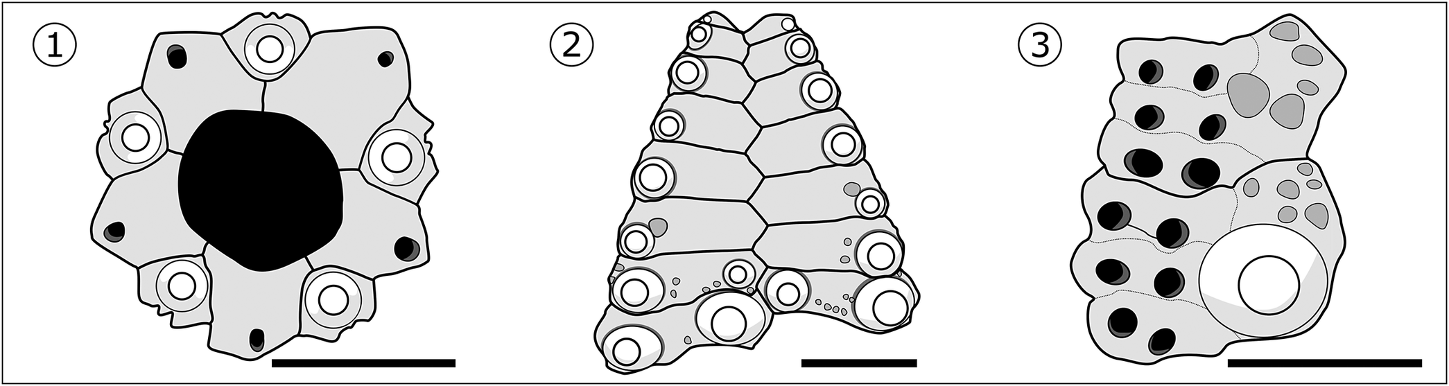
Arbacia punctulata (Lamarck, Reference Lamarck1816), details of test plating: (1) UF 112190, apical system; (2) UF 112190, aboral plates of interambulacrum; (3) USNM PAL 805809, aboral plates of ambulacrum. Scale bars = 10 mm (1, 2); 5 mm (3).

Figure 3. Long description
The figure consists of three panels labeled 1 through 3 from left to right.
* Panel 1 shows the apical system of U F 112190. At the center is a large, dark, pentagonal periproct. Surrounding this core are five genital plates. One plate in the upper right is larger and covered in fine stippling, representing the madreporite. Each plate contains a small circular pore near its outer edge. A 10 mm scale bar is positioned at the bottom right.
* Panel 2 displays the aboral plates of the interambulacrum of U F 112190. The structure is triangular, tapering toward the top. It consists of two vertical columns of interlocking hexagonal plates. Each plate features a prominent circular tubercle at its center, with smaller secondary tubercles scattered around the base. A 10 mm scale bar is at the bottom right.
* Panel 3 illustrates the aboral plates of the ambulacrum of U S N M P A L 805809. The structure shows two large primary tubercles on the right side, each surrounded by a smooth circular area. To the left of these are vertical rows of smaller, dark circular pore pairs. A 5 mm scale bar is located at the bottom right.
Holotype
One known syntype MNHN EcEs 5001. This specimen is broken and mixed in the same sample with another echinoid species, probably Caenocentrotus gibbosus (L. Agassiz in L. Agassiz and Desor, Reference Agassiz and Desor1846).
Occurrence
Fossil occurrences are known from Florida (USA), South Carolina (USA), Virginia (USA), and French Guiana. Upper Pleistocene Anastasia Formation (Herrera et al., Reference Herrera, Portell and Means2006; Osborn et al., Reference Osborn, Portell and Mooi2020), Upper Pleistocene Canepatch Formation (Ciampaglio et al., Reference Ciampaglio, Osborn and Weaver2009), Upper Pleistocene Wando Formation, Late Pleistocene of Virginia, Upper Pleistocene Norfolk Formation, and Late Pleistocene (130–115 Ka; Antoine et al., Reference Antoine, Wieringa, Adnet, Aguilera and Bodin2024) of French Guiana.
Remarks
Based on morphology (Figs. 2, 3), the fossil material studied from Florida, South Carolina, and Virginia seems to be closely related to specimens of A. punctulata from the eastern coast of the USA (Courville et al., Reference Courville, Mongiardino Koch, Jossart, Moreau and Mooi2025).
Arbacia improcera (Conrad, Reference Conrad1843a)
Morphology of Arbacia improcera (Conrad, Reference Conrad1843a): (1–4) USNM PAL 474032: (1) apical view; (2) oral view; (3, 4) oblique views; (5) UF 40402, oblique view; (6, 7) UF 21408: (6) apical view; (7) lateral view; (8, 9) UF 28205: (8) apical view; (9) oblique view; (10) UF 10086, oblique view. Scale bars = 10 mm.

Figure 4. Long description
A collection of ten photographs of sea urchin fossils. Each specimen features a hemispherical or dome-shaped test covered in rows of small, rounded tubercles.
* Panel 1 shows specimen U S N M P A L 474032 from the top apical view, revealing a central circular opening and radiating patterns.
* Panel 2 shows the same specimen from the bottom oral view, featuring a large central aperture.
* Panels 3 and 4 provide oblique side views of the same specimen, highlighting the height of the dome and the vertical rows of tubercles.
* Panel 5 is an oblique view of specimen U F 40402.
* Panel 6 shows specimen U F 21408 from the apical view, with a clearly defined central apical disc.
* Panel 7 is a lateral profile of the same specimen, showing a flatter, low-dome shape.
* Panel 8 shows specimen U F 28205 from the apical view.
* Panel 9 is an oblique view of the same specimen.
* Panel 10 shows specimen U F 10086 in an oblique view, partially embedded in or attached to a piece of light-colored matrix rock.
White scale bars representing 10 mm are positioned at the bottom right of most panels.
Arbacia improcera (Conrad, Reference Conrad1843a): (1) UF 101103, apical view; (2) USNM PAL 474029, apical view; (3) USNM PAL 474017, apical view; (4–7) UF 238568: (4) apical view; (5) oral view; (6, 7) lateral views; (8) UF 205196, apical view; (9) UF 232267, oblique view; (10) UF 110409, oblique view; (11) UF 289215, apical view. Scale bars = 10 mm.

Figure 5. Long description
A multi-panel display of fossilized sea urchin tests.
* Panel 1: Apical view of U F 101103, showing a circular test with a central hole and radiating rows of small tubercles.
* Panel 2: Apical view of U S N M P A L 474029, featuring a pinkish hue on the lower half and a handwritten catalog number.
* Panel 3: Apical view of U S N M P A L 474017, a light cream-colored specimen with distinct vertical catalog numbering.
* Panel 4: Apical view of U F 238568, showing a slightly weathered surface.
* Panel 5: Oral view of U F 238568, displaying the underside with a large central opening and a white label.
* Panels 6 and 7: Lateral views of U F 238568, showing the low, dome-like profile and the bumpy texture of the tubercles along the sides.
* Panel 8: Apical view of U F 205196, a larger, more encrusted specimen with a prominent central apical opening.
* Panel 9: Oblique view of U F 232267, showing the test from an angled perspective to highlight the curvature.
* Panel 10: Oblique view of U F 110409, similar to panel 9 but with a more yellowish tint.
* Panel 11: Apical view of U F 289215, showing a well-preserved symmetrical arrangement of ambulacral and interambulacral areas.
White scale bars representing 10 mm are placed near the bottom right of most panels to indicate size.
Arbacia improcera (Conrad, Reference Conrad1843a), intraspecific diversity and details of test plating: (1) UF 21408, apical system; (2, 3) USNM PAL 474032: (2) aboral plates of interambulacrum; (3) aboral plates of ambulacrum; (4–6) USNM PAL 642133: (4) apical system; (5) aboral plates of interambulacrum; (6) aboral plates of ambulacrum. Scale bars = 10 mm (2, 5); 5 mm (1, 3, 4, 6).

Figure 6. Long description
A multi-panel scientific illustration containing six numbered panels arranged in two rows.
Panel 1 shows the apical system of specimen U F 21408. At the center are four triangular periproctal plates forming a closed diamond shape, surrounded by a ring of five larger genital plates and five smaller ocular plates. One genital plate features a dense cluster of small pores. A 5 mm scale bar is at the bottom right.
Panel 2 displays the aboral plates of the interambulacrum for specimen U S N M P A L 474032. It shows a vertical column of interlocking trapezoidal plates with circular tubercles along the outer edges. A 10 mm scale bar is at the bottom right.
Panel 3 shows a close-up of the aboral plates of the ambulacrum for specimen U S N M P A L 474032, featuring two large primary tubercles with central pits and numerous smaller secondary pores. A 5 mm scale bar is at the bottom right.
Panel 4 illustrates the apical system of specimen U S N M P A L 642133. It features a large, dark, asymmetrical central opening surrounded by five large genital plates, each containing a single distinct pore. A 5 mm scale bar is at the bottom right.
Panel 5 shows the aboral plates of the interambulacrum for specimen U S N M P A L 642133. Similar to panel 2, it depicts a tapered column of plates with small circular tubercles concentrated at the base and along the margins. A 10 mm scale bar is at the bottom right.
Panel 6 provides a detail of the aboral plates of the ambulacrum for specimen U S N M P A L 642133, showing one large, prominent tubercle at the bottom right and several smaller pores and tubercles scattered across the plate surface. A 5 mm scale bar is at the bottom right.
Arbacia improcera (Conrad, Reference Conrad1843a), lowermost upper Goose Creek Limestone, South Carolina, USA (TD ranging 42–48 mm): (1) UF 356362, oblique view; (2, 3) UF 356359: (2) apical view; (3) lateral view; (4) UF 356361, lateral view; (5, 6) UF 356360: (5) apical view; (6) oblique view; (7) UF 356370, apical view. Scale bars = 10 mm.

Figure 7. Long description
A collection of seven photographs of fossilized echinoid tests, each accompanied by a white 10 mm scale bar.
* Panel 1 shows specimen U F 356362 in an oblique view, highlighting the rounded, dome-like shape and the bumpy texture of the primary tubercles.
* Panel 2 shows specimen U F 356359 in an apical view, looking directly down at the top of the test. Five distinct ambulacral segments radiate from the central apical system like a star.
* Panel 3 shows the same specimen, U F 356359, in a lateral view, emphasizing the height and the concentration of large tubercles around the midsection.
* Panel 4 shows specimen U F 356361 in a lateral view, displaying a slightly flatter profile compared to panel 3.
* Panel 5 shows specimen U F 356360 in an apical view. The five-fold symmetry is very clear, with rows of small pores visible along the ambulacral paths.
* Panel 6 shows the same specimen, U F 356360, in an oblique view, showing the transition from the top surface to the side.
* Panel 7 shows specimen U F 356370 in an apical view, featuring a more weathered or calcified surface texture while maintaining the radial symmetry.
All specimens exhibit a characteristic pattern of raised nodes (tubercles) and fine pore pairs arranged in vertical columns.
Reference Conrad1843a Echinus improcerus Conrad, p. 310.
Reference Stefanini1911 Psammechinus improcerus; Stefanini, p. 705.
Reference Clark and Twitchell1915 Coelopleurus improcerus; W.B. Clark and Twitchell, p. 180, pl. 84, figs. 4a–c.
Reference Clark and Twitchell1915 Coelopleurus sloani W.B. Clark in W.B. Clark and Twitchell, p. 181, pl. 84, figs. 5a, b.
Reference Cooke1941 Arbacia sloani; Cooke, p. 11.
Reference Cooke1941 Arbacia improcera; Cooke, p. 11, pl. 1, figs. 7–9.
Reference Cooke1959 Arbacia improcera; Cooke, p. 20, pl. 4, figs. 1–3.
Reference Cooke1959 Arbacia sloani; Cooke, p. 21, pl. 4, figs. 12–14.
Reference Kier1963 Arbacia improcera; Kier, p. 14, pl. 1, figs. 1–5.
Reference Kier1963 Arbacia crenulata Kier, p. 11, pl. 1, figs. 1–15, text figs. 3–7.
Reference Kier1972 Arbacia improcera; Kier, p. 8, pl. 1.
Reference Kier1972 Arbacia sloani; Kier, p. 8, pl. 2, fig. 1.
Reference Kier1983 Arbacia improcera; Kier, p. 501, pl. 1, fig. 6.
Reference Ciampaglio, Osborn and Weaver2009 Arbacia improcera; Ciampaglio et al., fig. 2.
Reference Osborn, Portell and Mooi2020 Arbacia improcera; Osborn et al., figs. 14, 15.
Type specimens
The material, probably originally housed at ANSP according to Cooke (Reference Cooke1941), has been lost (A. Martinez-Melo, collection manager of Invertebrate Paleontology, ANSP, personal communication, January 2023). Specimen USNM PAL 166487, figured by W.B. Clark and Twitchell (Reference Clark and Twitchell1915), Cooke (Reference Cooke1959), and Kier (Reference Kier1963), is herein designated as the neotype (see below).
Materials
More than 110 complete tests, ranging 8–48 mm TD. A list of studied material is given in Supplementary Data 1.
Emended diagnosis
Arbacia with tubercle on each ocular and genital plate usually, but not always completely resorbed during ontogeny; single, often discontinuous perradial column of primary tubercles aborally in interambulacrum; interambulacral granules often present interradially; numerous interambulacral plates (N = 13 at TD = 20 mm); ambulacral tubercles poorly developed aborally.
Occurrence
Known from Florida, Georgia, South and North Carolina, and Virginia, USA. The specimen described by Conrad (Reference Conrad1843a) came from James River, near Smithfield, Virginia. Kier (Reference Kier1963) listed additional localities within the Tamiami Formation (Osborn et al., Reference Osborn, Portell and Mooi2020). The designated neotype comes from the middle to upper Pliocene Yorktown Formation at J.T. Williams’ marl pit, Smith Creek, 0.5 mi (0.8 km) below Suffolk, Virginia. Type material derives from the Middle to Upper Pliocene Yorktown Formation of Virginia. Additional specimens can be found in the lower Goose Creek Limestone and the lowermost upper Goose Creek Limestone of North Carolina, the Upper Pliocene Tamiami Formation of Florida, and the Pliocene of Georgia.
Description
Test shape
Viewed from above, test circular to subpentagonal in outline (Figs. 4.1, 4.6, 4.8, 5.1, 5.8). Test of medium size, rarely exceeding 30–40 mm TD (without spines), exceptionally reaching 48 mm TD (see Remarks). TH relative to TD variable, usually high, close to 0.5 (mean ratio TH/TD = 49%, ranging 36–63%; N = 56). Test flattened orally.
Apical system
Ratio of apical system diameter to TD decreasing from 47% TD (TD = 9 mm) to 30% TD (TD = 30 mm), usually close to one-third of TD in large specimens (Figs. 4.1, 4.6, 4.8, 5.1–5.4, 5.8, 6.1, 6.4). Apical system always dicyclic, all ocular plates exsert with ocular plates I, IV, and V closer to periproct margin. Periproct elongated in direction 1-IV (Figs. 4.1, 5.1–5.3, 6.1, 6.4). Madreporic plate (genital 2) slightly larger than other genital plates (Figs. 4.1, 5.1–5.3, 6.1, 6.4). Periproct covered with four anal plates (Figs. 4.6, 6.1). Gonopores small and usually positioned in middle of genital plates. Gonopores open in very small specimens (TD = 8.5 mm). Ocular and genital plates with one tubercle often not resorbed during ontogeny (Figs. 4.1, 4.6, 4.8, 6.1). Epistroma well developed, forming strong punctation, often organized in latitudinal ridges on genital plates (Figs. 4.6, 5.1).
Peristome
Relatively large, > 50% TD (mean ratio PD/TD = 50%, ranging 45–66% TD; N = 52). Buccal notches shallow. Tags short and triangular (length = width, spanning < 2 tubercles).
Ambulacra
Narrow, < 50% of width of interambulacra. At ambitus, each ambulacral plate with one large, imperforate, noncrenulated tubercle positioned perradially. Areoles well developed over most of ambulacral plate. Ambulacral tubercles same size as interambulacral tubercles at ambitus, decreasing in size adapically (Figs. 4.3, 4.9, 4.10, 5.7, 5.9). Aborally, tubercles forming two columns, sometimes discontinuous, strongly or weakly developed. Presence of small, poorly developed granules between tubercles perradially. Large granules present on perradial side of inner ambulacral pore, sometimes forming longitudinal ridge in ambulacra (Fig. 6.6). Ambulacral plates trigeminate throughout, of arbacioid type, forming more complex compound plating adorally, slightly widening to form short, narrow phyllodes. Pores rounded and conjugated aborally; neural canal not visible (Figs. 6.3, 6.6).
Interambulacra
Plates wider than high at ambitus (H/L ratio of plates ranging 0.2–0.3), with two, sometimes three, large, imperforate, noncrenulate tubercles, all similar in size (Figs. 4.4, 4.7, 5.6, 5.9, 5.10, 6.5). Number of interambulacral plates ranging 10 (at TD = 10 mm) to 14 (at TD = 35 mm). Plate height increasing adapically, ranging 1 mm at ambitus to 2.5 mm near apex (for TD > 30 mm). Tubercles usually reduced in size above ambitus, forming often-dicontinuous adradial column of tubercles aborally (Figs. 5.2–5.4, 5.8–5.11, 7.5). At no more than one or two plates above ambitus, another inner tubercle, rarely two, of same size, usually abutting interradial suture (Figs. 5.6, 5.9, 5.10, 6.2, 6.5). The outer column sometimes bordered by inner column of granules progressively weakened and elongated adapically (Figs. 4.1, 4.3–4.10, 6.3). All tubercles surrounded by several tiny granules. Areoles of tubercles large at ambitus, small above ambitus. Interradial naked area large, extended from apex to ambitus (Figs. 4.1, 4.6, 4.8, 5.1–5.4, 5.6–5.11, 6.2, 6.5). Epistroma well developed, consisting of strong punctation, often forming longitudinal ridges from apex to ambitus.
Remarks
Redefinition of type material
The original description by Conrad (Reference Conrad1843a, p. 310) showed membership in Arbacia, but species diagnostic features were not precise: “Depressed or discoidal, anus suboval; 5 v-form granulated lines diverge from the anus, about 6 tubercles on each single line; between these 2 shorter tuberculated lines; interstices wide and without lines or tubercles; whole surface minutely granulated; periphery profoundly tuberculated, the tubercles subequal; on the base the tubercles are smaller and numerous.”
The type material has never been figured. According to Cooke (Reference Cooke1941), it was probably originally housed at ANSP, but inquiries indicate that it has been lost. Despite extensive search efforts in the expected repositories, no original type specimen could be located. Accordingly, and to clarify the taxonomic status of the nominal species, a neotype is designated here in accordance with Article 75 of the ICZN (1999). The specimen (USNM PAL 166487) is housed in a recognized scientific institution with permanent collections and is accessible for study.
Only specimen USNM PAL 166487 originates from a locality near the type locality (near Smithfield, Virginia, in the Yorktown Formation). It was collected at J.T. Williams’ marl pit on Smith Creek, ~ 0.5 mi [0.8 km] downstream from Suffolk, Virginia, ~ 20 mi (32 km) south of the original type locality along the James River above Smithfield. In contrast, most other specimens from the Yorktown Formation were collected as spoil from the PCS Phosphate (Lee Creek) Mine near Aurora in Beaufort County, North Carolina. Given its geographic proximity to the type locality and its morphology, USNM PAL 166487 is the most appropriate candidate for designation as the neotype.
Specimens from the lowermost UGCLS
A large sample of A. improcera was collected from the lowermost part of the UGCLS in South Carolina. Specimens of this sample (Fig. 7) depart from the typical form of A. improcera by their size, being much larger (between 42 and 48 mm TD) and in having the first column of interambulacral tubercles always alternating in size (Fig. 7).
Comparison with other species of Arbacia
This species differs from all other Arbacia except A. rivuli and A. waccamaw by the presence of an interambulacral inner tubercle aborally that is the same size as the primary tubercles and abutting the interradial suture, and a tubercle on each ocular and genital plate that is not always completely resorbed during ontogeny. It differs from A. rivuli and A. waccamaw in having a higher number of interambulacral plates (10 at TD = 20 mm in A. waccamaw, and no more than 9 at TD = 20 mm in A. rivuli, whereas usually 12 at TD = 20 mm in A. improcera).
Arbacia rivuli Cooke, Reference Cooke1941
Arbacia rivuli Cooke, Reference Cooke1941, holotype, USNM PAL 498887, Lower Pleistocene Waccamaw Formation, South Carolina, USA: (1) apical view; (2) oblique view. Scale bar = 10 mm.

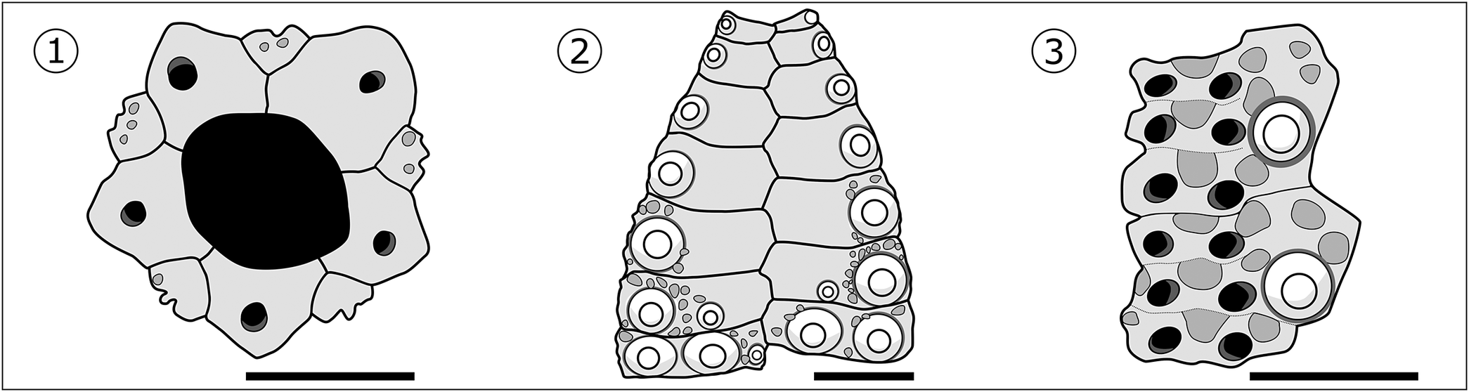
Arbacia rivuli Cooke, Reference Cooke1941, details of test plating: (1) UF 203039, apical system; (2) UF 203037, aboral plates of interambulacrum; (3) USNM PAL 805813, aboral plates of ambulacrum. Scale bars = 10 mm (1, 2); 5 mm (3).

Figure 9. Long description
A multi-panel line drawing containing three numbered sections.
* Panel 1 shows the apical system of U F 203039. At the center is a large, dark, irregular pentagonal opening. Surrounding this core are five primary plates. Each plate contains a circular pore. The top-right plate is distinguished by a dense stippled texture. A 10 mm scale bar is located at the bottom right.
* Panel 2 displays the aboral plates of the interambulacrum of U F 203037. The plates are arranged in two vertical columns that taper toward the top. Each plate features a prominent circular tubercle with a central ring. A 10 mm scale bar is at the bottom right.
* Panel 3 illustrates the aboral plates of the ambulacrum of U S N M P A L 805813. It shows two columns of plates. The left column contains pairs of dark, oval-shaped pores connected by dashed lines. The right column features a single large circular tubercle at the bottom and smaller pores above it. A 5 mm scale bar is at the bottom right.
Arbacia rivuli Cooke, Reference Cooke1941, UF 350877, basal bed of the Lower Pleistocene Waccamaw Formation, Intracoastal Waterway, northeast of Grand Strand Airport, North Myrtle Beach, Horry County, South Carolina, USA: (1) apical view; (2) oral view; (3) lateral view; (4, 5) oblique views. Scale bars = 10 mm.

Figure 10. Long description
A multi-panel photo set of a fossilized sea urchin test.
* Panel 1 at the top-left shows the apical view. At the center is a circular opening surrounded by five smaller pores. Radiating outward are five double-rows of small pores alternating with sections of larger rounded bumps. A 10 mm scale bar is located at the bottom right of this panel.
* Panel 2 at the top-center shows the oral view. It features a large, jagged circular opening at the center revealing a dark interior. The surrounding surface is covered in dense, rounded tubercles.
* Panel 3 at the top-right shows the lateral view. The fossil has a low, dome-like profile with a flat base. The surface texture of tubercles and pore rows is visible across the curve. A 10 mm scale bar is at the bottom right.
* Panel 4 at the bottom-left shows an oblique view. The perspective is angled to show both the top and side, highlighting the three-dimensional arrangement of the pore rows and the height of the tubercles.
* Panel 5 at the bottom-right shows a second oblique view from a slightly different angle, emphasizing the symmetry of the five-fold radial segments. A 10 mm scale bar is positioned at the bottom center between panels 4 and 5.
Reference Cooke1941 Arbacia rivuli Cooke, p. 10, pl. 1.
Reference Cooke1959 Arbacia rivuli; Cooke, p. 20, pl. 4, figs. 10, 11.
Holotype
USNM PAL 498887 (Fig. 8). Specimen is complete, preserved without spines, but with all diagnostic characters recognizable. Only one ambulacrum and one interambulacrum are completely prepared. The apical system is complete but not entirely visible. The specimen dimensions are TD = 32.5 mm; TH = 18.5 mm; PD = 16.5 mm.
Materials
More than 72 tests ranging 10–50 mm TD. A list of material studied is available in Supplementary Data 1.
Emended diagnosis
Arbacia in which ocular plates have one large tubercle (TD = 40 mm); number of interambulacral plates very low (N = 11 to 12 plates at TD = 40 mm); second column of interambulacral inner tubercle abutting interradius suture aborally; ambulacral tubercles irregularly developed aborally.
Occurrence
Arbacia rivuli occurs only at the type locality (Horry County, South Carolina) at the boundary between the uppermost Pliocene (upper Goose Creek Limestone) and the lowermost Pleistocene Waccamaw Formation. The type specimen comes from the Intracoastal Waterway canal, 4.5 mi (7.2 km) southwest of Little River, Horry County, South Carolina (USNM station 13377). Waccamaw Formation, Early Pleistocene.
Description
Test shape
When viewed from above, test circular to subpentagonal in outline at ambitus. Test often reaching 50 mm TD, without spines. TH to TD ratio variable, usually > 0.5 (mean ratio TH/TD = 52%; N = 39), flattened orally. No spines preserved.
Apical system
Approximately one-third TD, apical system diameter to TD with mean ratio of 29% (N = 40). Apical system usually dicyclic (i.e., with all ocular plates exsert), plates V occasionally insert or very close to periproct margin (> 30% specimens). Periproct elongated in direction 1-IV (Figs. 8.1, 9.1, 10.1). Gonopores small, circular, positioned close to outer margin of genital plates. Gonopores open in small specimens (TD = 15 mm). Each ocular plate bearing one large tubercle (Figs. 8.1, 9.1, 10.1), even in large specimens (TD = 40 mm). Genital plates free of tubercles. Ocular plates with few granules that likely bore pedicellariae, similar to those found in extant species. Epistroma well developed, forming punctation on genital plates (Figs. 8.1, 10.1).
Peristome
Large, close to 50% of TD (mean ratio PD/TD = 47.7%; N = 25). Buccal notches shallow (Fig. 10.2). Smooth tags very long (length < width, spanning > 3 tubercles). Perignathic girdle and auricles not visible.
Ambulacra
Narrow, less than half width of interambulacra. At ambitus, each ambulacral plate with one large, imperforate, noncrenulate tubercle positioned adradially (Fig. 9.3). Areole occupying most of ambulacral plate at ambitus. Ambulacral tubercles same size as interambulacral tubercles at ambitus, quickly decreasing in size adapically. Tubercles large and poorly developed aborally, rarely reaching upper second half of aboral surface. Small, poorly developed granules between tubercles perradially (Figs. 8.2, 9.3, 10.3). Ambulacral plates always trigeminate, of arbacioid type aborally, forming more complex compound plates adorally, slightly widening to form short, narrow phyllodes (Fig. 10.2). Pores round, conjugate aborally, with neural canal in perradial pores not developed. On oral side, one perradial spheridial pit near peristomial edge.
Interambulacra
Plates almost as wide as their height at ambitus (mean H/L ratio 0.8), with to two, occasionally three, large, imperforate, noncrenulate primary tubercles, similar in diameter toward interradius (Figs. 8.2, 9.2, 10.4, 10.5). Number of interambulacral plates 11 or 12 at TD = 30 mm, never exceeding 12 or 13 (TD = 46 mm), with five or six plates above ambitus (TD > 40 mm). Plates highest above ambitus (Figs. 10.4, 10.5), with plate height increasing from 2.5 to 3.5 mm aborally (TD > 30 mm). Tubercles of outer column close to adradial suture, of same size, and continuous from ambitus to apex. Inner tubercles of same size as outer ones, abutting interradial suture (Figs. 8.2, 9.2, 10.4, 10.5). Second inner tubercles well developed aborally, usually three to four plates above ambitus (Figs. 8.2, 9.2, 10.4, 10.5). Large tubercles surrounded by numerous, tiny granules, areoles of tubercles well developed at ambitus and aborally. Naked area restricted to most adapical part of the test, forming narrow and poorly extended area without tubercles (Figs. 8.1, 9.2, 10.1, 10.5). Epistroma well developed, usually consisting of small punctuation. Lack of strong separation between outer row of interambulacral tubercles and naked area.
Remarks
Intraspecific variation
Characters are constant within this species. Large ocular tubercles are always well developed, each one covering almost all of the ocular plate surface. Phyllodes are always narrow and short, almost undeveloped, with several pores comprised between 8 and 15 in number. Aborally, the interradial column of interambulacral tubercles is variably developed among specimens. In the studied specimens, variation ranges from one to five aboral plates free of interradial tubercles, only one column of tubercles being present adradially. Patterns of the epistroma are variable: in most specimens, the epistroma shows a radial pattern but some specimens are also covered with fine and randomly distributed punctations.
Comparison with other species of Arbacia
This species differs from all other Arbacia spp., except for A. waccamaw, by the presence of large tubercles on the ocular plates in large adult specimens. It differs from A. waccamaw and A. improcera in having a much lower number of interambulacral plates and a second column of interambulacral tubercles in the interradial area.
Arbacia waccamaw Cooke, Reference Cooke1941
Arbacia waccamaw Cooke, Reference Cooke1941, holotype, USNM PAL 498888, Lower Pleistocene Waccamaw Formation, South Carolina, USA: (1) apical view; (2) oblique view. Scale bar = 10 mm.

Arbacia waccamaw Cooke, Reference Cooke1941, UF 105636, details of test plating: (1) apical system; (2) aboral plates of interambulacrum; (3) aboral plates of ambulacrum. Scale bars = 10 mm (2); 5 mm (1, 3).

Figure 12. Long description
The illustration consists of three panels labeled 1 through 3 from left to right.
Panel 1 shows the apical system. At the center is a large, black, circular periproct. Surrounding this core are five large genital plates arranged in a ring. Each plate features a circular gonopore near its outer edge. Small, dark ocular plates are wedged between the genital plates at the perimeter. A scale bar at the bottom right represents 5 mm.
Panel 2 displays the aboral plates of the interambulacrum. The plates are arranged in two vertical columns that meet at a central zigzag suture line, forming a triangular shape that widens toward the base. Each plate contains a large, circular primary tubercle near the outer margin. A scale bar at the bottom right represents 10 mm.
Panel 3 illustrates the aboral plates of the ambulacrum. It shows two columns of plates. The left side features pairs of small, dark pore pairs arranged vertically. The right side shows larger plates with a single, very large primary tubercle at the bottom and smaller secondary tubercles and granules above it. A scale bar at the bottom right represents 5 mm.
Arbacia waccamaw Cooke, Reference Cooke1941, Lower Pleistocene Caloosahatchee Formation, Florida, USA: (1–4) UF 105636: (1) apical view; (2) oral view; (3, 4) lateral views; (5–8) UF 14131: (5) apical view; (6) oral view; (7, 8) lateral views. Scale bars = 10 mm.

Figure 13. Long description
The photo displays two fossilized echinoid tests against a black background.
Top row (Specimen U F 105636):
- Panel 1 (Top-Left): Apical view showing a circular, dome-shaped test with five radiating ambulacral areas. The surface is covered in small, rounded tubercles. A central hole marks the apical system.
- Panel 2 (Top-Center): Oral view showing the underside. A large circular opening at the center contains a white paper label with the text U F 105636. The perimeter is scalloped.
- Panels 3 and 4 (Top-Right): Two lateral views showing the low, rounded profile of the test and the vertical distribution of the tubercles. A white scale bar representing 10 mm is positioned below panel 1.
Bottom row (Specimen U F 14131):
- Panel 5 (Bottom-Left): Apical view of a lighter-colored specimen, showing similar pentameral symmetry and tuberculation.
- Panel 6 (Bottom-Center): Oral view showing a central depression with the handwritten identifier U F 14131.
- Panels 7 and 8 (Bottom-Right): Two lateral views illustrating the hemispherical shape and the rows of primary and secondary tubercles. A second white scale bar representing 10 mm is positioned below panel 5.
Reference Cooke1941 Arbacia waccamaw Cooke, p. 10, pl. 1, figs. 1–3.
Holotype
USNM PAL 498888 (Fig. 11). Specimen almost complete, preserved without spines. TD = 32 mm; TH = 17 mm.
Materials
More than 18 complete tests ranging 12–41 mm TD. A list of material studied is available in Supplementary Data 1.
Emended diagnosis
Arbacia in which ocular plates have one large tubercle, even in large specimens (TD = 40 mm); ambulacral and interambulacral tubercles largest at ambitus, much smaller aborally; interradial interambulacral tubercles present at ambitus only, on no more than two plates aborally; ambulacral tubercles irregularly developed aborally.
Occurrence
Known from North and South Carolina, and Florida. Type material is from the Intracoastal Waterway canal, 4.5 mi (7.2 km) southwest of Little River, Horry County, South Carolina, USA (USNM station 13377). Type material from the Lower Pleistocene Waccamaw Formation of South Carolina (Horry County, type locality). This species is also known from the Waccamaw Formation of North Carolina (Brunswick and Columbus counties) and the uppermost UGCLS of South Carolina. This species is here recognized for the first time in the Lower Pleistocene Caloosahatchee Formation of Florida (Hendry County; Fig. 13).
Description
Test shape
Test circular when viewed from above, rarely subpentagonal in outline (Figs. 11.1, 13.1, 13.5). Test typically large, often reaching 40 mm TD, TH relative to TD variable, usually < 0.5, flattened orally (N = 18).
Apical system
Approximately one-third TD, mean ratio of apical system diameter to TD 30% (N = 16). Apical system with ocular plate V often inserts. Oculars I and IV close to periproct (Figs. 12.1, 13.1, 13.5). Periproct elongated in direction 1-IV. Gonopores small, circular, and positioned in outer margin of genital plates. Gonopores open in small specimens (TD = 15 mm). Each ocular plate bearing one large tubercle (Figs. 11.1, 12.1, 13.1, 13.5), even in large specimens (TD = 40 mm). Genital plates free of tubercles. Epistroma well developed, forming punctation on genital plates.
Peristome
Large, occupying > 50% of TD. Buccal notches shallow (Fig. 13.2, 13.6) Tags very long (length > width, spanning > 3 tubercles). Perignathic girdle and auricles not visible.
Ambulacra
Narrow, less than half width of interambulacra. At ambitus, each ambulacral plate with one large, imperforate, noncrenulate tubercle positioned adradially (Figs. 11.2, 12.3, 13.3, 13.7). Areole occupying most of ambulacral plate. At ambitus and adorally, tubercles so large that pore pairs perforate bosses (Fig. 13.3). Ambulacral tubercles same size as interambulacral tubercles at ambitus, quickly decreasing in size above ambitus (Figs. 11.2, 13.3, 13.7). Aborally, tubercles large but poorly developed, rarely reaching second half of aboral surface. Presence of small but poorly developed granules between tubercles perradially. Ambulacral plates always trigeminate, of arbacioid type, forming more complex compound plates adorally, slightly widening to form short, very narrow phyllodes. Pores rounded, close, conjugating aborally, with neural canals not developed in perradial pores.
Interambulacra
Wide, with two, occasionally three, large imperforate, noncrenulate primary tubercles per plate at ambitus (Figs. 11.2, 12.2, 13.4, 13.8). Tubercles similar in size toward interradius. Number of interambulacral plates never exceeding 13 or 14, even in large specimens (TD > 40 mm), with six or seven plates above ambitus (Figs. 13.1, 13.5). Plates wider than their height at ambitus (plate H:L ratio 0.25). Plates higher above ambitus, plate height increasing from 1.5 to 2.5 mm aborally (TD > 40 mm). Aborally, outer column of tubercles continuous, of same size, extending from ambitus to apex (Figs. 11.2, 12.2, 13.1, 13.5). In no more than one or two plates above ambitus, another inner tubercle, rarely two, of same size usually abutting interradial suture (Figs. 11.2, 12.2, 13.4, 13.8). Large tubercles surrounded by several tiny granules. Naked area extending down to ambitus, forming large area without tubercles (Figs. 11.2, 12.2, 13.1, 13.5). Epistroma well developed, consisting of strong punctation, often forming longitudinal ridges.
Remarks
Comparison with other species of Arbacia
This species differs from all other Arbacia spp., except for A. rivuli and A. improcera, by the presence of large tubercles on the ocular plates in large, adult specimens. It differs from A. rivuli in having a higher number of interambulacral plates and in lacking a second column of interambulacral tubercles close to the interradial suture aborally. It differs from A. improcera in having a lower number of interambulacral plates, longer tags, and in lacking tubercles on the genital plates.
Arbacia percaribbeana new species
Arbacia percaribbeana n. sp., holotype, UF 353522, Upper Pliocene Tamiami Formation, Quality Materials Quarry, Placida, Charlotte County, Florida, USA: (1) apical view; (2) oral view; (3) lateral view; (4) detail of interambulacrum; (5) detail of ambulacrum; (6) apical system. Scale bar = 10 mm (1–3); 5 mm (4–6).

Figure 14. Long description
A multi-panel photo containing six numbered views of a fossilized sea urchin.
Panel 1 shows the apical view, a circular top-down perspective. At the center is a small circular opening surrounded by five radiating ambulacral segments and five wider interambulacral segments with a granular texture.
Panel 2 shows the oral view, the underside of the specimen. It features a large, recessed central opening with a jagged perimeter, surrounded by a heavily tuberculated surface.
Panel 3 shows the lateral view, a side profile of the dome-shaped test. The base is flat with rounded protrusions, and the surface curves upward to a peak, showing vertical suture lines.
Panel 4 is a close-up detail of the interambulacrum, highlighting the hexagonal plates and the zig-zagging suture line running vertically through the center.
Panel 5 is a close-up detail of the ambulacrum, showing rows of small pores and larger rounded tubercles arranged in vertical columns.
Panel 6 provides a high-magnification view of the apical system, focusing on the central opening and the surrounding genital plates and pore structures.
White scale bars are present below panels 2 and 4, and to the right of panel 6, representing 10 millimeters for panels 1 through 3 and 5 millimeters for panels 4 through 6.
Arbacia percaribbeana n. sp., details of test plating: (1) USNM PAL VGO-13, apical system; (2, 3) UCMP 38200: (2) aboral plates of interambulacrum; (3) aboral plates of ambulacrum. Scale bars = 10 mm (3); 5 mm (1, 2).

Figure 15. Long description
A multi-panel figure containing three numbered line drawings.
Panel 1 shows the apical system of U S N M P A L V G O-13. At the center is a large, dark, irregular circular opening. Surrounding this center are five large, interlocking plates. Each plate contains a single small, dark circular pore. A scale bar at the bottom right represents 5 mm.
Panel 2 shows the aboral plates of the interambulacrum of U C M P 38200. It depicts a tapered, vertical column of interlocking hexagonal plates. The plates are arranged in two columns meeting at a central vertical zigzag suture. Large circular primary tubercle scars are located on the outer edges of the lower plates, while smaller granules are scattered near the sutures. A scale bar at the bottom right represents 5 mm.
Panel 3 shows the aboral plates of the ambulacrum of U C M P 38200. It features a vertical arrangement of irregular plates. On the left side, there is a vertical series of paired, dark oval pore pairs. On the right side, larger circular tubercle scars are visible. The plates have a mottled appearance with varying shades of gray. A scale bar at the bottom right represents 10 mm.
Arbacia percaribbeana n. sp., paratype, UF 356363, Upper Pliocene Tamiami Formation, Quality Materials Quarry, Placida, Charlotte County, Florida, USA: (1) apical view; (2–5) oblique views. Scale bars = 10 mm.

Figure 16. Long description
A multi-panel photo series of a fossilized sea urchin paratype.
Panel 1 at the top-left shows a top-down apical view. The circular test is light tan and covered in small tubercles. A dark, irregular opening is at the center. Numerous cylindrical spines radiate outward from the perimeter of the test, partially embedded in a sandy matrix. A white 10 mm scale bar is in the bottom-right corner.
Panel 2 at the top-right provides an oblique view from above. The test appears dome-shaped, rising from the matrix. Spines are seen projecting downward and outward from the sides of the dome. A white 10 mm scale bar is in the bottom-right corner.
Panels 3, 4, and 5 at the bottom show close-up oblique views of the test surface and spine attachments.
* Panel 3 focuses on the granular texture of the plates and the base of several thick, vertical spines.
* Panel 4 shows the apical opening at the top of the dome with plates radiating downward toward the spines.
* Panel 5 highlights the density of the spines as they emerge from the lower portion of the test.
Each of these three bottom panels includes a vertical white 10 mm scale bar on the right side.
Morphology of Arbacia percaribbeana n. sp.: (1) USNM PAL 805806, apical view; (2) USNM PAL 805807, oblique view; (3) UF 168836, paratype, lateral view; (4–7) UF 230367, paratype: (4) apical view; (5) oral view; (6, 7) lateral views; (8) UF 232264, paratype, oblique view; (9) NHMUK E 82828-9, oblique view; (10) USNM PAL 805815, oblique view. Scale bars = 10 mm.

Figure 17. Long description
A collection of ten high-resolution photographs of fossilized sea urchins against a black background. Each panel contains a white scale bar representing 10 mm.
* Panel 1: Top-left. Apical view of specimen U S N M P A L 805806, showing a circular, flattened test with five radiating ambulacral segments and a central opening.
* Panel 2: Top-center. Oblique view of specimen U S N M P A L 805807, highlighting the domed shape and rows of small tubercles.
* Panel 3: Top-right. Lateral view of specimen U F 168836, showing the low-profile, hemispherical side view.
* Panel 4: Middle-left. Apical view of specimen U F 230367, showing clear pentameral symmetry and distinct plates.
* Panel 5: Middle-center. Oral view of specimen U F 230367, featuring a paper label with the specimen number near the central peristome.
* Panels 6 and 7: Middle-right. Two lateral views of specimen U F 230367, showing the distribution of primary and secondary tubercles along the sides.
* Panel 8: Bottom-left. Oblique view of specimen U F 232264, showing the transition from the apical surface to the lateral margin.
* Panel 9: Bottom-center. Oblique view of specimen N H M U K E 82828-9, showing a slightly weathered surface with visible plate boundaries.
* Panel 10: Bottom-right. Oblique view of specimen U S N M P A L 805815, showing the apical system and the fine texture of the ambulacral and interambulacral areas.
Reference Linder, Durham and Orr1988 Arbacia sp. indet. Linder et al., p. 8, table 1.
Type specimens
UF 353522 (Fig. 14). The holotype was collected from a weakly indurated series of pale to dark gray sandy calcarenite of the Tamiami Formation in the Quality Material Quarry in Charlotte County, Florida, USA. Specimen complete (TD = 30 mm; TH = 13 mm). Paratypes are UF 168836, 230081, 230367, 232264, and 356363.
Referred specimens
More than 35 complete tests from Florida, 25 from Venezuela and the Lesser Antilles. A complete list of referred material is available in Supplementary Data 1.
Diagnosis
Arbacia with very small ambulacral tubercles aborally; to four interambulacral primary tubercles at ambitus; inner interambulacral tubercles decreasing in size toward interradius at ambitus and positioned close to adoral plate margin.
Occurrence
Known from Florida (USA), Curaçao, Trinidad and Tobago, and Venezuela (Falcón State and Cubagua). Cocuiza Member, San Gregorio Formation, Early Pleistocene of Falcón State, Venezuela; Springvale Formation, Middle Pliocene (Piacenzian) of Trinidad; Balanus Bed (Bed 2) of the Rockly Bay Formation, Middle Pliocene of Tobago; Savarese Unit D, Seroe Domi Formation, Pliocene of Curaçao; Tamiami Formation, Late Pliocene of Florida, USA.
Description
Test shape
Test circular in outline at ambitus (Figs. 14.1, 16.1, 17.1, 17.4). Test ranging 21–42 mm TD without spines, TH relative to TD variable, usually < 0.5 (mean ratio TH/TD = 44.7%, ranging 37–52%; N = 13), flattened orally.
Apical system
Approximately one-third of TD (Figs. 14.1, 16.1, 17.1, 17.4), mean ratio of apical system diameter to TD 32% (N = 13). Apical system dicyclic, all ocular plates exsert, with ocular plates I, IV, and V closer to periproct margin (Figs. 14.1, 14.6, 15.1, 17.1, 17.4). Periproct oval, with maximum diameter in direction 1-IV. Gonopores opened in smallest observed specimens (TD = 21 mm). Gonopores small, circular, and positioned in middle of genital plates. Ocular and genital plates free of tubercles (Figs. 14.6, 15.1, 16.1, 16.4). Ocular plates with few granules close to adoral margin. Epistroma well developed, forming radiating ridges on ocular and genital plates.
Peristome
Large, close to 50% TD (mean ratio PD/TD 48.5%; N = 13) (Figs. 14.2, 17.5). Buccal notches shallow. Tags very short (length < width, spanning < width of one tubercle) (Figs. 14.2, 17.5).
Ambulacra
Ambulacrum narrow, less than half width of interambulacrum. At ambitus, each ambulacral plate with one small, imperforate, noncrenulate tubercle positioned adradially (Figs. 14.5, 15.3, 16.3, 16.4, 17.6). Ambulacral tubercles same size as interambulacral tubercles at ambitus, rapidly decreasing in size adapically (Figs. 14.3, 17.2, 17.3, 17.6–17.10). Tubercles small and well developed aborally, forming two columns of tubercles to apical system on aboral surface (Figs. 14.5, 17.2, 17.4, 17.6, 17.8). Well-developed granules between tubercles perradially. Ambulacral plates always trigeminate, of arbacioid type, forming more complex plating adorally, slightly widening to form short, narrow phyllodes. Pores rounded, spaced, strongly conjugate aborally, with neural canal in perradial pores reduced. One large granule on each side of inner ambulacral pore, almost forming longitudinal ‘ridge’ on ambulacrum (Fig. 15.3).
Interambulacra
Plates wider than high at ambitus, with usually three. up to four (TD = 40 mm) large, imperforate, noncrenulate primary tubercles (Figs. 14.3, 15.2, 17.2, 17.6–10). Number of interambulacral plates usually between 13 and 14 (TD > 40 mm), with six to seven plates above ambitus. Plates highest above ambitus, with plate height increasing from 1 to 2 mm aborally (TD > 30 mm). Outer column of tubercles similar in size aborally and at ambitus, slowly decreasing in size adapically (Figs. 14.1, 14.4, 14.5, 15.2, 16.1–16.5, 17.1, 17.2, 17.8–17.10). At ambitus, each plate with to two smaller inner tubercles (Figs. 15.2, 17.8–17.10). Inner tubercles much smaller than outer ones, abutting adoral margin of plates, and close to outer row of primary tubercles (Figs. 14.3, 14.4, 15.2, 17.8–17.10). Three or four tubercles at ambitus, this number reduced to only one tubercle on third or fourth plate above ambitus (at TD = 35 mm; Fig. 14.3). Exception observed in one specimen out of > 60 (UF 232264), in which additional secondary tubercles can be present aborally to primary tubercles (Fig. 17.8). Large tubercles with well-developed areoles at ambitus and aborally; areoles surrounded by numerous, tiny granules. Interradial naked area without tubercles extending from ambitus to apex, forming large and well-extended area without tubercles (Figs. 14.1, 15.2, 16.1, 16.2). Epistroma well developed, consisting of strong punctations (Fig. 14.4).
Spines
Spines with shaft finely striated found articulated with tubercles on tests. Ambital spines sturdy and long, but shorter than TD, circular in cross section, with large, oblique bulge at base. Spines connected with small aboral inner interambulacral tubercles very short (< 10 mm; Figs. 16.1–16.5).
Etymology
The species name is derived from the distribution of the species across the Caribbean region, including Venezuela and Florida.
Remarks
Taxonomic attribution
The presence of a well-developed epistroma, a single sphaeridium situated perradially in a pit near the peristome, and imperforate, noncrenulate tubercules support the assignment of the new species to the order Arbacioida. The compound, arbacioid-like ambulacral plating, tubercles similar in size in both ambulacra and interambulacra at the ambitus, and the pronounced difference in tuberculation between the oral and aboral side (aboral tubercles highly reduced in size and number) are diagnostic of the family Arbaciidae. The presence of a periproct slightly elongated in direction 1-IV, ambulacra protruding to form ‘ambulacral lips,’ buccal notches shallow and wide, straight pore zone with pores not arranged in arcs of three, ambulacral tubercles forming a regular series throughout, a large peristome (> 30% TD), interambulacra with numerous primary tubercles arranged in horizontal and vertical series orally, and a single sphaeridial pit near the peristomial edge in each interambulacrum are diagnostic of the genus Arbacia (Courville et al., Reference Courville, Poulin, Saucède, Mooi, Lessios, Martinez Salinas and Diaz2023c).
Comparison with other species of Arbacia
Arbacia percaribbeana n. sp. can be distinguished from other North American Atlantic species of Arbacia (A. improcera, A. waccamaw, A. rivuli, and A. punctulata), as well as from A. lixula; Arbacia quyllur Courville et al., Reference Courville, González, Mourgues, Poulin and Saucède2023b; Arbacia ballenensis Courville et al., Reference Courville, Mooi, Poulin and Saucède2023a; and Arbacia nigra (Molina, Reference Molina1782); by the presence of small interradial tubercles situated near the adoral suture of interambualcral plates above the ambitus. In addition, A. percaribbeana n. sp. is distinguished from A. nigra, Arbacia terraeignotae Courville et al., Reference Courville, González, Mourgues, Poulin and Saucède2023b, and Arbacia larraini Courville et al., Reference Courville, González, Mourgues, Poulin and Saucède2023b from Chile by the presence of trigeminate ambulacral plates, and from Arbacia dufresnii Blainville, Reference Blainville and Levrault1825 by a much lower number of interambulacral plates. Compared to Arbacia spatuligera (Valenciennes, Reference Valenciennes and du Petit-Thouars1846), A. percaribbeana n. sp. has a lower number of inner interambulacral tubercles that are restricted to the ambitus. Compared to Arbacia stellata (Blainville, Reference Blainville and Levrault1825), A. percaribbeana n. sp. has a higher number of inner interambulacral tubercles at the ambitus. Based on these morphological differences, this taxon is new, because it does not match any diagnosis of known Arbacia species.
Associated echinoid fauna
In addition to A. percaribbeana n. sp., the Cocuiza member of the San Gregorio Formation (Falcón State, Venezuela) yielded five other echinoid species: the two spatangoid species Moira atropos Lamarck, Reference Lamarck1816, also documented from the Middle Pleistocene Bermont Formation of Florida (Portell and Oyen, Reference Portell and Oyen2002), and Agassizia excentrica A. Agassiz, Reference Agassiz1869, and three scutelloids (Mellitidae), Mellitella falconensis (Cooke, Reference Cooke1961) from the ‘Late Miocene,’ Encope secoensis Cooke, Reference Cooke1961, from the ‘Pliocene,’ and the Recent Encope cf. E. emarginata Leske, Reference Leske1778.
Discussion
Taxonomic revision of Plio-Pleistocene Arbacia
This revision of Arbacia from the Plio‑Pleistocene of the western Atlantic clarifies a century of taxonomic confusion, thereby providing a more robust framework for future evolutionary and biogeographic analyses. In examining hundreds of specimens from major institutional collections, we describe A. percaribbeana n. sp. from southern Florida, Venezuela, and the Lesser Antilles, and we confirm the synonymy of Arbacia sloani, Arbacia cf. A. sloani (see Weaver et al., Reference Weaver, Richard and Chandler2006), and Arbacia crenulata, all with A. improcera (Table 2), whereas upholding A. waccamaw and A. rivuli as morphologically distinct, stratigraphically constrained species.
Comparison of original diagnostic characters and justification for the synonymization of Arbacia sloani (W.B. Clark in W.B. Clark and Twitchell, Reference Clark and Twitchell1915) and Arbacia crenulata Kier, Reference Kier1963 with Arbacia improcera (Conrad, Reference Conrad1843a)

Table 2. Long description
The table consists of three columns and four rows of data.
Row 1: Taxon is Arbacia improcera (Conrad, 1846). Diagnostic characters include a depressed or discoidal test, suboval anus, five V-shaped granulated lines diverging from the anus, and a strongly tuberculated periphery. Taxonomic conclusion is the Reference species.
Row 2: Taxon is Arbacia improcera (Conrad, 1846) in W. B. Clark and Twitchell, 1915. Diagnostic characters include a large 35 mm circular depressed test and narrow ambulacra with two rows of primary tubercles. Taxonomic conclusion states it is consistent with A. improcera, with differences reflecting ontogenetic variation.
Row 3: Taxon is Arbacia sloani (W. B. Clark in W. B. Clark and Twitchell, 1915). Diagnostic characters include a moderate-sized 27 mm circular elevated test and interambulacral median area without tubercles. Taxonomic conclusion states characters overlap broadly with A. improcera and elevation is insufficient for species separation.
Row 4: Taxon is Arbacia crenulata Kier, 1963. Diagnostic characters include crenulated plate ornamentation. Taxonomic conclusion states it is conspecific with A. improcera as ornamentation is not taxonomically informative.
Previous records of Arbacia from the West Indies are largely based on poorly preserved or nondiagnostic material, and several records are best regarded as uncertain or misidentified. In many cases, the specimens lack crucial characters needed for confident attribution to the genus (e.g., phyllodes, trigeminate plates, or the organization of ambulacral and interambulacral tubercles). This greatly undermines their taxonomic value, supporting the rejection of historical occurrences in Jamaica specifically. The fossil record currently provides only limited evidence for the presence of Arbacia in the region, except for the Upper Pleistocene Arbacia-like spines from Cuba, and for presence in the southern Lesser Antilles, including Curaçao and Tobago, and the coast of Venezuela, where A. percaribbeana n. sp. occurs.
This taxonomy, built on detailed ontogenetic series and exceptionally preserved material, greatly improves the reliability of species‑level identifications and establishes Arbacia as a model group for testing macroevolutionary and paleobiogeographic hypotheses in the western Atlantic, and, more broadly, across the Americas, including the eastern Pacific.
Biostratigraphic distribution of Arbacia species
The revised taxonomy reveals a coherent Plio-Pleistocene succession of Arbacia spp. in strata of the southeastern USA, with A. improcera in the Late Pliocene, A. waccamaw and A. rivuli in the earliest Pleistocene, and A. punctulata in Late Pleistocene to Holocene deposits (Fig. 18). Because these taxa occur in relatively narrow stratigraphic intervals and in well‑dated formations, such as the Yorktown, Goose Creek Limestone, Waccamaw, Caloosahatchee, Tamiami, and San Gregorio (Ward and Blackwelder, Reference Ward and Blackwelder1980; Missimer, Reference Missimer, Zullo, Harris, Scott and Portell1993; Klaus et al., Reference Klaus, Meeder, McNeill, Woodhead and Swart2017; Osborn et al., Reference Osborn, Portell and Mooi2020; Carrillo‑Briceño et al., Reference Carrillo-Briceño, Sánchez, Scheyer, Carrillo and Delfino2021), they provide a set of valuable biostratigraphic markers that complement existing litho‑ and biostratigraphic schemes. Known geographic and temporal distributions are reviewed in Table 3. The absence of Arbacia from Miocene deposits of the eastern USA, despite their rich and well‑studied echinoid faunas, is consistent with molecular evidence for a Pliocene origin of modern Atlantic species (Lessios et al., Reference Lessios, Lockhart, Collin, Sotil and Sanchez–Jerez2012; Courville et al., Reference Courville, Mongiardino Koch, Jossart, Moreau and Mooi2025).
Correlation of formations in the eastern USA that contain fossil remains of Arbacia, based on occurrences revised in the text. * = dredged sediments.

Figure 18. Long description
A stratigraphic table organized by geographical location across the top and geological time on the left.
Geographical columns from left to right: south Florida, north Florida, Georgia, South Carolina, North Carolina, and Virginia.
Geological epochs on the vertical axis:
* Pleistocene (top half): Formations include Anastasia (south Florida), Wando and Canepatch (South Carolina), Norfolk (Virginia), and Caloosahatchee (south Florida) and Waccamaw (South and North Carolina). North Florida and Georgia are labeled No Arbacia recorded.
* Pliocene (bottom half): Formations include Tamiami (south Florida), Intracoastal, Jackson Bluff, and Lower Nashua (north Florida), Upper Pliocene deposits near Brunswick asterisk (Georgia), upper and lower Goose Creek (South Carolina), and Yorktown (North and Virginia).
Arbacia species column on the far right:
* Corresponding to the Pleistocene: A. punctulata (pink), A. waccamaw (light tan), and A. rivuli (dark tan).
* Corresponding to the Pliocene: A. improcera (light teal) and A. percaribbeana n. sp. (dark teal).
Color coding links formations to species: pink for A. punctulata, tan for A. waccamaw and A. rivuli, and teal for A. improcera and A. percaribbeana.
Comprehensive checklist of fossil species occurrences including countries, geological formations, and their revised ages. Type localities marked by an asterisk (*)

Table 3. Long description
The table contains five columns: Species, Geographic unit, Geological formation, Age, and Reference.
* Arbacia rivuli Cooke, 1941: Found in South Carolina/North Carolina asterisk (USA) at the boundary U G C L S / Waccamaw. Dated to Early Pleistocene. Reference: Cooke, 1941.
* Arbacia waccamaw Kier, 1963:
- Florida (USA) in Caloosahatchee formation; Early Pleistocene; Reference: This work.
- South Carolina/North Carolina asterisk (USA) in uppermost U G C L S; latest Pliocene; Reference: Ciampaglio and Osborn, 2011.
- South Carolina/North Carolina asterisk (USA) in Waccamaw formation; Early Pleistocene; Reference: Cooke, 1941.
* Arbacia improcera (Conrad, 1843):
- Virginia asterisk (USA) in Yorktown formation; Late Pliocene; Reference: Kier, 1972.
- Georgia (USA); Pliocene; Reference: This work.
- Florida (USA) in Tamiami Intracoastal; Late Pliocene; Reference: Osborn et al., 2020.
- South Carolina/North Carolina (USA) in Yorktown (spoil); Late Pliocene; Reference: Conrad, 1843.
- South Carolina/North Carolina (USA) in L G C L S lowermost U G C L S; Late Pliocene; Reference: Ciampaglio and Osborn, 2011.
* Arbacia percaribbeana n. sp.:
- Venezuela in San Gregorio; Early Pleistocene ?; Reference: This work.
- Venezuela in Cubagua Group; Late Miocene to early Pleistocene; Reference: This work.
- Trinidad in Springvale; Middle Pliocene; Reference: This work.
- Tobago in Rockly Bay Balanus Bed (Bed 2); Middle Pliocene; Reference: This work.
- Curaçao in Seroe Domi Savarese Unit D; Pliocene; Reference: This work.
- Florida (USA) asterisk in Tamiami; Late Pliocene ?; Reference: This work.
* Arbacia punctulata (Lamarck, 1816):
- Florida (USA) in Anastasia; Late Pleistocene; Reference: Osborn et al., 2020.
- South Carolina (USA) in Wando; Late Pleistocene; Reference: This work.
- French Guiana; Late Pleistocene; Reference: Antoine et al., 2024.
Extinction patterns
The fossil record of Arbacia indicates that Plio-Pleistocene diversity in the western Atlantic, with several co‑occurring species in restricted intervals, exceeded the present‑day situation in which only a single species, A. punctulata, remains in the region. This decline parallels the well‑documented Plio-Pleistocene collapse of Caribbean reef coral diversity and other benthic groups, which has been linked to Northern Hemisphere glaciation, steepening environmental gradients, global sea‑level falls, increased oligotrophy, and the loss or fragmentation of shallow‑water shelf habitats (e.g., Pandolfi and Greenstein, Reference Pandolfi, Greenstein, Johnson and Marshall2007; Budd et al., Reference Budd, Klaus and Johnson2011).
Within Arbacia, species with narrow geographic and stratigraphic ranges, such as A. rivuli, appear to have been particularly vulnerable, being confined to a thin interval at the Pliocene-Pleistocene transition in the Waccamaw Formation, whereas more widespread taxa, such as A. improcera and in particular A. punctulata, exhibit broader environmental and geographic tolerances. These patterns suggest that the Plio-Pleistocene crisis disproportionately affected habitat‑specialist and regionally restricted echinoids. Arbacia provides a useful complement to studies of corals, mollusks, and other taxa for reconstructions of combined tectonic (isthmian closure) and climatic (glacial-interglacial) forcing that reshaped the biogeography and long‑term biodiversity of Caribbean and western Atlantic onshore marine environments (Crame and Rosen, Reference Crame and Rosen2002; Iturralde-Vinent, Reference Iturralde-Vinent2006).
Biogeographic distribution
The distribution of A. percaribbeana n. sp., extending from Venezuela to southern Florida without reaching the more northern localities of North and South Carolina, Virginia, or Georgia, represents the widest distribution known for a fossil species of Arbacia. This observation is nevertheless nuanced, because fossil Arbacia taxa are rare outside the eastern Atlantic and several species are known only from a few geographically proximal occurrences (Courville et al., Reference Courville, Mooi, Poulin and Saucède2023a, Reference Courville, Heijens, Guillaumot, Poulin and Saucède2024). Moreover, the observed distribution of A. percaribbeana n. sp. coincides with the present-day northern and southern boundaries of the Tropical Northwestern Atlantic Province (TNAP), which is characterized by high species-level endemism and by abiotic constraints that define its boundaries (Spalding et al., Reference Spalding, Fox, Allen, Davidson and Ferdaña2007).
We hypothesize that the establishment of this province was linked to Atlantic oceanographic rearrangements following the final emergence of the Isthmus of Panama during the Late Pliocene (O’Dea et al., Reference O’Dea, Lessios, Coates, Eytan and Restreo-Moreno2016). In this context, the broad and relatively homogeneous distributions of echinoderms within the TNAP have already been interpreted as being induced by marine currents (Miloslavich et al., Reference Miloslavich, Díaz, Klein, Alvarado and Díaz2010). Two extant taxa display even wider distributions: A. punctulata ranges from Massachusetts to French Guiana, with only Recent genetic structuring, supporting the species’ broad initial distribution (Courville et al., Reference Courville, Mongiardino Koch, Jossart, Moreau and Mooi2025); and A. lixula with a distribution at least twice as broad, extending from the Macaronesian islands to the eastern Mediterranean, without any genetic structuring (Courville et al., Reference Courville, Mongiardino Koch, Jossart, Moreau and Mooi2025). These two examples illustrate the strong dispersal capacity of species within Arbacia and their ability to maintain gene flow across significant spatial scales.
Caribbean-Pacific connections and biogeographic implications
Morphological comparisons show that four western Atlantic species—A. improcera, A. waccamaw, A. rivuli, and A. punctulata—share a distinctive suite of traits, including a similar arrangement of tubercles in the interambulacral areas, with interambulacral tubercles not decreasing in size toward interradius or adapically, that sets them apart from extant Pacific species of Arbacia (Table 4). In contrast, A. percaribbeana n. sp., documented from the Late Pliocene to Early Pleistocene of Florida (USA), Curaçao, Trinidad and Tobago, and Venezuela, combines characters that closely match those of Pacific Arbacia, particularly in the development and distribution of inner interambulacral tubercles (Courville et al., Reference Courville, Mooi, Poulin and Saucède2023a, Reference Courville, González, Mourgues, Poulin and Saucèdeb, Reference Courville, Mongiardino Koch, Jossart, Moreau and Mooi2025). This pattern is consistent with the existence of at least two lineages: a predominantly North Atlantic lineage (including A. improcera, A. waccamaw, A. rivuli, and A. punctulata) and a lineage of A. percaribbeana n. sp. with stronger Pacific affinities. The Caribbean-Florida-Venezuela distribution could reflect past biogeographic connections across the Central American Seaway. Similar transatlantic to Pacific dispersal events (Stehli and Webb, Reference Stehli and Webb2013; Leigh et al., Reference Leigh, O’Dea and Vermeij2014) are well documented throughout the Neogene in scallops such as Euvola Dall, Reference Dall1898; Nodipecten Dall, Reference Dall1898; and Spathochlamys Waller, Reference Waller1993 (Waller, Reference Waller2007), the chionine clade of venerid bivalves (Roopnarine, Reference Roopnarine2001), and the strombid genus Persististrombus Kronenberg and Lee, Reference Kronenberg and Lee2007. A comparable pattern was also reported in a fossil crab species (Persephona Leach, Reference Leach and Leach1817) from the same correlate of the Upper Pliocene Tamiami Formation (Ossó et al., Reference Ossó, Kendrew and Luque2018) as A. percaribbeana n. sp., further suggesting that currents could have facilitated dispersal from the Atlantic to the Pacific at that time. These hypotheses provide a testable framework for future phylogenetic work that integrates fossil morphology with molecular data from living species and highlight the Caribbean as a potential corridor for past dispersal and diversification within Arbacia.
Morphological differences in Pliocene-Pleistocene Atlantic and Caribbean species of the genus Arbacia. Iamb. = interambulacral; tub. = tubercles

Table 4. Long description
The table consists of six columns and six rows including the header.
Columns from left to right are:
1. Species.
2. Ocular tubercles.
3. Iamb. tub. reducing in size toward interradius.
4. Iamb. tub. decreasing in size adapically.
5. Iamb. inner tubercle abutting interradius suture aborally.
6. Inner interradial tubercles developed aborally.
Data rows:
- A. percaribbeana n. sp.: Absent, Yes, Yes, No, No.
- A. improcera (Conrad, 1843): Sometimes, No, No, No, No.
- A. waccamaw Kier, 1963: Large, No, No, Yes, No.
- A. rivuli Cooke, 1941: Large, No, No, Yes, Yes.
- A. punctulata (Lamarck, 1816): Absent, No, No, No, Yes.
Acknowledgments
We thank C. Kammerer (NCMNS) for providing high-quality digital images of comparative material. For facilitating access to collections under their care, we are sincerely grateful to T. Ewin and H. Carter (NHMUK), A. Dineen (UCMP), and N. Drew and K. Hollis (USNM). Thanks are due to BIOGEOSCIENCES and Millennium Institute BASE ICN2021_002 for financial support. Additionally, R. Mooi received support from National Science Foundation (NSF) grant DEB 2036298, and some specimens used in this research were made available for study due to NSF collection improvement grants (DBI 0645865, 1055588, 1458151) received by R.W. Portell. For donation of specimens to UF utilized in this study and/or assistance in the field with R.W. Portell, we thank S. Bird, E. Bradley, D. Bryant, D. Burns, T. Cale, L. Campbell, D. Campbell, M. Campbell, J. Carter, G. Cummings, B. Davis, T. Dexter, C. Dimmick, S. Donovan, J. DuBar, L. Dunnam, R. Echols, W. Fallow, B. Fite, R. Green, B. Hale, A. Handgen, D. Harper, G. Hecht, J. Herrera, M. Hunter, L. Ivany, A. Kerner, C. Kirby, A. Kittle, A. Klompmaker, G. Lobisch, B. Lyons, T. McCleskey, H. Means, M. Miller, C. Oyen, B. and A. Peterson, G. Robinson, K. Schindler, G. Schmelz, H. and M. Schriner, N. Schutt, G. and W. Stephens, T. Scott, C. Thompson, J. and M. Toomey, H. and E. Vokes, J. Waldrop, and P. Whisler. We thank J. Thompson and an unidentified reviewer for their insightful and helpful suggestions.
Author contribution
Conceptualization: EC, TS, EP; data curation: EC, RWP; investigation: EC, RM, AO, RWP; supervision: TS, EP, RM, RWP; writing (original draft): EC, TS; writing (review and editing): RM, RWP, AO, TS, EP.
Competing interests
The authors declare no conflicts of interest.
Data availability statement
All specimens examined during this study are listed in Supplementary Material 1 (https://doi.org/10.5061/dryad.d7wm37qh3) and are available in the respective collections indicated therein.