Children with CHDs surgically corrected in infancy show impairment in neurocognitive abilities and increased risk of attention deficiency and hyperactivity at school age. Reference Sarrechia, Miatton and Francois1-Reference Gaynor, Nord and Wernovsky3 Poor academic outcomes are evident in children with all types of CHD when compared with healthy peers. Reference Oster, Watkins, Hill, Knight and Meyer4 Underlying this observed neurodevelopmental delay is evidence that brain development is altered during foetal life. Reference Limperopoulos, Tworetzky and McElhinney5-Reference Miller, McQuillen and Hamrick8 A leading cause of brain dysmaturity is believed to be decreased cerebral oxygen delivery secondary to the altered cardiac anatomy and resultant intrauterine hypoxaemia. Reference Sun, Macgowan and Sled9,Reference Licht, Wang and Silvestre10 Until recently, in utero investigations of foetal hypoxaemia have been extremely limited. The development of a system mimicking the intrauterine environment (Extra-uterine Environment for Neonatal Development system) in sheep now allows in-depth study of the impact of chronic foetal hypoxaemia on foetal brain development. Reference Partridge, Davey and Hornick11-Reference McGovern, Lawrence and Baumgarten14 With the use of this model, we have shown that chronic foetal hypoxaemia at a level found in CHD leads to white matter hypervascularity, decreased neuronal density, and impaired myelination; without histopathological evidence of necrosis or apoptosis. Reference Lawrence, McGovern and Mejaddam12 The developmental disturbances observed in this model are similar to those seen in premature infants as well as children with CHD. Reference Volpe15,Reference Volpe16 However, in children born with CHD, it remains unknown whether the observed white matter changes are the result of cellular injury or altered developmental pathways.
Serum biomarkers can provide information regarding the health of brain cells. Myelin Basic Protein is a component of the myelin sheath of axons and is one of the most abundant proteins in the white matter. Elevations of Myelin Basic Protein are seen in children with traumatic brain injury, and have been correlated with mortality and unfavourable neurologic outcome in children after cardiac arrest. Reference Prout, Wolf and Fink17,Reference Topjian, Lin and Morris18 Glial Fibrillary Acidic Protein is a neurofilament found in astrocytes and ependymal cells. Glial Fibrillary Acidic Protein is released from glial cells as a result of cellular damage and is highly correlated with brain injury. Reference Lumpkins, Bochicchio, Keledjian, Simard, McCunn and Scalea19,Reference Mondello, Kobeissy, Vestri, Hayes, Kochanek and Berger20 Glial Fibrillary Acidic Protein serum levels are increased in neonates with ischaemic encephalopathy and are found to be predictive of abnormal brain imaging after ischaemic encephalopathy. Reference Ennen, Huisman and Savage21
While Glial Fibrillary Acidic Protein and Myelin Basic Protein have been used to assess histological evidence of acute hypoxia injury in foetal sheep, to our knowledge, no studies have investigated temporal changes in circulating levels of these biomarkers in a model of chronic hypoxaemia alone Reference Back, Riddle, Dean and Hohimer22 In this study, we used the EXTEND system to investigate serum biomarkers of white matter (Myelin Basic Protein) and astrocyte injury (Glial Fibrillary Acidic Protein) in the setting of chronic foetal hypoxaemia similar to that of children born with complex CHD to determine if chronic foetal hypoxaemia results in acute brain injury.
Materials and methods
Animal experiments
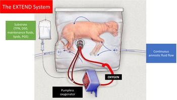
In this pilot study, the EXTEND system was used to support lambs in a model mimicking intrauterine life and foetal physiology (Fig 1). Reference Partridge, Davey and Hornick11 Fourteen premature lambs were delivered by caesarean section and cannulated via the umbilical vessels at 105−115 days of gestation, a period which roughly corresponds to the brain development observed in a preterm human infant born between 28 and 30 weeks of gestation. Reference Lawrence, Hennessy-Strahs and McGovern23 After cannulation the animals were transitioned to a sterile fluid-filled environment and connected to a low-resistance pumpless oxygenator (Maquet Quadrox-ID Pediatric Oxygenator: Maquet Cardiopulmonary AG, Rastatt, Germany) and supported by the EXTEND system for up to 21 days. The EXTEND system and corresponding surgical procedures have previously been described in detail by Partridge and colleagues. Reference Partridge, Davey and Hornick11
The EXTEND System. Fetal sheep cannulated via the umbilical vessels, attached to a pumpless, low-resistance oxygenator circuit and incubated in a sterile, fluid environment with continuous fluid exchange and substrate infusion.

All fetuses were maintained in a normoxemic environment defined as oxygen delivery 20−25 ml/min/kg for 24 hours post-cannulation. After this initial period, the fetuses were assigned into one of the two groups (normoxemia or hypoxaemia). Normoxemia was defined as oxygen delivery 2025 ml/kg/min, and hypoxaemia was defined as oxygen delivery 14−16 mL/kg/min, similar to the oxygen delivery in fetuses with CHD. The reduction in oxygen delivery was achieved by gradually decreasing the oxygen supply to the oxygenator, as previously described. Reference Lawrence, McGovern and Mejaddam12,Reference McGovern, Lawrence and Baumgarten14 Oxygen delivery was monitored continuously throughout the run (LabChart 7, ADInstruments Inc., Colorado Springs, CO, United States of America) and calculated using an estimated weight, umbilical blood flow (HXL Tubing Flowsensor, Transonic Systems Inc., Ithaca, NY, USA), post-oxygenator oxygen saturation, and haematocrit concentration (M2-Sensor, Spectrum Medical, Gloucester, United Kingdom). Oxygen delivery was calculated using the following equation:
 $${\rm{Oxygen}}\;{\rm{Delivery}} = {{\matrix{
{{\rm{umbilical}}\;{\rm{blood}}\;{\rm{flow}} \times (1.39 \times {\rm{Hemoglobin}}} \hfill \cr
{ \times {\rm{postoxygenator}}\;{\rm{oxygen}}\;{\rm{saturation}})} \hfill \cr
} } \over {{\rm{Daily}}\;{\rm{bodyweight}}}}$$
Body weight was measured at the beginning and end of each study. The estimated daily weight was calculated assuming an exponential growth rate. Reference Partridge, Davey and Hornick11 Total parental nutrition and lipids (intralipid 20%) were provided to all animals in both groups. Dextrose was administered to target a blood glucose between 20 and 30 mg/dl and TrophAmine (B. Braun Medical, Bethlehem, PA, USA) titrated to keep the blood urea nitrogen between 20 and 30 mg/dl.
In addition to the aforementioned parameters, the following physiologic factors were monitored and recorded continuously throughout the entire time in the EXTEND system: Pre-oxygenator pressure (systolic/diastolic and mean; mmHg), post-oxygenator pressure (systolic/diastolic and mean; mmHg), prepost oxygenator pressure difference (mmHg), heart rate (bpm), temperature (°C), and oxygenator sweep gas input and output. Blood samples from the umbilical artery and vein were taken at 6-hour intervals to measure pH, lactate, electrolytes, haemoglobin, and oxygen saturation. After being supported by the EXTEND system for up to 21 days, all animals were humanely euthanised with the use of pentobarbital sodium (117 mg/kg) and phenytoin sodium (15 mg/kg) (Euthasol Solution, Virbac, Westlake, TX, USA).
Eight gestational age-matched controls were delivered via caesarian section from time-dated ewes.
Serum analysis
Umbilical arterial blood was sterilely collected from the pre-oxygenator lines on the first day after surgery (baseline), and then serially on a weekly basis (days 7, 14, and 21). Umbilical arterial blood from control fetuses was assessed at baseline (n = 4) and day 14 (n = 4). The blood was immediately centrifuged at 3000 rpm for 15 min at 4 °C, aliquoted to subsamples, and stored at −80 °C. Myelin Basic Protein and Glial Fibrillary Acidic Protein were analysed with the use of enzyme-linked immunosorbent assay (MyBioSource Inc., San Diego, CA, United States of America). Serum sample analysis was conducted in a central laboratory (Translational Core Lab at The Children’s Hospital of Philadelphia). All samples were assayed in duplicates with the mean concentration used for analysis. The detection range was 0.15610 ng/mL and 0.56 ng/mL for Glial Fibrillary Acidic Protein and Myelin Basic Protein, respectively.
Statistical analyses
Data analysis proceeded in two distinct phases: a descriptive phase and an inferential phase. In the descriptive phase, parametric as well as non-parametric measures of central tendency, variability, and association were calculated, with a particular emphasis on weekly serum levels, umbilical blood flow, and oxygen delivery levels. Experimental groups were compared at baseline to demonstrate equivalence between groups using a non-parametric Wilcoxon rank-sum test (i.e., gestational age at cannulation, umbilical blood flow, and oxygen delivery). Second, in the inferential phase, Myelin Basic Protein and Glial Fibrillary Acidic Protein weekly serum levels were compared between groups across all weekly time points, using median point estimates and 95% distribution-free confidence intervals. Finally, oxygen delivery levels were correlated with Myelin Basic Protein and Glial Fibrillary Acidic Protein levels and tested for statistical significance using Spearman’s Rho correlation coefficients. In all cases, non-parametric tests, point estimates, and confidence intervals were deemed most appropriate given the small, non-normative nature of the data. All analyses were conducted using SAS v9.4 (SAS, Cary, North Carolina, United States of America).
Results
Animal groups
A total of 14 animals were included in the study. Non-biomarker data from some of these animals were been reported in previous papers. Reference Lawrence, McGovern and Mejaddam12,Reference Lawrence, McGovern and Mejaddam13,Reference Lawrence, Hennessy-Strahs and McGovern23,Reference McGovern, Hornick and Mejaddam24 Serum levels of the seven animals supported under hypoxemic conditions were compared to the serum levels of the seven animals maintained under normoxemic conditions. Characteristics of the two groups are shown in Table 1. The distribution of sex was equal with three males in each group. There was no statistically significant difference in gestational age between the two groups at cannulation (p = 0.57), and no statistically significant difference between the two groups in umbilical blood flow during the run (p = 0.90). After induction of hypoxaemia at 24 hours post-cannulation, the hypoxemic animals experienced significantly lower oxygen delivery throughout the remainder of the study compared to the normoxemic group (median) 15.4 [14.0 (minimum);17.9 (maximum)] versus 19.8 [16.2;25.5], p = 0.02).
Sample characteristics by group (n = 14)

Serum assessment
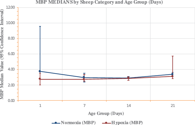
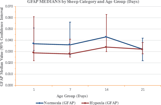
Weekly serum levels of Myelin Basic Protein and Glial Fibrillary Acidic Protein for the hypoxemic and normoxemic animals are shown in Table 2. Overall, no differences were found in Myelin Basic Protein levels throughout the study period when comparing the hypoxemic animals to the normoxemic animals, based on overlapping 95% confidence intervals (Fig 2). Similarly, no statistically significant differences were found in weekly serum Glial Fibrillary Acidic Protein levels at any time point during the run between animals exposed to hypoxaemia and animals exposed to normoxemia (Fig 3).
Serum level of MBP in both groups. All data are shown as mean ± 95% confidence intervals.

Serum level of GFAP in both groups. All data are shown as mean ± 95% confidence intervals.

Weekly MBP and GFAP levels by group (n = 14)

GFAP = Glial Fibrillary Acidic Protein (ng/ml); MBP = Myelin Basic Protein (ng/ml) . CI0.95 = Distribution-free 95% confidence interval.
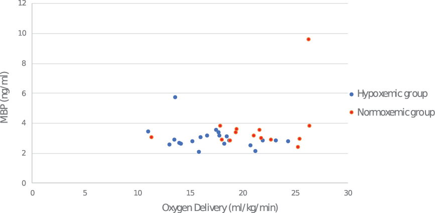
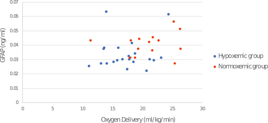
In Figures 4 and 5, Myelin Basic Protein and Glial Fibrillary Acidic Protein levels are shown as a function of oxygen delivery in each animal regardless of group. No statistically significant correlation was observed between serum levels of Myelin Basic Protein and oxygen delivery. Likewise, no statistically significant correlation was observed between serum Glial Fibrillary Acidic Protein levels and oxygen delivery.
Correlation of MBP Levels and DO2. The Correlation Coefficient (R) = 0.036, p = 0.8.

Correlation of GFAP Levels and DO2. The Correlation Coefficient (R) = 0.28, p = 0.1.

In the gestational age-matched control group, Myelin Basic Protein levels were 4.48 (4.09; 4.72) at baseline (n = 4) and 4.62 (4.01; 5.27) at day 14 (n = 4). In the same group, Glial Fibrillary Acidic Protein levels were undetectable at baseline (n = 4) and at day 14 (n = 4).
Discussion
In this model of chronic foetal hypoxia, we have previously demonstrated brain dysmaturity without histopathological evidence of increased cellular necrosis or apoptosis. Reference Lawrence, McGovern and Mejaddam12 In the current study, we found that circulating levels of Glial Fibrillary Acidic Protein and Myelin Basic Protein, biomarkers used to assess brain injury, are not elevated in this model of chronic foetal hypoxaemia. This finding extends our previous findings and supports the hypothesis that chronic foetal hypoxaemia as seen in children with CHD leads to brain dysmaturity, including impaired myelination through an alteration in the normal developmental pathway rather than being caused by direct cellular injury.
Most models of intrauterine hypoxaemia utilise either acute, transient hypoxaemia (umbilical cord occlusion) or maternal hypoxaemia (which may impact the placenta and substrate delivery). Reference Wood and Keller-Wood25 With the use of the EXTEND system, we have been able to investigate in utero hypoxaemia in a setting of chronic hypoxaemia, similar to the conditions of children with CHD. The EXTEND system does not mimic all of the intrauterine abnormalities potentially present in a foetus with CHD (placental insufficiency, abnormal flow patterns, impaired substrate delivery). However, it does provide an exceptional model to investigate the impact of changes in a single factor (oxygen delivery) while holding other parameters constant.
Impaired brain development and neurologic injury is well documented in children with CHD. Reference Limperopoulos, Tworetzky and Newberger6,Reference Licht, Shera and Clancy7,Reference Limperopoulos, Tworetzky and McElhinney26,Reference Miller, McQuillen and Hamrick27
These brain abnormalities are present in neonates even before surgical repair. The pattern of developmental disturbances has been described by Volpe as the “encephalopathy of CHD” and is similar to the brain injury seen in premature infants,. Reference Volpe15,Reference Volpe16 Preoperative cerebral blood flow is low in infants with CHD and impaired cerebral oxygen delivery is thought to be a cause of the observed brain dysmaturity. Reference Sun, Macgowan and Sled9,Reference Licht, Wang and Silvestre10
We have previously investigated the effect of chronic intrauterine hypoxaemia on neurodevelopment. With the use of the EXTEND system, we found that sustained hypoxaemia led to altered cerebrovascular resistance and loss of brain mass. Reference McGovern, Lawrence and Baumgarten14 Using the same model, we found histopathological evidence of brain dysmaturity (without increased cellular death) characterised by white matter hypervascularity, impaired myelination, and decreased neuronal density when exposed to chronic foetal hypoxia, consistent with the findings in children with CHD. Reference Lawrence, McGovern and Mejaddam12 However, it is unknown if these changes are driven by cellular injury with subsequent leakage of serum biomarkers or if brain dysmaturity is caused by disruption of normal developmental pathways.
In the present study, the animals were kept under the same conditions as animals used in our previous studies demonstrating histological changes in the brain when exposed to chronic foetal hypoxaemia.
We observed no statistically significant differences in circulating serum levels of Myelin Basic Protein or Glial Fibrillary Acidic Protein at any time point between the normoxemic and hypoxemic groups. No elevation of Myelin Basic Protein was observed at any time point in animals exposed to the EXTEND system compared to gestational age-matched controls representing the normally developing fetus. In the control group, Glial Fibrillary Acidic Protein was undetectable, whereas this biomarker was detectable, albeit at very low values in the animals exposed to the EXTEND system.
Traumatic and ischaemic white matter injury is associated with elevated Myelin Basic Protein levels and release of Glial Fibrillary Acidic Protein from glial cells as a result of cellular injury. Serum levels of Glial Fibrillary Acidic Protein have been shown to be associated with traumatic brain injury in children and serum levels of Myelin Basic Protein have been shown to be associated with mortality and unfavourable neurologic outcome in children after cardiac arrest. Reference Lumpkins, Bochicchio, Keledjian, Simard, McCunn and Scalea19,Reference Mondello, Kobeissy, Vestri, Hayes, Kochanek and Berger20,Reference Wilkinson, Simic and Frndova28
Combining our findings of stable biomarkers of Glial Fibrillary Acidic Protein and Myelin Basic Protein even under hypoxaemia with the histopathological evidence from the above-mentioned previous study, we suggest that the cause of brain dysmaturity, and possible later neurocognitive challenges, is an alteration of normal neurodevelopmental pathways and not driven by the same cellular damage as seen in acute brain injury.
A limitation of the present study is the restricted amount of serum available for analysis, limiting us from studying other potential biomarkers.
Conclusion
Using the EXTEND system, we have previously shown that brain development is altered in the setting of chronic hypoxaemia at a level consistent with CHD. The finding of no difference in serum Myelin Basic Protein and Glial Fibrillary Acidic Protein levels between normoxemic and hypoxemic animals provides additional evidence supporting the hypothesis that brain dysmaturity with impaired myelination as previously seen in fetuses with chronic foetal hypoxaemia is caused by disruption of normal developmental pathways rather than caused by direct cellular injury.
Acknowledgements
None
Financial support
This work was funded by The Department of Surgery at CHOP and the Daniel M. Tabas Endowed Chair in Pediatric Cardiac Surgery.
Conflict of interest
Alan W. Flake holds multiple patents related to the EXTEND technology and is a Clinical Advisor for Vitara Biomedical Inc.. Marcus Davey is listed as an inventor on EXTEND device patents.
Ethical Standards
The authors assert that all procedures contributing to this work comply with the ethical standards of the relevant national guides on the care and use of sheep and has been approved by the Institutional Animal Care and Use Committee (IACUC) at The Children’s Hospital of Philadelphia.