Implications
Heat stress causes huge economic losses to the pig industry worldwide because of reduced performance and suppressed porcine immune function. We hypothesized that Toll-like receptors (TLRs) may play a role in the immune dysfunction of heat-stressed pigs. We investigated the expression profile of TLR2, TLR4 and the concentration of acute inflammatory cytokines in Bama miniature pigs subjected to heat stress both in in vivo and in vitro models. Heat stress increased the expression of both TLRs and the concentration of IL-12, but not of IL-2, IL-8 and IFN-γ. We concluded that the dysregulation of cytokines may be correlated with immunosuppression in heat-stressed pigs.
Introduction
Global warming and high stocking density increase the chances of heat stress in pigs. Many stressors that pigs are subjected to are associated with immunosuppressive effects that increase disease susceptibility (Hermann et al., Reference Hermann, Amy Tovar, Michael Beck, Allen and Sheridan1993; Hyun et al., Reference Hyun, Ellis, Riskowski and Johnson1998). The total economic losses across animal classes because of heat stress are reported to average $2.4 billion annually in the United States (St-Pierre et al., Reference St-Pierre, Cobanov and Schnitkey2003). Understanding the stress-associated mechanisms of immune system function and the increased susceptibility of livestock to disease is now more important than ever, because the majority of emerging animal diseases is zoonotic and, therefore, threaten public health.
Toll-like receptors (TLRs), which are part of the IL-1 family, were characterized in mammals by their ability to recognize pathogen-associated molecular patterns and to initiate appropriate immunes response (O’Neill, Reference O’Neill2006). In addition to their role in pathogen detection and defense, TLRs act as sentinels of tissue damage and mediate inflammatory responses to aseptic tissue injury (Marsh et al., Reference Marsh, Williams-Karnesky and Stenzel-Poore2009). Once TLRs are activated by either pathogens or host-derived ligands, they induce downstream signals that lead to cytokine and chemokine production, thereby initiating inflammatory responses (Marsh et al., Reference Marsh, Williams-Karnesky and Stenzel-Poore2009).
Many studies have shown that the TLR signaling pathway is activated under heat stress in mammals (Eicher et al., Reference Eicher, Sartin, Schwartz, Lay and Cheng2004; Zhou et al., Reference Zhou, An, Xu, Liu and Cao2005), although the conclusions are conflicting and difficult to reconcile into universally applicable theories. In the present study, we hypothesized that heat stress leads to increased expression of TLR2 and TLR4 and induction of inflammatory factors. We investigated TLR2 and TLR4 expression in peripheral blood mononuclear cells (PBMC) of Bama miniature pigs under heat stress both in in vitro and in vivo models. Immune parameters and inflammatory factor, such as IL-12, IL-2, IL-8 and IFN-γ, were analyzed with the aim of elucidating their roles in immunosuppression in pigs under heat stress.
Material and methods
Animals and housing
Eighteen Bama miniature pigs (aged 3 to 4 months) were obtained from the Bama Miniature Pig Breeding Farm of Guangxi Zhuang Autonomous Region of China. Before the experiments, pigs were allowed an acclimatization period of ~30 days. Sets of three littermates were kept together in nursery pens (floor space, 1.5 m2 per pig). All pigs were given free access to water, and feed was supplied three times per day. The diet was formulated according to the recommended nutrient allowances for this breed of pigs (online resource, http://www.86zizhu.cn). Experimental procedures were approved by the Guangdong Ocean University Animal Care and Use Committee.
In vivo experimental design and blood sample collection
Nine Bama miniature pigs were assigned to the control group and maintained with an air temperature was 28±3°C and relative humidity of ~90%. The other set of pigs were assigned to the heat stressor treatment group that were maintained at 35±1°C with a relative humidity of ~90% (HEAT). Stressors were imposed for 21 consecutive days. The body temperature, heart rate and respiratory rate were detected by traditional methods on the morning of days 1, 7, 14 and 21 after the start of treatment. Blood samples were collected by anterior vena cava puncture at the same time-points. For blood sample collection, pigs were held in a supine position, and 15 ml of blood was collected into vacutainers containing heparin (143 USP) by anterior vena cava puncture. Whole blood (5 ml) was used for total white blood cell (WBC) counts, differential percentages, red blood cell (RBC) indices and cell isolation. The remainder of the sample was used for the isolation of total RNA and plasma.
Leukocyte counts and RBC indices
Whole blood (diluted 18,050-fold) was used to assay total WBC numbers, leukocyte differential counts and RBC indices using a BC-2300 hematology analyzer (Mindray Bio-Medical Electronics Co. Ltd, Shenzhen, China) according to the manufacturer’s instructions. For leukocyte differential counts, 1.02 ml of erythrocyte lysis buffer (M-50 LEO I; Mindray Bio-Medical Electronics Co. Ltd) was added to 20 μl of whole blood, followed by the addition of 0.32 ml erythrocyte lysis buffer (M-50 LEO II; Mindray Bio-Medical Electronics Co. Ltd). For leukocyte counts, 1.02 ml of erythrocyte lysis buffer only (M-50 LBA) was added. RBC indices included RBC counts, mean corpuscular volume (MCV), mean corpuscular hemoglobin (MCH), mean corpuscular hemoglobin concentration (MCHC) and red cell distribution width (RDW).
PBMC isolation, culture and heat shock
PBMCs were isolated from whole blood (5 ml) of control pigs by density gradient centrifugation using LTS-1077 (density=1.077 g/ml; TBD, Tianjin Haoyang Biological Manufacture Co., Ltd, Tianjin, China) according to the manufacturer’s protocol. Isolated PBMCs were washed twice in Hank’s media, counted and resuspended in culture medium (RPMI 1640; GIBCO,Shanghai, China) at a concentration of 2×10⁵/ml. Then, 0.5 ml of cell suspension was added to two 24-well tissue culture plates. One culture plate was incubated at 42°C (95% air, 5% CO2, 100% humidity) for 1 h and then incubated at 37°C for 9 h (heat-treated cells). The control cells were incubated at 37°C (95% air, 5% CO2, 100% humidity) for 10 h without any treatment (control cells). Cells and culture supernatants were collected at 1, 3, 6 and 9 h after treatment began for subsequent analysis.
Analysis of TLR2/4 expression by qRT-PCR assay
To explore the changes in TLR2 and TLR4 mRNA expression, total RNA was extracted from 1 ml of PBMC suspension isolated from peripheral blood (in vivo) or digested from tissue culture plates (in vitro) using commercial RNAiso plus kits (TaKaRa Biotechnology Co., Ltd., Dalian, China) following the manufacturer’s recommendations. The yield of total RNA (10 to 20 μg) was resuspended with DEPC-treated distilled water and stored at −70°C for later use. For cDNA synthesis, total RNA was reverse transcribed with a PrimeScriptTM RT reagent kit (TaKaRa Biotechnology), according to the manufacturer’s instructions. In brief, 2 μl of RNA (100 ng) was added to a reaction mixture containing 2 μL PrimeScriptTM Buffer, 0.5 μl PrimeScriptTM RT Enzyme Mix I, and 0.5 μl random primers (50 μM).
To normalize the expression of TLR2/4, TATA-box-binding protein (TBP) and β-2-microglobulin (B2M) were selected as reference genes. The primers used for amplification of TLR2, TBP and B2M have been described previously (Ju et al., Reference Ju, Yong, Xu, An, Xu, Jiao and Liao2011b), whereas the primers used for amplification of TLR4 were as follows: forward primer (5′-TTCATACAGAGCCGATGGTG-3′) and reverse primer (5′-TCCTGCTGAGAAGGCGATA-3′) and were synthesized by GeneCore (Shanghai GeneCore BioTechnologies Co., Ltd, Shanghai, China). qRT-PCR reactions were performed in an iCycler IQ Real-Time PCR Detection System (Bio-Rad, Hercules, CA, USA). The 25 μl reaction mixture contained 12.5 μl SYBR Premix EX TaqTM (2×; TaKaRa Biotechnology), 0.5 μl forward primer (20 μmol/l), 0.5 μl reverse primer, 2 μl reverse-transcription product and 9.5 μl distilled water. The following experimental protocol was used: a quantification program consisting of 40 cycles of 95°C for 4 s, 15 s at the annealing temperature (55°C) and 10 s at 72°C, ending with a melting program ranging from 60°C to 95°C with a heating rate of 0.5°C/5 s and continuous fluorescence measurement. The results were exported from the iCycler IQ Real-Time PCR Detection System into Microsoft Excel files. The equation EΔ quantification cycle (Cq, Ref Cq – Cq sample), where Ref Cq is the mean value of reference genet Cq value for the primer pair and Cq sample is the Cq value for each amplification, was used to calculate a relative gene expression value.
Cortisol assay
Animal cortisol radioimmunoassay kits (Beijing North Institute of Biological Technology, Beijing, China) were used to determine cortisol levels in all individual plasma samples (50 µl) according to the manufacturer’s instructions. Radioactivity was determined using a gamma counter (Cobra 5005; Perkin Elmer Life and Analytical Sciences, Beijing, China). The cortisol levels for individual assays were calculated by interpolation from the standard curves using RiaSmart software (PerkinElmer Life and Analytical Sciences). Multiple tubes of low- and high-level quality control samples were prepared and kept frozen for use in each assay. Standards ranged from 0 to 500 ng/ml and the detection limit was 0.625 ng/ml. Intra-assay and inter-assay coefficients of variation were <8%.
Analysis of cytokines levels in plasma and culture supernatants
Plasma and culture supernatants were stored at −80°C before analysis. Cytokines were measured using ELISA kits for porcine IL-12 (P1240; R&D Systems Inc., Minneapolis, MN, USA), IL-8 (R&D Systems), IL-2 (F25070; Shanghai Xitang BioTec Co., Ltd, Shanghai, China) and IFN-γ (ab113353; Abcam, Cambridge, UK) according to the manufacturer’s protocols. A fourfold dilution of plasma samples was carried out before the assay, and individual samples of diluted plasma and intact supernatant were used in subsequent analyses. Plates were read using a microplate reader (BioTek Instrument Inc., CA, USA) at a wavelength of 405 nm. A standard curve comprising dilutions of recombinant cytokine was prepared and used to estimate the concentration in the samples.
Statistical analysis
Statistical analyses were performed using SAS version 8 (SAS Institute Inc., Cary, NC, USA). A linear mixed-effects model was used to analyze these variables using the mixed procedure of SAS. The main fixed effects included were day (four levels) and treatment (two levels). Data from day 0 were used as a covariate to compensate for baseline variation. Random effects of litter and pen were included in the model. The model had a repeated structure for day, which allowed the incorporation of heterogeneity of variances across days. Residuals were tested for departures from assumptions. Results of qRT-PCR analysis, leukocyte counts, RBC indices and cytokine concentrations are expressed as the mean±s.e.m., and the changes were displayed by the ratio of heat-stressed pigs to control in figures for facilitation of viewing. Differences in the immunological changes observed between the control and heat stress animals were analyzed by Student’s t-test for independent pairs. P<0.05 was considered to indicate statistical significance.
Results
Physiology indices and cortisol levels induced by heat stress
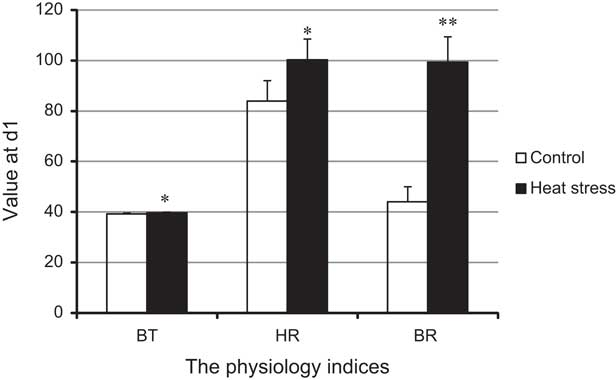
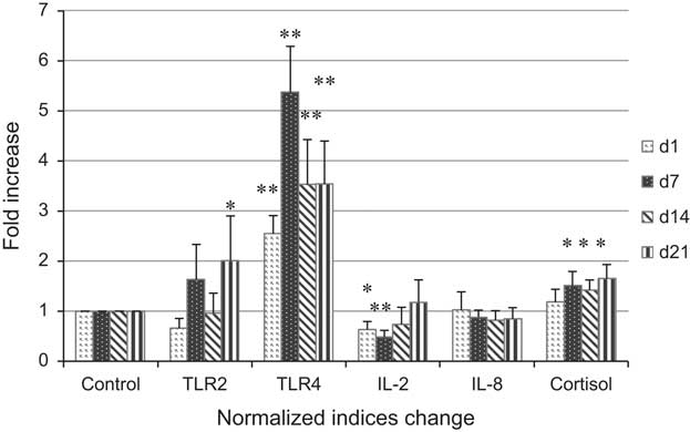
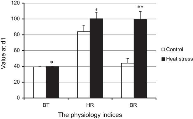
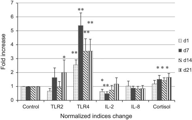
We measured the body temperature, heart rate and respiratory rate of pigs at day 1. The results showed that there was an increased body temperature of 0.5°C to 1°C in heat-stressed pigs compared with that in control pigs, and the heart and respiratory rates were increased ~1.2-fold and 2.3-fold, respectively (Figure 1). Analysis of plasma cortisol levels in Bama miniature pigs revealed that the levels increased with the duration of heat stress (Figure 2). Although there was no significant difference in the cortisol levels of control and heat-stressed pigs on the first day (P>0.05), at subsequent time points, cortisol levels were significantly higher in heat-stressed pigs compared with those in the control animals (P⩽0.05).
The effect of heat stress on the body temperature, heart rate and respiratory rate at day 1. We found that heat stress increased the body temperature (BD), heat rate (HR) and respiratory rate (RR) of pigs significantly. The asterisk ‘*’ and double-asterisk ‘**’ denote a significant difference between stressed pigs and control pigs on the same day (P<0.05 and 0.01, respectively). The unit of the above indices are centigrade (°C), beats per minute and breaths per minute, respectively.
The effect of heat stress on the expression of Toll-like receptors (TLRs) and cytokines in pigs by in vivo study. We detected the concentration of cortisol, IL-2 and IL-8 in the plasma as well the expression of TLR2 and TLR4 mRNA in the peripheral blood mononuclear cells (PBMC) of pigs. The asterisk ‘*’ and double-asterisk ‘**’ denote a significant difference between stressed pigs and control pigs on the same day (P<0.05 and 0.01, respectively).
Leukocyte counts and RBC indices
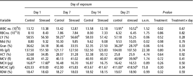
The effects of heat stress on leukocyte counts and RBC indices assayed over time are presented in Table 1. There was no difference between heat-stressed pigs and controls in terms of the number of RBCs and the percentage of neutrophils (Mid%). However, the number of WBC and the percentage of granulocytes (eosinophils+basophils) (Gran%) decreased significantly in heat-stressed pigs at day 21. The percentage of lymphocytes was also reduced markedly in heat-stressed pigs at 7 days (P⩽0.05). With regard to RBC indices, there were no differences between heat-stressed and control pigs in terms of the percentages hematocrit (HCT), RDW and hemoglobin (Hb). It is notable that MCH and MCHC were increased in heat-stressed pigs at days 1 and 21, respectively (P⩽0.05). MCV was decreased in heat-stressed pigs at day 21 (P⩽0.05).
Leukocyte cell counts and RBC indices in Bama miniature pigs under heat stressFootnote 1

Hb=hemoglobin; HCT=hematocrit.
a,bMeans with different superscripts within a row and day are different (P⩽0.05).
1 The leukocyte cell counts and red blood cell (RBC) indices in Bama miniature pigs under heat stress and control pigs are listed. The leukocyte cell counts include the number of white blood cells (WBC), the percentage of lymphocytes (LY, %), the percentage of neutrophils (Mid%) and the percentage of eosinophil+basophil granulocytes (eosinophils+basophils) (Gran%). The RBC indices include the counts of RBC, mean corpuscular volume (MCV), mean corpuscular hemoglobin (MCH), mean corpuscular hemoglobin concentration (MCHC) and red cell distribution width (RDW). The above variables were detected in heat-stressed pigs on day 1, day 7, day 14 and day 21.
The effect of heat stress on TLRs and acute inflammatory cytokines in vivo
The changes in TLR2/4 expression and the concentration of cytokines are shown in Figure 2. Compared with controls, heat stress upregulated the expression of TLR2 in PBMC at days 7 and 21, although the difference was significant only at the later time-point (P<0.05). Levels of TLR4 mRNA were higher in the heat-stressed pigs than in the control pigs at each time-point (P<0.01). The plasma concentrations of IL-2 and IL-8 in control pigs and heat-stressed pigs were also detected in the present study (Figure 2). It was observed that IL-2 concentrations in heat-stressed pigs were significantly lower than those in control pigs at days 1 and 7 (P<0.01). However, there was no difference in the levels of IL-8 detected in the two groups at any time-point.
The effect of heat stress on TLRs and inflammatory cytokines in vitro
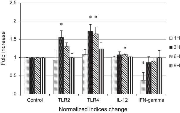
Although our in vivo study indicated that heat stress upregulates the expression of TLRs and some inflammatory cytokines, many factors may influence the results of in vivo experiments. Therefore, further studies were conducted in vitro to investigate the effects of heat stress on PBMC under more controlled conditions. Figure 3 shows the changes in TLR2 and TLR4 expression in PBMC under conditions of heat stress. There was no significant difference in PBMC TLR2 mRNA levels between control and heat-stressed pigs at 1, 6 and 9 h after treatment, except at 3 h, where TLR2 mRNA levels were higher in the heat-stressed group than those in the control group (P<0.05). However, TLR4 mRNA expression was significantly increased in heat-stressed PBMC at 3 h, and peaked at 6 h (P<0.05), with levels returning to normal by 9 h after the start of treatment.
The effect of heat stress on the expression of Toll-like receptors (TLRs) and cytokines in pigs by in vitro study. We detected the effect of heat shock on the concentration of IL-12 and IFN-γ in the supernatant of peripheral blood mononuclear cells (PBMC) subjected to heat stress for 1 h and recovery for 9 h, as well the expression of TLR2 and TLR4 mRNA in the PBMC. The asterisk ‘*’ denotes a significant difference between heat shocked and control PBMC at the same time-point (P⩽0.05).
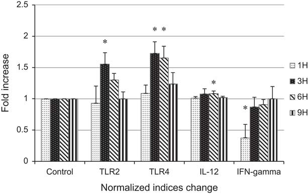
Inflammatory cytokines were also detected in the in vitro model of heat stress (Figure 3). We found decreasing levels of IFN-γ in control PBMC over the period of the experiment, but in heat-stressed PBMC, IFN-γ levels were initially decreased followed by increased levels with the period of heat stress (P<0.05). It is notable that IFN-γ levels in heat-stressed PBMC decreased significantly at 1 h compared with control PBMC (P<0.05). In contrast, there was no significant difference in IL-12 levels in control PBMC over the period of the experiment, although IL-12 levels were higher in heat-stressed PBMC than those in the control group at 6 h after the start of the treatment (P<0.05).
Discussion
In the present study, we demonstrated that heat stress regulates the expression of TLRs, not only in an in vivo Bama miniature pig model, but also in in vitro analyses of PBMC. Moreover, the inflammatory cytokines thought to be related to the activation of TLRs were also shown to fluctuate in response to heat stress. Our results indicated that the TLR signaling pathway is involved in heat stress responses correlated with immunosuppression induced at high temperatures.
Stressors activate the hypothalamic–pituitary–adrenal axis, resulting in elevated corticotrophin-releasing hormone, which induces the release of ACTH and other peptides. Elevated ACTH stimulates the release of glucocorticoids from the adrenal cortex into the blood of stressed farm animals (Hicks et al., Reference Hicks, McGlone, Whisnant, Kattesh and Norman1998). Although many stressors have been reported to increase plasma cortisol concentrations in pigs (Becker et al., Reference Becker, Nienaber, Christenson, Manak, DeShazer and Hahn1985; Parrott and Misson, Reference Parrott and Misson1989), conflicting results have been reported by other researchers. For example, lower cortisol levels were detected in heat-stressed pigs compared with cold-stressed and transportation-stressed pigs, with statistically similar values for cortisol compared with control pigs (Hicks et al., Reference Hicks, McGlone, Whisnant, Kattesh and Norman1998, Sutherland et al., Reference Sutherland, Niekamp, Rodriguez-Zas and Salak-Johnson2006). Furthermore, heat stress did not increase plasma cortisol in cows with the exception of a slight elevation on the first day of heat stress (Farooq et al., Reference Farooq, Samad, Shehzad and Qayyum2010). However, in the present study, cortisol levels were found to be elevated in stressed pigs from day 7 to day 21. This difference may be due to the intensity of stress or the breed of animal used in the study.
High environmental temperature and specific physical circumstances can cause unusual physical effects (Kelley, Reference Kelley1985; Ju et al., Reference Ju, Yong, Xu, An and Xu2011a). For example, the percentage of leukocytes in the blood is altered as a result of smoking, by the time of day and during menstruation (Kauffmann et al., Reference Kauffmann, Neukirch, Annesi, Korobaeff, Dor and Lellouch1988). We detected the baseline immune parameters in heat-stressed pigs in earlier studies, such as the number of WBC and cell differential counts by using the Neubauer chamber method (Ju et al., Reference Ju, Yong, Xu, An and Xu2011a). However, in the present study, the hematology analyzer was used showing that the number of WBC and the percentage of granulocytes (eosinophils+basophils) decreased significantly in heat-stressed pigs. These findings are in accordance with a previous study in which the percentages of basophils and eosinophils, leukocytes and monocytes were reduced by glucocorticoids in stressed animals (O’Connor et al., Reference O’Connor, O’Halloran and Shanahan2000). This inhibition is mediated at the level of cytokine gene transcription, and by inhibition of the production of arachidonic acid-derived proinflammatory substances (Panes et al., Reference Panes, Perry and Granger2009). However, rise of blood neutrophil concentrations in the presence of glucocorticoids because of impaired endothelial cell adhesion, and consequently impaired diapedesis and migration to inflammatory sites (Doukas et al., Reference Doukas, Hechtman and Shepro1988). Increased neutrophil counts were not detected in the present study, which may be owing to differences in stress-intensity or plasma glucocorticoid levels in the animals used in this study compared with those used in prior research.
MCH refers to the concentration of Hb in each RBC. The capacity of RBC to carry oxygen is reduced with the MCH value, leading to iron deficiency anemia. Heat stress can disrupt normal physiological pathways and cause erythrocyte destruction (Lee et al., Reference Lee, Roussel and Beatty1976). MCH and MCHC, but not HCT and Hb, were elevated in heat-stressed pigs in the present study. These findings were in conflict with prior reports in that MCH was not affected by heat stress in pigs (Hicks et al., Reference Hicks, McGlone, Whisnant, Kattesh and Norman1998) or by cyclic heat stress in chicken (Marchini et al., Reference Marchini, Nascimento, Silva and Guimares2011). These conflicting findings may result from the difference of experimental animal species.
The innate immune system recognizes invading microbes and triggers a defense response in the host aimed at clearing the invading pathogen. TLRs play an important role in the recognition of microbial components and activation of innate immunity (Inohara et al., Reference Inohara, Ogura, Fontalba, Gutierrez, Pons, Crespo, Fukase, Inamura, Kusumoto and Hashimoto2003). TLR2 and TLR4 are implicated in the recognition of various bacterial cell wall components, such as lipopolysaccharide (LPS), but TLR2 plays a major role in Gram-positive bacterial recognition. When combined with an agonist, TLR2 induces the activation of NF-κB via a MyD88-dependent/independent pathway and stimulates the production of many proinflammatory cytokines, such as TNF-α, IL-6, IL-12, IFN-α and IFN-γ (Zhang and Ghosh, Reference Zhang and Ghosh2001; Mostefaoui et al., Reference Mostefaoui, Bart, Frenette and Rouabhia2004). We hypothesized that the response of the TLR signaling pathway to heat stress may play a role in stress-induced immune suppression.
Heat stress upregulates the expression of TLR2 and TLR4 in PBMC and increases the response of PBMC to lipoteichoic acid or LPS (Zhou et al., Reference Zhou, An, Xu, Liu and Cao2005). TLR2 and TLR9 also rise in RPMI 8226 cells under heat stress, and IL-6 increases when these cells are stimulated with CpG-containing oligonucleotides (Chen et al., Reference Chen, Wang, An, Zhou, Zhang and Cao2005). These observations are consistent with the present study in that TLR2 and TLR4 were elevated in PBMC in vivo and in vitro, which was in accordance with our previous study (Ju et al., Reference Ju, Yong, Xu, An, Xu, Jiao and Liao2011b). In contrast, Eicher et al. (Reference Eicher, Sartin, Schwartz, Lay and Cheng2004) reported that TLR2 was not elevated at 12 h and 24 h after the start of heat stress in pigs, although the authors postulated that TLR2 plays a role in the regulation of heat stress based on a slight increase in expression compared with control animals. Furthermore, heat shock did not alter the expression of TLR4 on the surface of Kupffer cell in vitro (Sun et al., Reference Sun, Chen, Du and Pan2005), suggesting that the inhibitory effect of a heat shock response on the LPS-induced NF-κB activation and subsequent cytokine production is not a result of downregulation of TLR4 expression. However, restrictive stress induced a reduction in lymphocyte counts and a shift in the Th1/Th2 balance via the TLR4 signaling pathway: a phenomenon that was abolished by TLR4 silencing (Zhang et al., Reference Zhang, Miao, Hanley, Stuart, Sun, Chen and Yin2008). These contradictory conclusions may be because of the differences in experimental materials and treatment conditions, although the involvement of the TLR4 signaling pathway in heat-stressed pigs is indicated.
The stimulus of CpG induces the expression of TNF-α, IL-6 and IL-12 in pig PBMC (Kamstrup et al., Reference Kamstrup, Verthelyi and Klinman2001), and LPS also results in the elevation of IFN-γ, TNF-α, IL-1, IL-10 and IL-12 (Reddy and Wilkie, Reference Reddy and Wilkie2000). However, in stressed animals, glucocorticoids inhibit the function of virtually all inflammatory cells by altering the transcription of cytokine genes (Williams, Reference Williams1990). IL-2 is secreted by activated T cell and plays an important role in the proliferation of CD8+ T cells (Schwartz, Reference Schwartz1990). Glucocorticoids inhibit lymphocyte proliferation and activation by directly inhibiting IL-2 and IL-2 receptor production, and by indirectly reducing lymphocyte IL-2 synthesis through reduced monocyte IL-1 synthesis (Ross et al., Reference Ross, Bacon and Camp1990). In addition, neutrophil-attracting factors, such as IL-1, IL-8 and leukotriene B4, are reduced in the presence of glucocorticoids. However, in the present study, IL-2 was decreased, and IL-8 levels were not altered in heat-stressed pigs compared with the control pigs, indicating that the regulation of cytokines is a complex process in heat-stressed pigs than that only glucocorticoids treated.
The balance of Th1/Th2 influences host immune function and the shift of Th1 to Th2 is a marker of immunosuppression. Heat stress or oxidative stress upregulates the expression of IL-15 via the activation of NF-κB (Wang et al., Reference Wang, Seidl, Whittall, Babaahmady and Lehner2010). In pigs under transportation stress, serum concentrations of IL-2, IL-6 and IL-10 were highest at 1 h, whereas concentrations of IL-6 and IL-10 were significantly decreased at 4 h (Lv et al., Reference Lv, Zhang and Zhao2011). In the present study, IL-12 was elevated in heat-stressed pigs, whereas IFN-γ levels were increased. These results did not correspond to those of Li et al. (Reference Li, Wang, Shao, Zhang, Feng, Wang and Chang2001) who showed that IL-12 was inhibited via the inhibition of NF-κB in murine macrophages. It is notable that the reports of activation of NF-κB in response to heat stress are inconsistent. Thus, reports of the changes in proinflammatory and anti-inflammatory cytokine production mediated by NF-κB were also inconsistent. It is difficult to reconcile these reports into a universal theory of a heat stress-induced shift from Th1 to Th2 that plays an important role in host immune suppression. Further studies are required to elucidate the role of the TLR signaling cascade in the expression of T-box expressed in T cells (T-bet) and GATA binding protein 3 (GATA-3), two key transcription factors of Th1 and Th2 responses, respectively.
Conclusion
A period of 21 consecutive days of heat stress elevated the expression of TLRs in PBMC of pigs, not only in an in vivo model but also in in vitro experiments. The inflammatory cytokine IL-12 was upregulated, whereas IL-2 and IFN-γ were reduced in heat-stressed pigs compared with control pigs. The activation of TLRs and the dysregulation of cytokine expression in heat-stressed pigs may be associated with immune suppression, resulting in greater susceptibility to antigenic challenge in Bama miniature pigs.
Acknowledgments
This research was supported by grants from the National Natural Science Foundation of China (No. 31101862), grants from China Postdoctor Science Foundation and Guangdong Ocean University Doctor Seed Grant (No. 0712107).