Introduction
Ascaridoid nematodes are well-known parasites of wild and captive reptiles. Larval stages migrate through the host tissue, potentially causing damage to organs, whereas adults in the alimentary canal often burrow into the mucosal lining (Jacobson, Reference Jacobson2007). Mortality of reptiles due to ascaridoid infections has been reported in various captive specimens (Jacobson, Reference Jacobson2007).
Healthy reptiles can harbour a number of parasites with no apparent ill effect; however, if placed under stress, disease can become an issue in captive reptiles (Rataj et al., Reference Rataj, Lindtner-Knific, Vlahović, Mavri and Dovč2011; Reese et al., Reference Reese, Kinsella, Zdziarski, Zeng and Greiner2004). A compounding factor in parasite transmission can be the mixing of host species in a tank, either consecutively or concurrently, potentially leading to a naïve host species acquiring a novel parasite that may be harmful. This is especially true if the parasite has a lack of host specificity which can also lead to the potential for transmission among species in a collection (Reese et al., Reference Reese, Kinsella, Zdziarski, Zeng and Greiner2004).
Despite crested geckoes, Correlophus ciliatus Guichenot (Diplodactylidae), being a common pet in many parts of the world, very few parasites have been recorded from them (Brusso, Reference Brusso2013), and there are no records of parasites from wild crested geckoes. Crested geckoes are native to just three islands in New Caledonia (Brusso, Reference Brusso2013), and since their introduction to the pet trade in 1994 (Brusso, Reference Brusso2013), crested geckoes are now exclusively traded from captive-bred populations.
In this paper, we determine the species of nematode present in the captive crested geckoes which were ill and subsequently died, and discuss the potential sources of the infection within the gecko population. A redescription and genetic characterisation are provided for the nematodes found in the present study.
Materials and methods
History of the captive population of crested geckoes
In 2015, 10 crested geckoes were legally sourced from Canada (from a breeder of New Caledonian species of reptiles only; n = 4) and Germany (from a pet shop with a variety of reptile species; n = 6), and subsequently kept at the Ocean Park Aquarium in Hong Kong (no permit was required). The geckoes were randomly separated into two groups of one male and four females each, and each group was housed in a separate terrarium. In 2016, the crested geckoes were combined into a single terrarium and a group of wild-caught Madagascan mossy geckoes, Uroplatus sikorae Boettger (Gekkonidae), were placed into the other. The mossy geckoes all died within a few weeks of acquisition; examination by the owner (WL) did not show any obvious signs of parasitic infection and the cause of death remains unknown. The terrarium, and its furniture, was washed with water prior to the return of the crested geckoes. The second terrarium has only ever housed crested geckoes; however, crested geckoes have been moved between the two terraria over the remaining time period for breeding purposes. All geckoes were provided with water ad lib and commercially available dry foods specifically formulated for geckoes (Repashy™ and Pangea™). In addition, their diet was occasionally supplemented with shop-bought crickets and roaches bred in-house.
Collection of nematodes
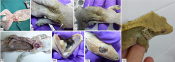
In the year following their return to the terrarium, the five crested geckoes contained in the terrarium that had briefly housed the Madagascan mossy geckoes became moribund and died over a short period of time. The frozen carcasses of these crested geckoes were presented to the Ocean Park Conservation Foundation Veterinary Hospital (OPCFVH), Hong Kong, in July 2018. A post-mortem examination showed that the internal organs were mildly autolysed, preventing a sound histological examination. A number of large nematodes were recovered from the coelomic cavity, under the skin and penetrating organs (liver and alimentary canal) as well as emerging from the eye socket, tympanum and skin (see Fig. 1).
Correlophus ciliatus infected with Hexametra angusticaecoides. (a) Deceased gecko with adult H. angusticaecoides penetrating through the ventral skin surface. (b and c) Gecko with nematodes under the skin. (d) Dissected gecko with larval and immature H. angusticaecoides within the coelomic cavity. (e) Migrating nematode in the mouth cavity. (f) Migrating nematode in the eye socket. (g) A moribund gecko with a H. angusticaecoides emerging from the tympanum.

One of the geckoes from the second terrarium also showed signs of infection, with two nematodes observed moving under the skin (Fig. 1b and c). The nematodes from this gecko were surgically removed and the gecko was treated with fenbendazole (25 and 50 mg/kg), then with pyrantel (10 mg/kg); fecal examination remained positive and a further two nematodes appeared under the skin were also removed surgically. The gecko was then treated with levamisole per os (10 mg/kg) as a single dose. The gecko itself was initially negatively affected by the levamisole and was inappetent and listless for 10–15 days but has subsequently recovered and appears to be in full health at the time of writing. The remaining geckoes in the second terrarium have not shown any signs of infection or illness.
Over 50 nematodes were collected from the geckoes; all of which were collected externally to the alimentary canal. However, given the state of autolysis in the geckoes at the time of post-mortem, and the positive fecal examinations, the possibility of some of the nematodes having escaped from the alimentary canal cannot be excluded. All nematodes were preserved in 90% ethanol.
Morphological identification
A small piece of the mid body of each nematode was excised for molecular study and the rest of the nematode was cleared in lactophenol for morphological examination. The anterior and posterior part of each nematode was examined under a light microscope. Illustrations were made using a microscope equipped with a camera lucida. Photos were taken using an AmScope MU900 eyepiece camera. All measurements are in micrometres, unless stated otherwise. Mean measurements are given, followed by the range in parentheses. Specimens have been deposited in the Australian Helminthological Collection, South Australian Museum, Adelaide, South Australia, under accession numbers AHC48786 and AHC48787.
One male and one female were subjected to scanning electron microscopy (SEM). They were washed and dehydrated overnight in a series of graded ethanol solutions (70, 80, 90, 95% and absolute ethanol). After three additional overnight washes in absolute ethanol, the specimens were critical point dried using a tousimis Autosamdri-931 (USA). Samples were then mounted on a 12 mm carbon tab (ProSci Tech, Thuringowa, QLD, Australia) and sputter coated with gold using a K550X Sputter Coater (Quorum Technologies, UK). The specimens were examined under a JEOL (Peabody, Massachusetts, USA) NeoScope SEM with accelerating voltage set at 10 kv.
DNA extraction, PCR and sequencing
Genomic DNA was isolated from all individual nematodes using Qiagen kit and eluted into 45 μl of water. The internal transcribed spacer (ITS) region (including ITS1, 5.8 and ITS2) was amplified using the primer sets SS1: 5′-GTTTCCGTAGGTGAACCTGCG-3′ (forward) and NC2: 5′-TTAGTTTCTTTTCCTCCGCT-3′ (reverse), and cycling conditions in accordance with Shamsi and Suthar (Reference Shamsi and Suthar2016). An aliquot (4 μl) of each amplicon was examined on a 1.5% w/v agarose gel, stained with GelRed™ and photographed using a gel documentation system. PCR amplicons were sent to the Australian Genome Research Facility (Queensland) for sequencing.
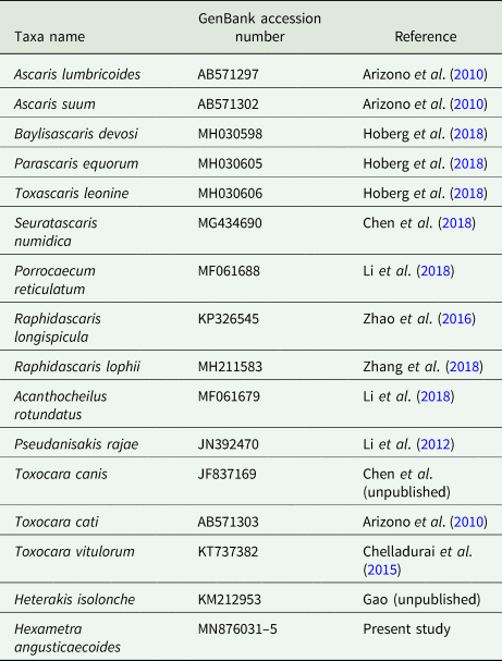
ITS sequences of Hexametra angusticaecoides Chabaud & Brygoo, Reference Chabaud and Brygoo1960 (Ascarididae) were generated in the current study, while ITS sequences from additional nematode species were obtained from GenBank (Table 1). Very few molecular sequences for ascarids from reptiles are available on GenBank, thus ITS sequences were obtained for other members of the Ascarididae (Table 1). Sequences were aligned with Geneious alignment algorithm by using Geneious version 11.1.4 (Kearse et al., Reference Kearse2012), and then were double checked with all variable sites in the original trace files for confirmation. Alignments were then truncated to 865 characters, based on the shortest sequence for ITS. Heterakis isolonche von Linstow, 1906 (Heterakidae), a species belonging to the superfamily Heterakoidea but also residing under the infraorder Ascaridomorpha, was used as an outgroup. Phylogenetic relationship among species was calculated by MrBayes 3.2 (Ronquist and Huelsenbeck, Reference Ronquist and Huelsenbeck2003) with the HKY + I + G model as indicated by JmodelTest 2.0 (Darriba et al., Reference Darriba, Taboada, Doallo and Posada2012).
GenBank accession numbers for ITS sequences used in this study

Results
Clinical findings
The geckoes presented with visible subcutaneous parasites (Fig. 1) that would occasionally emerge through the skin. The geckoes would sometimes lose appetite, appear uncomfortable and then die or sometimes simply be found dead with no prior evidence of distress.
Morphological findings
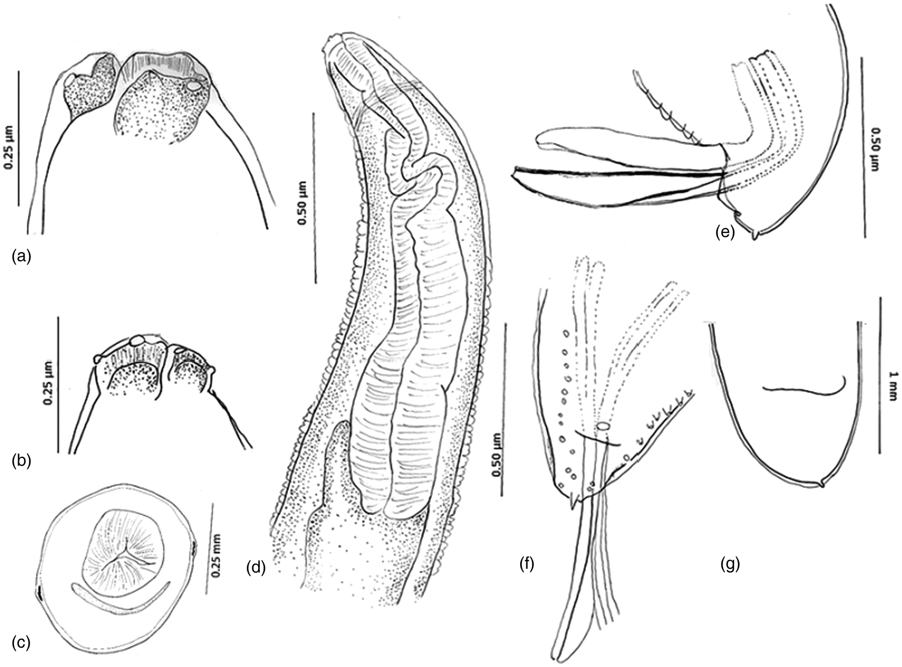
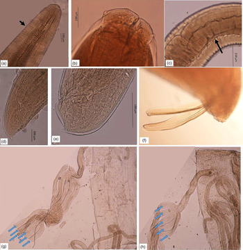
A total of 13 nematodes were available for examination including one larva. Descriptions of adults are based on three males and nine immature females (Figs 2–4), unless otherwise stated. None of the females contained eggs. All specimens were identified as belonging to H. angusticaecoides Chabaud & Brygoo, Reference Chabaud and Brygoo1960.
Line drawing of Hexametra angusticaecoides found in the present study: (a and b) lateral view of the labia in a female and a male, respectively, (c) cross-section below the nerve ring in a female, (d) lateral view of the anterior end showing the nerve ring, oesophagus and short intestinal caecum, (e) lateral view of the posterior end of the male showing a pair of spicules (left spicule was broken off) and a short mucron, (f) ventral view of the male tail showing the mucron, spicules, arrangement of the postcloacal papillae and partial view of the pre-anal papillae. Note the presence of the single median pre-cloacal papilla; (g) ventral view of the posterior end of the female tail being broad, short and simple.

Light micrographs of Hexametra angusticaecoides. (a) Dorsal view of the anterior end of the larva; arrow indicates the position of the excretory pore. (b) Ventral view of the anterior extremity showing subventral lips of an adult male specimen. (c) Intestinal caecum. (d) Lateral view of the posterior end of a male (note terminal mucron). (e) Lateral view of the posterior end of a female. (f) Distal tips of alate spicules. (g and h) Dissected uteri from an immature female. Only five uterine branches were present in one specimen (image h), whereas the diagnostic feature of six uterine branches was evident in the other specimen (image g; one branch is hidden in the photo).

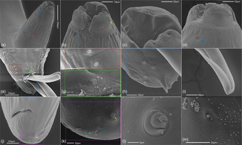
Scanning electron micrographs of a male and a female Hexametra angusticaecoides. (a) Ventral view of the anterior part of the male parasite showing labia, excretory pore (red arrow) and an unknown feature which has not been previously described by other authors (blue arrows); (b) Magnified view of the sub-ventral labium, note the absence of inter-labia, the presence of the dentigerous ridge on the cranial edge of the labia, the presence of papilla on the same level as the labial flange, and an amphid located slightly above the flange line in the opposite direction, blue arrow corresponds with the blue arrows in image (a), red and yellow arrows pointing at the labial papilla and amphid, respectively. (c) Magnified view of the image (b), note the dentigerous ridges do not exceed the cranial edge of the labium. Also note the lobular shape of the cranial side of the labia and the presence of a pore in the centre of each lobe. (d) Dorsal view of the dorsal labium. (e) Ventral view of the posterior end of an adult male. (f) Magnified view of the precloacal papillae. (g) Close up of the median ventral precloacal papilla located superomedially to the cloaca. (h) Tip of the tail in the male specimen showing mucron and postcloacal papillae. (i) Tip of the spicule. (j) Ventral view of the female posterior extremity: Phasmid (yellow arrow), mucron (green arrow) and a minute papilla (blue arrow). (k) Magnified view of image (j). (l) High magnification view of the phasmid. (m) High magnification view of a minute papilla-like structure in the tail of a female specimen.

Description: Anterior extremity with three large labia, including larger dorsal labium and two smaller subventral labia; all labia wider than long; two papillae on dorsal labium; subventral labia with one papilla and one amphid each; labia concave on their cranial side, with minute dentigerous border, uniform in size and confined to the cranial edge of labia (Fig. 4c); inter-labia and post-labial grove absent; excretory pore almost at the same level as nerve ring, slightly posterior to it; lateral alae present between nerve ring and anterior to the oesophagus–intestinal junction; oesophagus muscular, cylindrical, slightly wider at the oesophagus–intestinal junction; short intestinal caecum present; tail short with mucron in both sexes; mucron longer in younger nematodes, becoming shorter and almost invisible in older/larger specimens, particularly in females; tail short, very broad and rounded in females, and conical, bent ventrally and papillose in males; spicules almost equal and alated; six pairs of post-cloacal papillae and up to 84 pre-cloacal papillae; uterus divided into 5–6 branches (two female nematodes were dissected for the purpose of counting uterus branches, one having five uterus branches and the other six uterus branches).
Morphometrics of the specimens were as below:
Males: Total body length 46.43 mm (39.00–60.03 mm); body width 1.21 mm (1.08–1.38 mm); dorsal labium 107 (95–125) high, 168 (150–190) wide; sub-ventral labia 110 (100–130) high, 112 (100–125) wide; nerve ring 610 (480–700) from the anterior end; deirids visible in one specimen only, 700 from the anterior end; excretory pore visible in one specimen, 875 from the anterior end; oesophagus 3133 (2350–3750) long, i.e. 7% (6–8%) of the body length; intestinal caecum 650 (600–700) long; tail 207 (200–220) long (excluding mucron), i.e. 0.5% (0.4–0.5%) of the body length; tail length with mucron 247 (240–250); tail width at the level of cloaca 267 (200–300); spicules 1000 (1000–1000) and 1083 (1050–1100) long, i.e. 2% (2–3%) and 2% (2–3%), respectively, of the body length; number of pre-cloacal papillae 70 and 84 (n = 2).
Females: Total body length 43.40 mm (22.75–69.50 mm); body width 1.08 mm (0.65–1.70 mm); dorsal labium 107 (50–150; n = 8) high, 164 (100–250; n = 8) wide; sub-ventral labia 92 (65–150; n = 6) wide, 90 (50–160; n = 6) high; nerve ring 543 (375–800; n = 6) from the anterior end; excretory pore 639 (450–950; n = 7) from the anterior end; oesophagus 2352 (1750–3700) long, i.e. 6% (3–9%) of the body length; intestinal caecum 461 (250–650; n = 7) long; tail 278 (100–700) long (excluding mucron), i.e. 1% of the body length; tail length with mucron 325 (150–700; n = 8); tail width at the level of cloaca 422 (220–800); vulva 16.94 mm (16.13–17.75 mm; n = 2) from the anterior end, i.e. 43% (34–53%; n = 2) of the body length.
Fourth stage larva: Total body length 13.00 mm; body width 610; nerve ring 400 from the anterior end; excretory pore 450 from the anterior end; oesophagus 1650 long, 13% of the body length; intestinal caecum 250; tail 100 long (excluding mucron), 1% of the body length; tail length with mucron 170; tail width at the level of cloaca 200.
Molecular findings
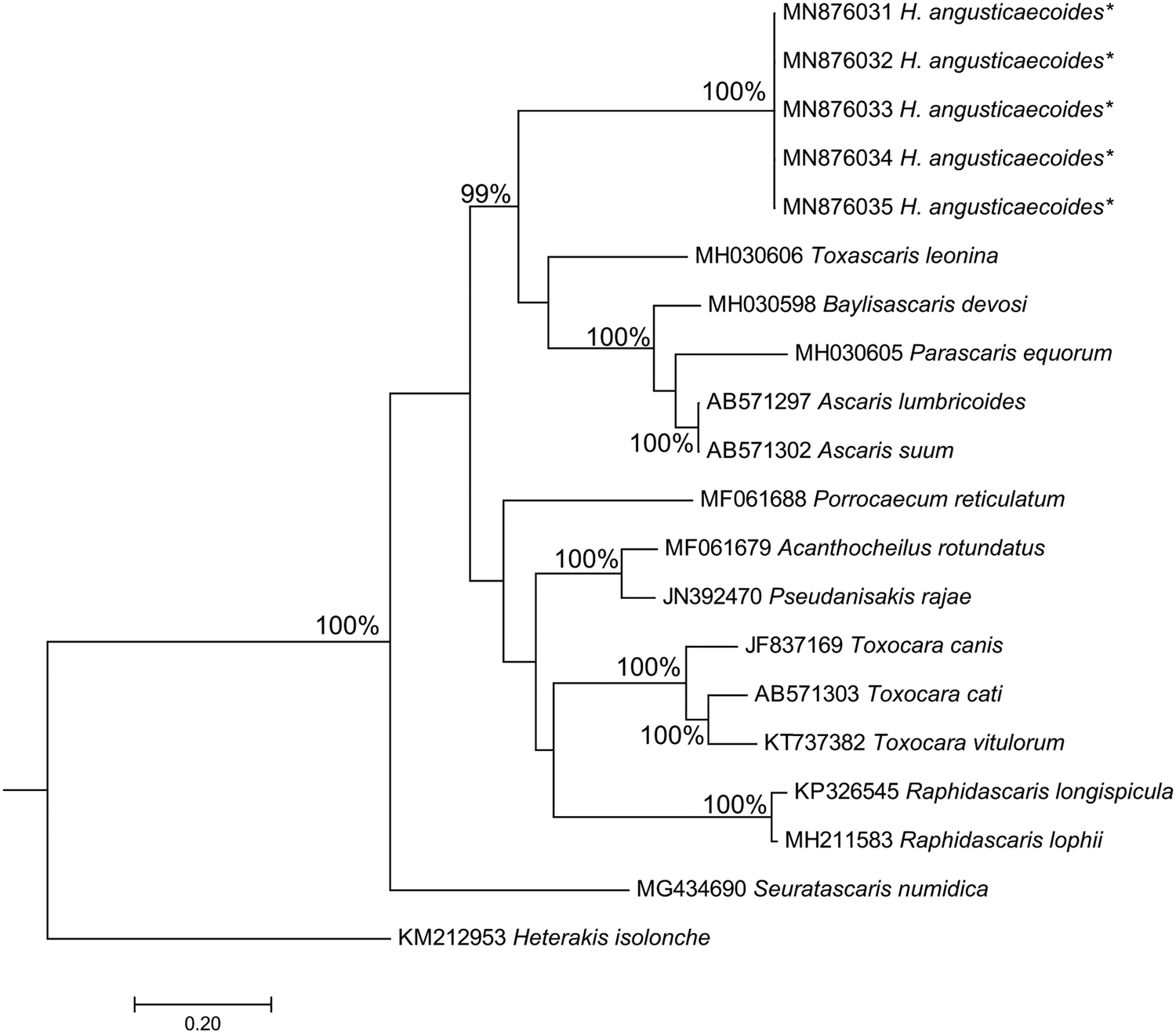
Its sequences were identical among all the males, females and larva found in the present study, confirming that only one species was involved in the infection. We found no ITS sequences for any Hexametra spp. in GenBank. The newly generated ITS sequences of H. angusticaecoides were deposited in the GenBank database under accession numbers MN876031–5. Our sequences formed a distinct group from other terrestrial ascaridoids (Fig. 5).
Phylogenetic analysis of ITS sequences for Hexametra angusticaecoides found in the present study, with Heterakis isolonche as an outgroup. Posterior probability values >90% are indicated above the branches. Sequences obtained in this study were denoted with * symbol. MrBayes best-fit model was used for analysis as suggested by JmodelTest. Branch supports lower than 85% are not shown on the Phylogenetic tree.

Discussion
This is the first report of a species of the genus Hexametra Travassos, 1920 from the crested gecko. Despite several reports of Hexametra spp. in South East Asia (Clarke et al., Reference Clarke, Chang and Cross1970; Sprent, Reference Sprent1978), this is the first report of Hexametra from Hong Kong. Given the fact that the geckoes were captive bred and housed in a terrarium that had previously housed a different species of gecko originally from Madagascar, where H. angusticaecoides was originally described from, it is unlikely that the crested gecko is a natural host, hence the migration of adult nematodes and the absence of gravid females in the present study. Infertility of female nematodes may have also been due to high level of mobility observed in adult nematodes, giving them no opportunity for mating and explaining the absence of the eggs in adult females despite the presence of adult males in one of the geckoes. The high level of mobility observed among these nematodes was also an important factor in the pathogenicity and fatality caused by these parasites. Since the crested geckoes were fed on a diet specifically formulated for crested geckoes, it is unlikely that malnutrition, which is known to make hosts more vulnerable to infection with parasites and to increase the severity of the clinical signs (Urquhart et al., Reference Urquhart, Armour, Duncan, Dunn and Jennings1996), would have played a role in the infection patterns seen in the present geckoes.
Determining the original source of the nematode infection is difficult, due primarily to the multiple sources of host animals, multiple species of hosts in the terraria and the lack of knowledge about the life cycles of most nematodes of reptilian hosts. Hexametra angusticaecoides is widespread in a number of reptilian species around the world (Sprent, Reference Sprent1978), so it is possible that the crested geckoes obtained the infection from the original captive-bred colony or pet shop from which they were acquired. However, this possibility would tend to be refuted as only geckoes within the terraria that also housed the Madagascan mossy gecko became heavily infected. This leaves two potential sources of infection: the Madagascan mossy gecko or the crickets/roaches fed to the crested geckoes. Chabaud et al. (Reference Chabaud, Brygoo and Petter1962) conducted a series of experimental infections with eggs and larvae of H. angusticaecoides, using wild-caught chameleons, Chamaeleo pardalis (Cuvier) (Chamaeleonidae), from Madagascar with an, unfortunately, unknown infection history. Due to the successful transmission of the parasite through various insects, tadpoles, mice and chameleons, Chabaud et al. (Reference Chabaud, Brygoo and Petter1962) concluded that the ‘physiological possibilities of the parasite are very broad’. They also found that in one chameleon, subcutaneous and coelomic larvae co-existed with intestinal adults. We believe that it is most likely that the Madagascan mossy gecko is the true original source of infection, as many species of Hexametra, including H. angusticaecoides, have been originally described from reptiles from Madagascar (Sprent, Reference Sprent1978). The fact that the Madagascan mossy geckoes that were kept in the terraria were wild caught would also support this. These geckoes died rapidly, but with no obvious signs of parasite infection. It is possible that the stress of capture and housing, in hosts that had a subclinical infection, was enough to cause the death of the hosts (Rataj et al., Reference Rataj, Lindtner-Knific, Vlahović, Mavri and Dovč2011).
Host mortality due to infection with ascaridoid nematodes has previously been reported (Smith, Reference Smith1999). Despite the common occurrence of Hexametra in lizards and snakes, there are few reports of associated mortality (Jacobson, Reference Jacobson2007). An infection of Hexametra boddaertii (Baird, 1860) was found in a wild-caught snake in Argentina; the snake died after a few days despite being in apparent good health (Peichoto et al., Reference Peichoto, Sanchez, Lopez, Salas, Rivero, Teibler, Toledo and Tavares2016). Over 100 nematodes were collected from the coelomic cavity (smaller worms) and the intestinal tract (larger worms); initial fecal examination had shown a heavy infection with ascaridoids but attempts to de-worm the snake were unsuccessful (Peichoto et al., Reference Peichoto, Sanchez, Lopez, Salas, Rivero, Teibler, Toledo and Tavares2016). Harmful effects of ascaridoid infections are difficult to assess due to the paucity of knowledge of symptomatology in reptiles. Harmful effects could be due to the size of the nematode in relation to the host (competing for nutrients and causing malnutrition) and/or causing physical damage through their attachment to the mucosal lining of the stomach and/or physical damage during larval migration (Jacobson, Reference Jacobson2007; Sprent, Reference Sprent, Hoff, Frye and Jacobsen1984). Clinical signs, when present, are non-specific; infected reptiles may be anorectic and may slowly lose weight (Jacobson, Reference Jacobson2007). A captive panther chameleon, Furcifer pardalis (Cuvier) (Chamaeleonidae), that died suddenly without signs of illness harboured H. angusticaecoides in its stomach and intestine; a veiled chameleon, Chamaeleo calyptratus Duméril & Bibron, was euthanized after manifesting respiratory distress and had 20–30 immature Hexametra specimens in its coelomic cavity (Jacobson, Reference Jacobson2007).
The fact that the terraria used in this study were not disinfected between housing of the two gecko species and the crested geckoes were returned to the terraria within a matter of days could have ensured that any nematode eggs in the terraria were able to survive and infect the new hosts. Chabaud et al. (Reference Chabaud, Brygoo and Petter1962) found that the eggs of H. angusticaecoides took approximately 15 days to become infective, with second stage larvae spontaneously hatching from the eggs. Additionally, the use of roaches and crickets as food items could have enabled the cycle to continue (see Chabaud et al., Reference Chabaud, Brygoo and Petter1962). The initial infection potentially occurred in 2016, so it would appear that the nematode is able to complete its life cycle within a captive situation, making it a potential problem in a captive colony if it became infected.
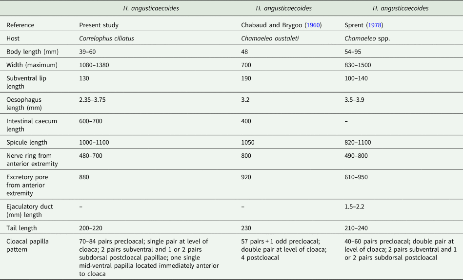
Morphological examination identified the nematodes as belonging to the genus Hexametra based on the keys of Hartwich (Reference Hartwich, Anderson, Chabaud and Willmott1974): female with six uterine branches, inter-labia absent and parasites of snakes and lizards. Sprent (Reference Sprent1978) stated that an intestinal caecum can be present or absent; it was present in the current specimens. Measurements of specimens and the presence of an intestinal caecum showed the specimens were H. angusticaecoides, based on the data provided previously (Chabaud and Brygoo, Reference Chabaud and Brygoo1960; Sprent, Reference Sprent1978) (Table 2). Although most of the measurements of the specimens in the present study fall within the range of measurements provided for H. angusticaecoides in previous studies (Table 2), the number of precloacal papillae and the single vs a double pair of papillae at the level of the cloaca were different and might cast doubt on the specific identification of species of Hexametra in general. Hexametra quadricornis (Wedl 1862) from snakes has 50–100 precloacal papillae (Petter, Reference Petter1968), but Sprent (Reference Sprent1978) believed that this parasite was unable to develop in lizards. While in experimental infections in snakes the larvae of H. quadricornis did develop into adults in the body cavity, they never reached the intestine and the adult females never possessed eggs (Sprent, Reference Sprent1978), which is similar to the case in our study.
Measurements of male specimens of Hexametra angusticaecoides collected from the crested gecko, Correlophus ciliatus, in the present study and comparative measurements obtained from the literature

All measurements in micrometres unless otherwise indicated.
Additionally, during the examination of the morphology of specimens, one female was found to only possess five uterine branches, with another specimen having six (Fig. 3). In addition, despite the overall similarity in measurements, the pattern of cloacal papillae in the male was highly variable (see Table 2). This suggests that there may be a level of morphological variation in the genus that should be considered in future descriptions. This has important implications for the taxonomy of the genus and the closely related Polydelphis Dujardin, 1845 (Ascarididae), which are separated by the number of uterine branches (six in Hexametra compared to four in Polydelphis; Sprent, Reference Sprent1978). Future researchers should be aware of the potential of morphological variation and ensure genetic sequences are included to enable better systematic differentiation of this group of nematodes, especially in relation to potential issues in captive hosts.
The molecular analysis for the specimens of H. angusticaecoides displayed no nucleotide variability in the ITS sequences, indicating that there was only the one species of nematode infecting the geckoes as both larval and adult stages. A comparison of ITS sequences with other species of Ascarididae from terrestrial hosts available in GenBank showed 41.2% nucleotide divergence to the closest representative, Toxoascaris leonine (Linstow, 1902). It must be noted, however, that there are very few full ITS sequences available for ascarid nematodes of terrestrial animals, making direct comparisons between closely related genera difficult. However, the sequences obtained here are extremely important and valuable for future research and diagnosis of infections with H. angusticaecoides. Specimens of H. angusticaecoides from the present study formed a sister group to the clade composed of other terrestrial ascaridoids, including T. leonina, Baylisascaris devosi, Parascaris equorum, Ascaris lumbricoides and Ascaris suum (Goeze, 1782), with the latter two taxa being considered conspecific by some authors (e.g. Leles et al., Reference Leles, Gardner, Reinhard, Iniguez and Araujo2012; Liu et al., Reference Liu, Wu, Song, Wei, Xu, Lin, Zhao, Huang and Zhu2012).
In conclusion, our study reinforces the importance of application of transmission-based precautions to prevent animal and human infection with potentially deadly ascaridoid parasites. In terms of the specific identity of the nematodes, although we considered our specimens as H. angusticaecoides, more specimens from other members of the genus should be examined both morphologically and by molecular tools to be able to more accurately comment on the taxonomic status of these taxa.
Financial support
This study was partially funded by Charles Sturt University A516 828–66770.
Conflict of interest
None.
Ethical standards
Ethical standards were maintained during the study.