INTRODUCTION
Driving performance significantly declines with advancing age. While this may in part reflect increased frailty and reductions in functional ability (Bedard, Guyatt, Stones, & Hirdes, Reference Bedard, Guyatt, Stones and Hirdes2002; Bunce, Young, Blane, & Khugputh, Reference Bunce, Young, Blane and Khugputh2012), cognitive impairment places older adults at an even greater risk of car accidents than their age-matched peers (Cooper, Tallman, Tuokko, & Beattie, Reference Cooper, Tallman, Tuokko and Beattie1993; Frittelli et al., Reference Frittelli, Borghetti, Iudice, Bonanni, Maestri, Tognoni and Iudice2009). Given the imperative to ensure both patient and community safety, it is important to understand how and under what conditions cognitive impairment is likely to impede driving ability.
Cognitive impairment in older people can be due to diverse etiologies and typically there is a continuum of cognitive decline, ranging from only subjective cognitive impairments to dementia. Within this spectrum, Mild Cognitive Impairment (MCI) is considered a pre-dementia syndrome whereby there is objective neuropsychological evidence of a marked decline in cognition, in the context of minimal or no functional impairment (Petersen, Reference Petersen2009; Winblad et al., Reference Winblad, Palmer, Kivipelto, Jelic, Fratiglioni, Wahlund and Petersen2004). Patients with MCI are at high risk of converting to dementia, with figures suggesting around 50% converting within a 5-year period.
Although many patients with MCI may drive safely, there is clearly heterogeneity in this sample. Factors such as the type of cognitive impairment (amnestic or non-amnestic) in addition to the number of cognitive domains impaired (single or multiple), even the criteria used in diagnosis, could possibly contribute to the mixed results observed in the literature (Frittelli et al., Reference Frittelli, Borghetti, Iudice, Bonanni, Maestri, Tognoni and Iudice2009; Kawano et al., Reference Kawano, Iwamoto, Ebe, Suzuki, Hasegawa, Ukai and Ozaki2012; Wadley et al., Reference Wadley, Okonkwo, Crowe, Vance, Elgin, Ball and Owsley2009; for review, see Olsen, Taylor, & Thomas, Reference Olsen, Taylor and Thomas2014). Importantly, it is patients with multi-domain MCI who are more likely to develop ongoing cognitive and functional decline when compared to those with single domain MCI (Mitchell & Shiri-Feshki, Reference Mitchell and Shiri-Feshki2009).
Meta-analytic studies surrounding neuropsychological tests and real-world driving ability have been conducted in healthy older adults (Mathias & Lucas, Reference Mathias and Lucas2009) as well as in older adults with dementia (Reger et al., Reference Reger, Welsh, Watson, Cholerton, Baker and Craft2004). These have shown that deficits in a range of cognitive domains, such as visuospatial ability, processing speed, and executive function (e.g., attention, task-switching), are prominently associated with driving performance. It appears that these cognitive deficits may be associated with driving performance in older adults with MCI as well (Dobbs & Shergill, Reference Dobbs and Shergill2013; Kawano et al., Reference Kawano, Iwamoto, Ebe, Suzuki, Hasegawa, Ukai and Ozaki2012). However, only a few such studies have been conducted in MCI, and the predictive ability of neuropsychological tests for driving ability is low (Dobbs & Shergill, Reference Dobbs and Shergill2013).
It is likely that comorbid medical conditions may contribute to deficits in driving capacity in some MCI patients. This in turn may contribute to the heterogeneity in the predictive ability of tests of cognition. A large body of concurrent literature in largely younger and middle-aged samples has recognized the detrimental effect of sleepiness and fatigue on driving performance (George, Findley, Hack, & Douglas McEvoy, Reference George, Findley, Hack and Douglas McEvoy2002; Horne & Reyner, Reference Horne and Reyner1999). In particular, sleep-related breathing disorders (sleep disordered breathing, SDB) have been strongly associated with an increased risk of motor vehicle accidents (MVAs) in younger and middle-aged samples (Turkington, Sircar, Allgar, & Elliott, Reference Turkington, Sircar, Allgar and Elliott2001; Young, Blustein, Finn, & Palta, Reference Young, Blustein, Finn and Palta1997). This may be due to daytime sleepiness and the negative effect SDB has on vigilance and cognition, in particular processing speed and executive function (Beebe & Gozal, Reference Beebe and Gozal2002; Naismith, Winter, Gotsopoulos, Hickie, & Cistulli, Reference Naismith, Winter, Gotsopoulos, Hickie and Cistulli2004; Turkington et al., Reference Turkington, Sircar, Allgar and Elliott2001).
The pathological mechanisms behind the negative behavioral consequences of SDB are generally proposed to involve an interaction between intermittent changes in blood gas composition (hypoxia or hypercapnia) and sleep fragmentation. Both are a result of recurrent apneas; however, each may elicit specific effects on the brain leading to behavioral deficits. There appears to be a differential relationship between physiological features of SDB and neuropsychological performance when examined in patients with MCI versus cognitively intact older adults. Our previous research suggests that SDB may be most detrimental in MCI subjects and sleep fragmentation more damaging for those with preserved cognition (Terpening et al., Reference Terpening, Lewis, Yee, Grunstein, Hickie and Naismith2015). Of note, levels of oxygen saturation were associated with poorer processing speed in both groups. As sleep fragmentation is common in later life (Almeida & Pfaff, Reference Almeida and Pfaff2005), hypoxia may have an added deleterious effect on performance and brain function in older adults and longitudinal evidence suggests it is associated with an increased risk of dementia (Yaffe et al., Reference Yaffe, Laffan, Harrison, Redline, Spira, Ensrud and Stone2011).
The prevalence of SDB increases with age (Launois, Pepin, & Levy, Reference Launois, Pepin and Levy2007), while sleep disturbance and SDB are evident in over two-thirds of MCI patients (McKinnon et al., Reference McKinnon, Terpening, Hickie, Batchelor, Grunstein, Lewis and Naismith2014; Naismith et al., Reference Naismith, Rogers, Lewis, Diamond, Terpening, Norrie and Hickie2011). However, there is a paucity of studies investigating the effect of SDB on driving performance in older adults (Vaz Fragoso, Van Ness, Araujo, Iannone, & Marottoli, Reference Vaz Fragoso, Van Ness, Araujo, Iannone and Marottoli2013). In particular, there are no studies specifically examining whether older adults who have cognitive deficits are at a greater risk of driving impairment if SDB is also present. From the limited evidence, it appears that SDB is highly prevalent in older drivers (Ancoli-Israel et al., Reference Ancoli-Israel, Kripke, Klauber, Mason, Fell and Kaplan1991; Vaz Fragoso, Araujo, Van Ness, & Marottoli, Reference Vaz Fragoso, Araujo, Van Ness and Marottoli2008). Of concern, these data highlight that presenting with SDB does not necessarily deter or influence the driving habits of these older adults, especially at night (Vaz Fragoso et al., Reference Vaz Fragoso, Araujo, Van Ness and Marottoli2008, Reference Vaz Fragoso, Van Ness, Araujo, Iannone and Marottoli2013). This is surprising given that SDB is associated with hypersomnolence, however, there is evidence that subjective ratings of sleepiness do not parallel objective deficits in vigilance (Banks & Dinges, Reference Banks and Dinges2007).
The problem may be partly exacerbated by concomitant cognitive impairment, particularly if meta-cognition or insight is affected. Greater understanding of these relationships is of critical clinical and major public health significance given the potential consequences of poor driving may lead to road traffic accidents and fatalities (Kowalski et al., Reference Kowalski, Love, Tuokko, MacDonald, Hultsch and Strauss2012; Wong, Smith, & Sullivan, Reference Wong, Smith and Sullivan2012).
Therefore, in this study, we aimed to explore the extent to which SDB and sleepiness contribute to the relationship between cognitive impairment and driving ability in older adults. We hypothesized that greater severity of SDB would negatively impact on performance on a simulated driving task in older adults, and that this influence would be more pronounced in patients with MCI.
METHODS
Participants and Screening
Nineteen older adults meeting Winblad criteria (Winblad et al., Reference Winblad, Palmer, Kivipelto, Jelic, Fratiglioni, Wahlund and Petersen2004) for multiple domain MCI [age range, 50–79 years; mean age, 67.8 years; standard deviation (SD), 7.3 years] were recruited from the Healthy Brain Ageing Clinic, a specialist assessment clinic for those concerned about declining cognition. Twenty-three age-matched healthy control participants (age range, 51–78 years; mean age, 63.3 years; SD, 8.1 years) were recruited via community advertisement. Exclusion criteria were: history of stroke; neurological disorder; head injury with loss of consciousness >30-min; medical conditions known to affect cognition (e.g., cancer); other psychiatric illness; Mini Mental State Examination Score (MMSE) <24, and/or diagnosis of dementia; shiftworkers; transmeridian travel in the previous 60-days; known or clinician suspected sleep disorder (including moderate to severe OSA if previously treated by CPAP, narcolepsy, and restless legs syndrome); use of medication known to affect sleep and/or melatonin secretion including beta-blockers, lithium, or benzodiazepines.
Patients taking sedative hypnotics were requested to have a 2-week washout period monitored by their treating physician. For ethical reasons, patients were not required to abstain from antidepressant medications. Control participants were screened according to the above exclusion criteria and were excluded if neuropsychological testing showed evidence of cognitive impairment. The study was approved by the University of Sydney Human Research and Ethics Committee, and all participants gave written informed consent before participation.
Procedures
All testing was conducted at the Brain and Mind Centre, Sydney, Australia. Medical assessment and neuropsychological testing was performed within the Healthy Brain Ageing Clinic. Participants then returned to the sleep laboratory within 2 weeks for further testing, which included a simulated driving assessment and overnight assessment of sleep in the laboratory. At all times, participants were kept in a controlled temperature and light environment and were requested to have abstained from caffeine, nicotine, and alcohol 48 hr before testing.
Neuropsychological and medical assessment
Participants were assessed by a medical practitioner who recorded a full medical and psychiatric history, and conducted a physical examination. The Cumulative Illness Rating Scale - Geriatric Version (CIRS-G, total score) (Miller et al., Reference Miller, Paradis, Houck, Mazumdar, Stack, Rifai and Reynolds1992) was used to assess medical burden.
As described previously (Duffy et al., Reference Duffy, Lagopoulos, Hickie, Diamond, Graeber, Lewis and Naismith2014), a Clinical Neuropsychologist administered a standardized neuropsychological test battery chosen for its capacity to examine broad aspects of cognition relevant to neurodegeneration. Scores were either expressed as raw scores, Z-scores, or percentile ranked, adjusted for age and education as appropriate, in accordance with normative data.
In this study, we used the Trail Making Test (Reitan, Reference Reitan1958) to investigate how processing speed and executive functioning, in particular set-shifting, were associated with driving performance. Both Trail Making Test Parts A (TMT-A) and B (TMT-B) were used, and scores were presented as Z-scores, normalized according to age and education status (Tombaugh, Reference Tombaugh2004), except where specified. In addition, a ratio of raw TMT-B over TMT-A scores was calculated. TMT-B/TMT-A has been shown to be more sensitive to cognitive flexibility than Trails A or B separately (Arbuthnott & Frank, Reference Arbuthnott and Frank2000), as it controls for the individual’s processing speed.
Visuospatial ability was assessed by the copy section of the Rey Complex Figure Task (RCFT) (Meyers & Meyers, Reference Meyers and Meyers1995) (maximum raw score=36). For reporting purposes, the MMSE (Folstein, Folstein, & McHugh, Reference Folstein, Folstein and McHugh1975) and years of education were recorded, and premorbid intellect (IQ) was estimated using the Wechsler Test of Adult Reading (Wechsler, Reference Wechsler1997).
A clinical diagnosis of multiple-domain MCI was obtained using Winblad’s criteria of cognitive decline of at least 1.5 SDs on neuropsychological tests in multiple cognitive domains, relative to age- and education-adjusted normative data (Winblad et al., Reference Winblad, Palmer, Kivipelto, Jelic, Fratiglioni, Wahlund and Petersen2004). MCI diagnoses were consensus rated by an old-age psychiatrist and two neuropsychologists, based on clinical profile, neuropsychological assessment, and when available with reference to structural magnetic resonance images. The broad clinical definition of MCI was categorized further into amnestic MCI (aMCI; n=8) and non-amnestic MCI (naMCI; n=11).
Self-reported measures
The Karolinska Sleepiness Scale (KSS) (Akerstedt & Gillberg, Reference Akerstedt and Gillberg1990) was used to obtain a measurement of subjective sleepiness of the participant at the time of driving [range=1 (very alert) - 9 (extremely sleepy-fighting sleep)]. Self-reported depressive symptom severity was measured by the 15-item Geriatric Depression Scale (GDS) (Yesavage et al., Reference Yesavage, Brink, Rose, Lum, Huang, Adey and Leirer1982).
Sustained vigilance measures
Before the driving assessment, subjects completed a 10-min Psychomotor Vigilance Test (PVT; Model PVT-192; CWE, Inc., Ardmore, PA) (Dorrian, Rogers, & Dinges, Reference Dorrian, Rogers and Dinges2005). The PVT is a portable mechanized reaction time test that has been shown to be extremely sensitive to sleep deprivation (Lim & Dinges, Reference Lim and Dinges2008). Patients were instructed on the device, and once a training phase had been completed, were left undisturbed in a room for the entirety of the test.
AusEdTM simulated driving performance
Participants completed a 30-min drive on the AusEdTM driving simulator (Woolcock Institute of Medical Research; Desai et al., Reference Desai, Wilsmore, Bartlett, Unger, Constable, Joffe and Grunstein2007) in the laboratory on the evening before the polysomnographic (PSG) overnight study. This task simulates driving on a rural road at night, in a non-stimulating environment, and is sensitive to driving deficits associated with circadian effects and sleep loss (Desai, Marks, Jankelson, & Grunstein, Reference Desai, Marks, Jankelson and Grunstein2006; Vakulin et al., Reference Vakulin, Baulk, Catcheside, Anderson, van den Heuvel, Banks and McEvoy2007, Reference Vakulin, Baulk, Catcheside, Antic, van den Heuvel, Dorrian and McEvoy2009).
After explanation of the instructions and controls, participants completed a 5-min practice run supervised by a researcher. The participants then completed a 30-min test run. This comprised a course of alternating 2-min winding (chicane) and 5-min straight driving periods. Participants were asked to drive in the center of the left hand lane, and to maintain their speed between 60 and 80 km/h. Speed was indicated by a speedometer depicted in the top left hand corner of the screen. Ten trucks were presented at random intervals during the task, upon which the participants were asked to remove their foot from the accelerator pedal and depress the brake pedal as quickly as possible, and then return their foot to the accelerator to continue driving. If the participant did not stop in time, or if they hit any obstacles on the screen, then this was logged as a “crash.”
The primary driving parameter of interest in this study was the “number of crashes” during the entire 30-min task. However, we also were interested in how neuropsychological and PSG measures were associated with mean “braking reaction time” in milliseconds, as well as the accumulated total “steering deviation” from the median lane position in centimeters and “speed deviation” from the required speed limit in kilometers per hr. Data from the first 6-min were excluded from the analysis, to reduce the effect of subject acclimatization to the task (Vakulin et al., Reference Vakulin, D’Rozario, Kim, Watson, Cross, Wang and Grunstein2016). The test was performed in a dark and quiet room, and in a position that was deemed to be comfortable and realistic for driving.
PSG sleep assessment
Nocturnal PSG recordings were collected at the Chronobiology and Sleep laboratory at the Brain and Mind Centre on an ambulatory recording system (Compumedics Siesta, Melbourne, Vic, Australia), which included electroencephalography (EEG), electrooculography (EOG), electromyography (EMG), and pulse oximetry. A subset of participants (MCI=13; controls=14) also had nasal airflow (using a nasal pressure transducer or thermistor) and respiratory effort (using thoracic and abdominal bands) measured, which enabled the Apnea-Hypopnea Index (AHI) to be calculated.
Sleep architecture was visually scored on a computer by an experienced sleep technician using standardized criteria (Rechtschaffen & Kales, Reference Rechtschaffen and Kales1968) with modifications for older participants (Webb & Dreblow, Reference Webb and Dreblow1982). SDB was not clinically diagnosed by a specialist, instead standard metrics of SDB were obtained: the main outcome variables in this study were the AHI (the number of respiratory events per hour of sleep), oxygen desaturation index (ODI; the number of oxygen desaturations greater than 3%), and the percentage of total sleep time spent below 90% oxygen saturation (time <90% SpO2). For descriptive purposes, time of sleep onset (24-hr clock time), time of sleep offset (24-hr clock time), total sleep duration (minutes), time spent in slow wave sleep (SWS), and rapid eye movement (REM) sleep were calculated. Additionally, we calculated the arousal index, defined as number of arousals per hour of sleep, as a more comprehensive variable of sleep fragmentation and depth.
Statistical Analysis
Data were analyzed using the Statistical Package for Social Sciences (SPSS version 22, Windows). Analyses used Pearson’s or Spearman’s correlations where appropriate based on normality of distribution of the data. Student’s t-tests, Mann-Whitney Z-test or Chi Squared tests were used to analyze these data using assumptions of equal or unequal variance where appropriate. All analyses were two-tailed and used an alpha level of .05.
RESULTS
Subject Characteristics
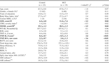
Demographic characteristics, neuropsychological test scores and sleep characteristics for the sample are presented in Table 1. There were no significant differences between MCI and healthy control subject groups for age, gender, years of education, premorbid IQ or total medical burden. While MCI patients and controls did not significantly differ on their level of depressive symptoms, a significantly greater proportion of MCI patients were taking an antidepressant medication (2 vs. 9; t=4.9; p=.03). As expected, MMSE scores were significantly lower for MCI patients. Compared with control subjects, patients with MCI showed significantly poorer executive functioning in the domain of set shifting (TMT-B), but did not have slower processing speed (TMT-A) or poorer visuospatial function (RCFT). Raw ratio scores for TMT-B over TMT-A showed that the MCI group exhibited significantly lower ratio scores than controls. There were no differences in AHI, oxygen saturation, or any sleep quality measures between the two groups.
Participant demographics, clinical characteristics, and respiratory metrics during sleep

Note. Data are represented as mean±SD or n (%). Bolded values indicate significant differences.
a Represents a χ2 statistic.
b Represents Mann-Whitney Z statistic.
c AHI only calculated in a subset of participants (Control: n=14; MCI: n=13).
MCI=mild cognitive impairment; MDE=major depressive episode; GDS=Geriatric Depression Scale; CIRS=Cumulative Illness Rating Scale; MMSE=Mini-Mental State Examination; KSS=Karolinska Sleepiness Scale; TMT=Trail-Making Test; RCFT=Rey Complex Figure Task; AHI=apnea-hypopnea index; SpO2=oxygen saturation; ODI=oxygen desaturation index.
AusEdTM driving performance between MCI and control groups
There was no difference in the number of crashes, or the level of steering and speed deviations between MCI and control participants. However, the MCI group had significantly longer braking reaction time than the control group on the driving simulator (Table 2). In either group, the only demographic characteristics that were associated with worse driving performance was age in the control group (r=0.55; p=.009) and estimated premorbid IQ in the MCI group (r=−0.60; p=.011). When the MCI group was divided into amnestic (n=8) and non-amnestic (n=11), there were no differences in driving performance, except that the non-amnestic subtype had greater steering deviation from the median lane position (t=2.27; p=.036).
Participant performance on the AusEdTM driving simulator

Note. Data are represented as mean±SD or n (%). Bolded values indicate significant differences.
a Represents a t-value.
Association between AusEdTM driving performance and neuropsychological functioning
For the MCI group only, a greater number of crashes and slower braking reaction time were associated with poorer set-shifting, as measured by the TMT-B. This was also evident for the TMT-ratio score (B/A), which suggests that the relationship is independent of processing speed and is a pure reflection of set-shifting. In addition, speed deviation on the AusEdTM simulator was associated with TMT-B in the control group. No associations were found between other cognitive tests (TMT-A and RCFT) and driving performance in either group.
Association between AusEdTM driving performance and SDB
Just before completing the AusEd TM simulator, the groups did not differ in terms of their ratings of subjective sleepiness (KSS). The level of subjective sleepiness did not correlate with any measure of driving performance in either group. The mean reaction time on the PVT in control subjects was significantly related to mean braking reaction time on the driving simulator (rho=0.64; p=.02), but this was not evident in MCI patients. However, mean PVT reaction time in the MCI group was significantly correlated with speed deviation during simulation (r=0.69; p=.03)
In the MCI group only, there were strong associations between several markers of driving performance and measures of SDB. Specifically, greater ODI was associated with greater number of crashes (r=0.66; p=.003), more steering deviation from the median lane position, (r=0.52; p=.026) and speed deviation (rho=0.51; p=.033). After controlling for age, both relationships with number of crashes and steering deviation remained significant, whereas the relationship with speed deviation became non-significant. In addition, time <90% SpO2 was associated with speed deviation in the MCI group, but did not correlate with any other measure of driving performance in either group and this association did not remain significant when controlling for age. The AHI was associated with a greater speed deviation (rho=0.60; p=.031), but did not correlate with any other metric of driving performance in this subset of the MCI group. Furthermore, the arousal index was positively correlated with a greater number of crashes on the AusEdTM simulator (rho=0.48; p=.037).
In the control group, neither ODI, AHI, nor time <90% SpO2 were associated with driving performance in any measure (all p>.05). Arousals were not related with any metric of driving performance.
To compare the nature of these relationships between MCI patients and controls, we performed Fisher r-to-z transformations on the bivariate correlation coefficients in both groups. It was found that the relationship between ODI and the number of crashes was significantly different between patients and controls (Z=−2.06; p=.020), as was relationship between the time <90% SpO2 and speed deviation (Z=−2.27; p=.012). The correlation between ODI and braking reaction time was not significantly different between the two groups (Z=−1.44; p=.069).
Combined effects of executive dysfunction and SDB on AusEdTM driving performance
To determine if the observed relationships between driving and ODI were due to poorer executive functioning, we ran partial correlations between ODI and the “number of crashes” on the driving simulator controlling for Z-scores on the TMT-B. The association remained significant and strong (partial r=0.60; p=.015). Furthermore, executive functioning did not correlate with any PSG measure of hypoxemia (ODI, AHI, time <90% SpO2, p>.050; data not shown).
As depressive symptom severity was significantly greater in the MCI group, we also re-ran the analyses controlling for scores on the GDS in addition to performance on the TMT-B. The correlation between ODI and the number of crashes remained significant (partial r=0.52; p=.047).
DISCUSSION
This study is the first to directly investigate the effects of SDB on driving ability in older adults with MCI. While the MCI patients showed slower reaction times, their overall performance on the driving simulator did not differ greatly compared with age-matched controls. However, our results show that measures of SDB (e.g., hypoxemia and sleep fragmentation) are associated with poorer driving, specifically within these cognitively impaired older adults.
Hypoxemia is one of the main consequences of SDB, and reductions in oxygen saturation are thought to be one of the potential mechanisms by which SDB negatively impacts on brain function (Engleman & Douglas, Reference Engleman and Douglas2004). However, such measures of oxygen have previously not been consistently associated with driving ability or neurobehavioral dysfunction (e.g., reduced cognitive function, excessive daytime sleepiness) in younger, less impaired cohorts (Beebe, Reference Beebe2005; Dempsey, Veasey, Morgan, & O’Donnell, Reference Dempsey, Veasey, Morgan and O’Donnell2010; Vakulin et al., Reference Vakulin, D’Rozario, Kim, Watson, Cross, Wang and Grunstein2016).
Although these results are promising, they are not definitive, nor can the effect of sleep fragmentation on driving be ruled out with this study. While several measures of hypoxemia were related to a range of driving performance measures, arousals during sleep were also related with the number of crashes. This is consistent with mixed evidence surrounding the relationship with SDB severity measures in younger and middle aged samples (Ellen et al., Reference Ellen, Marshall, Palayew, Molnar, Wilson and Man-Son-Hing2006) , and that there may be a complex relationship between sleep fragmentation and blood oxygen levels on neurobehavioral function in adults with SDB (D’Rozario et al., Reference D’Rozario, Cross, Vakulin, Bartlett, Wong, Wang and Grunstein2016).
While both MCI and control groups performed similarly on the driving simulator, the associations between measures of SDB and driving performance were only observed in patients with objectively diagnosed MCI and not cognitively intact control subjects. MCI is thought to signal early or “preclinical” stages of neurodegenerative disease and is likely to reflect alterations to underlying brain integrity (Duffy et al., Reference Duffy, Lagopoulos, Hickie, Diamond, Graeber, Lewis and Naismith2014; Mowszowski et al., Reference Mowszowski, Hermens, Diamond, Norrie, Hickie, Lewis and Naismith2012; Nickl-Jockschat et al., Reference Nickl-Jockschat, Kleiman, Schulz, Schneider, Laird, Fox and Reetz2012). Therefore, these results suggest that SDB may only be related to functional outcomes in patients with reduced brain integrity already. This in turn may be reflective of reduced cognitive resilience in MCI, leaving patients more susceptible to the negative effects of SDB such as pronounced driving performance decrements.
This is consistent with previous evidence, which has suggested that differences in brain resilience could modulate the neurobehavioral consequences of SDB (Alchanatis et al., Reference Alchanatis, Zias, Deligiorgis, Amfilochiou, Dionellis and Orphanidou2005). It is also plausible that SDB could influence the development of cognitive decline in some patients, with greater severity leading to poorer outcomes (Yaffe et al., Reference Yaffe, Laffan, Harrison, Redline, Spira, Ensrud and Stone2011). It is uncertain the extent to which the presence of SDB contributed to the cognitive deficits observed in this MCI cohort. SDB severity was not associated with neuropsychological performance metrics, and the level of SDB was not significantly different between MCI and their age-matched controls.
Executive functioning was more impaired in the MCI group, and was significantly associated with poorer driving performance, specifically a greater number of crashes and slower braking reaction speed. This is consistent with previous research examining the general older population and those with dementia (Bedard, Weaver, Darzins, & Porter, Reference Bedard, Weaver, Darzins and Porter2008; Dobbs & Shergill, Reference Dobbs and Shergill2013; Mathias & Lucas, Reference Mathias and Lucas2009).
Importantly, executive functioning is postulated to be the cognitive domain showing the greatest impairment in patients with SDB (Saunamaki & Jehkonen, Reference Saunamaki and Jehkonen2007), possibly due to the chronic effect of hypoxemia on “watershed” brain areas (Naegele et al., Reference Naegele, Thouvard, Pepin, Levy, Bonnet, Perret and Feuerstein1995; Naismith et al., Reference Naismith, Winter, Gotsopoulos, Hickie and Cistulli2004). The finding that tests of executive function are related to driving performance demonstrate that while driving requires the employment of a mixture of cognitive domains, decrements in performance are sensitive to deficits in executive functioning.
However, the level of executive function did not appear to have any direct influence on the relationship between hypoxia and the number of crashes in our MCI sample, as this relationship was still strong and significant after controlling for performance on the TMT. This suggests that, while executive functioning is significantly involved in driving ability, the hypoxic mechanisms that are part of SDB may be affecting driving through another pathway, or at least another pathway is involved. As driving is a complex behavior that requires the interaction of multiple cognitive abilities, adequate and safe driving ability may rely on the integrity of other cognitive domains in addition to or instead of executive functioning, such as certain aspects of attention (Reger et al., Reference Reger, Welsh, Watson, Cholerton, Baker and Craft2004; Silva, Laks, & Engelhardt, Reference Silva, Laks and Engelhardt2009).
It is also possible that the nature of the driving simulator itself may influence this. For example, the driving simulator used in this study was designed to emulate a long drive during night, and the limited stimulation involved in the task may require intact capacity in domains of sustained attention and vigilance. While slower reaction times on the PVT were related to braking reaction times in the control group, there was no such association in the MCI group. However, taking into account PVT performance, partial associations between oxygen saturation and driving were not significant, suggesting that sustained attention may modulate the relationship between SDB and driving. Further research should investigate which cognitive domains may be most important in the association between SDB and driving in older adults, particularly as sleepiness appeared to have little impact in these findings.
Of clinical significance, these results also show that a high number of our older sample experienced crashes (and other measures of poor driving ability) on the driving simulator task. Previously, in younger cohorts, it has been found that crashes were rare on the same simulator (Vakulin et al., Reference Vakulin, D’Rozario, Kim, Watson, Cross, Wang and Grunstein2016), yet in our sample, crashes occurred in 26% of controls and 42% of MCI patients. This may suggest that older adults are indeed at increased risk of driving accidents; however, it may alternatively indicate that crashes on the AusEdTM simulator are not indicative of real MVAs and may possibly over inflate the risk of real-life accidents. While the AusEdTM driving simulator has been validated and used in multiple previous studies of driving performance (Vakulin et al., Reference Vakulin, Baulk, Catcheside, Anderson, van den Heuvel, Banks and McEvoy2007, Reference Vakulin, Baulk, Catcheside, Antic, van den Heuvel, Dorrian and McEvoy2009, Reference Vakulin, D’Rozario, Kim, Watson, Cross, Wang and Grunstein2016), it is performed inside a laboratory without external cues and simulates a night time drive. This limits its robustness to be extrapolated to daytime driving. Regardless, these findings highlight the critical need for more research investigating the clinical predictors of driving ability (including SDB and cognitive functioning) so that effective screening and risk stratification measures can be developed.
Further investigations should account for the limitations of this study. The sample size was relatively small and whilst including only those with multiple-domain MCI, the MCI group comprised both amnestic and non-amnestic MCI subtypes. It is thus possible that driving ability may differ between subgroups and further research in a larger sample comparing MCI subtypes would be required to confirm the findings of this study. However, the MCI diagnosis in this study was thorough and our sample was matched in terms of medical and functional capacity. Also, the effects observed were all moderate to strong, and withheld after controlling for confounding variables such as age, performance on neuropsychological tests, and depressive symptoms.
There was a significant level of SDB present in this sample of older adults. However, these participants were not recruited based upon any characteristic of sleep, and it is known that the prevalence of SDB increases with age (Ancoli-Israel et al., Reference Ancoli-Israel, Kripke, Klauber, Mason, Fell and Kaplan1991), which may often go undetected with subjective screening measures (Wilson et al., Reference Wilson, Terpening, Wong, Grunstein, Norrie, Lewis and Naismith2014). Furthermore, SDB was not clinically diagnosed in this sample by a sleep-respiratory specialist, and it is possible that patients with clinically diagnosed SDB may exhibit greater functional deficits, particularly in driving. Regardless, both the MCI and control groups had comparable levels of SDB. Finally, sleep data were obtained after the driving simulator task. As such we were unable to assess the effect of sleep before the simulator, to determine whether poor sleep the prior night may be linked to performance. However, arguing against this possibility, sleepiness measures were not related to performance, and the indices of SDB that were used in this study have been shown to have minimal inter-night variability (Fietze et al., Reference Fietze, Dingli, Diefenbach, Douglas, Glos, Tallafuss and Witt2004).
CONCLUSION
This study has illustrated that SDB is related to driving performance specifically in a group of older adults with MCI, a group “at risk” of dementia. Importantly, while results suggest that measures of SDB may be an important consideration when screening for those who are no longer safe to drive, further research is now required to develop a comprehensive screening test to assess functional capacity related to driving in older populations that takes into consideration cognitive ability as well as SDB. Furthermore, investigations are now required to elucidate the cognitive domains by which SDB may impact on driving ability in these patients.
ACKNOWLEDGMENTS
Conflicts of Interest: The authors report no personal or financial conflicts of interest. Funding: This study was supported by an NHMRC Project Grant No. 632689.