Introduction
The ontogeny of fossil Lophotrochozoa in general and fossil brachiopods in particular may be reconstructed based on the relief and size of their first-formed shells, which begin to develop quite early in ontogeny. Extant representatives of the phylum Brachiopoda have different types of life cycles: Craniiformea and Rhynchonelliformea have floating lecitotrophic stages that lack a shell, while Linguliformea have floating juveniles that have a shell (Lüter Reference Lüter2001; Kuzmina et al. Reference Kuzmina, Malakhov and Temereva2019). These differences in life cycle are reflected in the properties of their juvenile shells. However, data on the correlation between the shell properties of extant juvenile brachiopods and their life cycles are insufficient. Although juvenile shells have been well studied for fossil brachiopods (e.g., Baliński Reference Baliński, Brunton, Cocks and Long2001; Balthasar Reference Balthasar2009; Ushatinskaya Reference Ushatinskaya2016), data on the juvenile shells of extant brachiopods are insufficient to support inferences on life cycles of the early Paleozoic taxa.
Extant brachiopods with calcium phosphate shells (superfamilies Linguloidea and Discinoidea) have two different life cycles (Yatsu Reference Yatsu1902; Lüter Reference Lüter2001) and differ in shell properties of the floating juvenile (Chuang Reference Chuang1977). The morphology, ornamentation, and microstructure of juvenile shells have been described only for the discinides (Williams et al. Reference Williams, Lüter and Cusack2001; Bitner et al. Reference Bitner, Logan and Gischler2008). Although the superfamily Linguloidea appeared in the early Cambrian, only two genera, Lingula Bruguière, Reference Bruguière1797 and Glottidia Dall, Reference Dall1870, have survived to the present. The microstructure and morphology of juvenile shells of lingulides remain completely unknown, because the published photographs of the juvenile lingulide shells do not provide any clear information on their structure (Paine Reference Paine1963; Chuang Reference Chuang1977; Collin et al. Reference Collin, Venera-Pontón, Driskell, Macdonald and Boyle2019) and descriptions in the literature are absent. The lingulides, however, may be regarded as a key group for understanding the evolution of brachiopod ontogeny, because they appeared in the early Cambrian and because many authors now infer that their life cycle, in which the shell forms at the planktonic stage, is ancestral for all other brachiopod life cycles (e.g., Ushatinskaya Reference Ushatinskaya2016; Zhang et al. Reference Zhang, Popov, Holmer and Zhang2018).
In this report, we provide new data on the boundary types, relief, and ornamentation of juvenile shells of the extant species Lingula anatina Lamarck, Reference Lamarck1801, from two localities in the Indo-Pacific. We then use the new data to reevaluate the various ways in which brachiopod life cycles have evolved.
Terminology
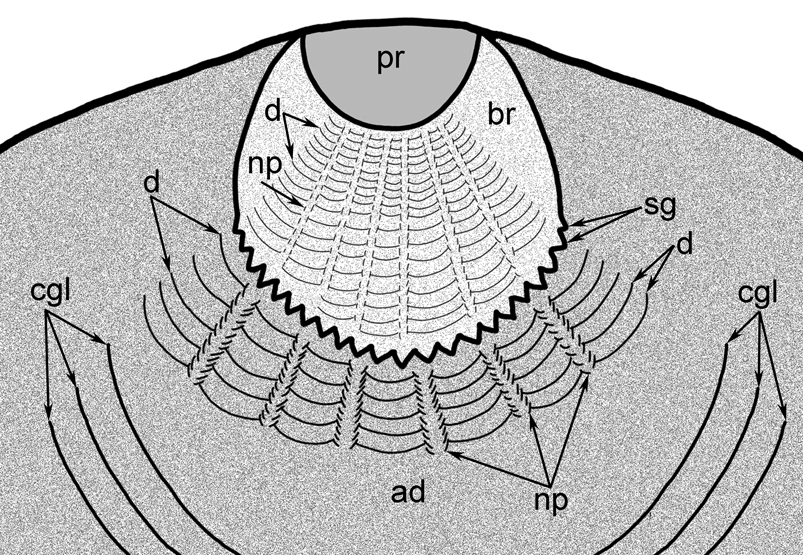
The juvenile shells of extant and fossil brachiopods are known to differ in shape, microstructure, relief, ornamentation, and size depending on developmental stage. The interpretation of their morphology and terms used for their description are still the subject of discussion. In the present paper, we use the terminology that we previously suggested for the description of growth stages of strophomenide shells (Madison and Kuzmina Reference Madison and Kuzmina2019b). We assume that the protegulum and brephic shell (which together constitute the “first-formed shell”) are homologues in all brachiopod taxa independent of the time of their formation: within the egg envelope, in the water column, or after settlement. The protegulum and brephic shell have characteristic features that differ in brachiopods with different life cycles (for details, see Madison and Kuzmina Reference Madison and Kuzmina2019b). The protegulum is secreted simultaneously by the whole mantle surface and therefore does not bear growth lines, that is, the marks of accretional growth (Beecher Reference Beecher1891; Chuang Reference Chuang1977; Zhang et al. Reference Zhang, Popov, Holmer and Zhang2018); the brephic shell bears faint growth lines; the adult shell bears well-developed growth lines and external ornamentation that is characteristic for the taxon (Fig. 1). The size of the protegulum and brephic shell and boundaries between them differ in brachiopods depending on their type of life cycle. Extant lingulides lack a true larval stage, and their juveniles hatch with an adult body plan and a thin organic protegulum (Freeman Reference Freeman1999; Lüter Reference Lüter2001). The lingulide juvenile swims for about 1 month and its brephic shell is secreted during the pelagic stage. The adult shell forms after settlement and differs superficially from the first-formed shell in having radial ornamentation. The neanic shell is in fact a transitional shell zone between the brephic and adult shell and is readily distinguished only in taxa with prominent adult ornamentation. The lingulide mantle therefore forms and grows at three developmental stages: within the egg envelope, at the pelagic stage, and after settlement. Until settlement, however, the shell remains totally organic and transparent (Paine Reference Paine1963; Collin et al. Reference Collin, Venera-Pontón, Driskell, Macdonald and Boyle2019).
Scheme of the ornamentation of the dorsal valve of juvenile Lingula anatina. Abbreviations: ad, adult shell; br, brephic shell; cgl, concentric growth line; d, drapes; np, nickpoints; pr, protegulum; sg, cog of anterior margin of brephic shell.

Here, we use the term “larva” for a stage characterized by the presence of larval structures such as ciliary bands and larval setae, and we use the term “juvenile” for a pelagic stage with the adult body plan and shell and a rudimentary lophophore.
The linguliformean brachiopods are also characterized by shell ornamentation of the drapes and nickpoints. The drapes are the anteriorly curved serial lines that are arranged in radial rows; the nickpoints are the small radial notches between the drapes. This ornamentation of the shell is thought to be generated by the muscles that control the adult setae (Williams and Holmer Reference Williams and Holmer1992; Williams Reference Williams and Williams1997).
Materials and Methods
Five specimens of L. anatina were collected in Nha-Trang Bay, South China Sea, Vietnam. All investigations were made with the permission and support of the Joint Vietnamese-Russian Tropical Research and Technological Centre (18 Hoang Quoc Viet Street, Caugiay District, Hanoi, Vietnam) and with the assistance of Academician Dmitry Pavlov, who directs the center, and Temir Britaev, who is a member of the center. Animals were collected, fixed, and then preserved in 4% formalin in filtered seawater. After fixation, the specimens were rinsed in distilled water and air-dried. Another five specimens of L. anatina were collected from Cebu Island, Philippines, and then air-dried without using any chemical agents. The 10 specimens were nearly adult shells, ranging in ventral length from 11 to 18 mm. All shells were photographed with a Leica MZ6 stereomicroscope (Leica Microsystems GmbH, Wetzlar, Germany) equipped with a digital camera and were then coated with an alloy of gold and palladium and examined with a scanning electron microscope (Hitachi S-405A and Tescan Vega II XMU SEM) before any chemical treatment and after etching with a collagenase/proteinase mixture. The valves were etched to remove the remains of periostracum that obscured the elements of the microornamentation. For collagenase/protease etching, the preparations were incubated at 37°C for 24 h in 10 ml of standard phosphate-buffered saline solution (pH = 7.4) containing collagenase type II (1 mg/ml) and 0.1 mg/ml protease activated by 10 μl of 10% CaCl2.
The material is currently stored at the Institute of Geology at Tallinn University of Technology (Estonia), collection no. GIT 587 (Philippines), and the Borissiak Paleontological Institute of Russian Academy of Sciences (Moscow), collection no. PIN 5783 (Vietnam).
Results
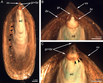
The shell of L. anatina is composed of two slightly convex valves (a dorsal valve and a ventral valve); the ventral umbone is more elongated posteriorly than the dorsal umbone. The posterior margin of the shell is conical. The muscular pedicle protrudes between the valves posteriorly. The anterior margin of both valves is broadly rounded (Fig. 2A). In fixed intact animals from Vietnam and the Philippines, three shell zones can be distinguished: the protegulum, the brephic shell (together, the protegulum and brephic shell constitute the first-formed shell), and the adult shell (Figs. 2B, 3A). These zones differ in ornamentation and relief and are separated by prominent borders. The size of the whole shell is not correlated with the size of its different zones (Table 1). All zones are similarly organized in dorsal and ventral valves, although the zone of the protegulum and brephic shell is more elongated in the ventral valve than in the dorsal valve (Fig. 2C).
Fixed complete shells of Lingula anatina. A, GIT 587/56, dorsal valve view; arrows point to some concentric growth lines. B, GIT 587/73, posterior region of the dorsal valve with protegulum and brephic shell. C, GIT 587/73, posterior region of the ventral valve, arrowheads point to several cogs of the zigzag margin of brephic shell. Abbreviations: ad, adult shell; br, brephic shell; dv, dorsal valve; pr, protegulum; pr + br, first-formed shell; vv, ventral valve.

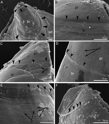
Protegula of Lingula anatina (scanning electron microscopy). The protegulum–brephic shell boundary is indicated by concave arrows; the anterior border of the brephic shell is indicated by arrows. A, GIT 587/76, unetched umbonal region of the ventral valve with protegulum and brephic shell. B, GIT 587/56, unetched dorsal protegulum. C, GIT 587/73, etched protegulum and part of brephic shell of the ventral valve. D,E, Protegulum–brephic shell boundary on the etched ventral valves (D, GIT 587/60; E, GIT 587/73). F, Distinct protegulum–brephic shell boundary on the etched ventral valve. Abbreviations: ad, adult shell; br, brephic shell; d, drapes, np, nickpoints; pr, protegulum.

Some morphological parameters of the studied shells of Lingula anatina. A plus sign (+) indicates the presence of a structure.

The Protegulum
Although all 10 of the studied shells were from nearly adult specimens, 7 had a well-pronounced protegulum (Fig. 3B–D). Unfortunately, the lateral valve margins often became internally curved during drying due to the retraction of the drying soft body, and thus the protegular width could not be precisely measured for all specimens. All measurements are given in Table 1.
On average, the protegula of the studied specimens are 200 μm wide and 50 μm long. The protegulum of the largest ventral valve, which is 18 mm long, is 210 μm wide and 90 μm long. The protegular surfaces lack any markings of accretional growth, that is, the protegulum does not bear growth lines. In the case of the unetched surface, the anterior protegular margin forms a ridge that in some cases slightly extends over the brephic shell surface (Fig. 3F). The protegulum–brephic shell boundary on the treated shell resembles a low ledge that in some cases is followed by a 2-μm-wide groove (Fig. 3E). The ridge is absent in some specimens; in those cases, the protegulum is slightly raised in relief relative to the brephic shell (Fig. 3B,D). In some specimens, the surface of the protegulum bears uneven concentric wrinkles that are similar to drapes (Fig. 3D).
The Brephic Shell
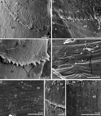
The brephic shell is a well-defined portion of the shell in all studied specimens, including treated and nontreated shells. In most cases, the brephic shell is a bit shorter in the dorsal than in the ventral valve (Table 1). The brephic shells are about 200 μm longer for the Philippine specimens than for the Vietnamese specimens. The morphology of the brephic shell is similar in all 10 specimens. In unetched specimens, the concentric lines are evident (Fig. 4A,B). In etched specimens, the margin of the brephic shell is notched (Fig. 4C). The cogs are located serially; each cog is about 10 μm wide at its base. The cogs are also evident in unetched specimens but are obscured by the primary layer (Fig. 4B). In all studied specimens, the surface of the brephic shell is covered with many thin drapes (Figs. 3E, 4D). The drapes are evident in all specimens independent of method of preparation and are grouped into radial rows, which run from the posterior boundary of the brephic shell to its anterior boundary (Figs. 3E, 4D). The drapes increase in width from the posterior to the anterior boundaries of the brephic shell: they are about 10–15 μm wide anterior of the protegulum (Fig. 3E), about 20 μm wide in the middle region (Fig. 4E), and about 30 μm wide at the anterior margin of the brephic shell (Fig. 4F). In some cases, the nickpoints are well pronounced between the rows of drapes (Fig. 4D). The ornamentation of the brephic shell therefore consists of faint growth lines and serial radial drapes with nickpoints.
Ornamentation of brephic and adult shells in Lingula anatina (scanning electron microscopy). A, PIN 5783/5, anterior margin of dorsal brephic shell. B, GIT 587/75, anterior margin of unetched ventral brephic shell. C, GIT 587/73, notched anterior margin of etched ventral brephic shell. D, GIT 587/73, drapes starting from the posterior border of etched ventral brephic shell. E, GIT 587/56, middle region of unetched ventral brephic shell. F, GIT 587/56, anterior margin of unetched ventral brephic shell. G, GIT 587/56, unetched ventral adult valve with evident drapes. Abbreviations: ad, adult shell; br, brephic shell; cg, cog of anterior margin of brephic shell; cl, concentric growth lines; d, drapes; np, nickpoints; pr, protegulum; scg, screened cogs of anterior margin of brephic shell.

The Neanic Shell
The neanic shell is not prominent due to the absence of prominent adult ornamentation; the radial multicostellate ornamentation starts almost immediately from the anterior margin of the brephic shell at the etched valves; on the unetched specimens, this zone is not readily distinguishable due to canopy-like anterior margin of the brephic shell. Because this shell region is difficult to observe in our material, we will not consider it in the present study.
The Adult Shell
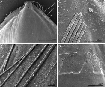
The adult shell is the largest part of the shell. This shell region differs from the brephic shell in the size of drapes, the presence of multicostellae and lateral radial plications, and more pronounced concentric growth lines (Figs. 2A, 4G). The width of drapes increases anteriorly from the anterior margin of the brephic shell up to 40 μm (Fig. 4G). In some studied specimens, the groups of setae adhered to the surface of the brephic and adult shells (Fig. 5A). These groups usually consist of 5–6 setae (Fig. 5C); each seta has a typical horsetail-like shape indicating that the seta belonged to the adult animal (Fig. 5B). After etching, these setae left well-pronounced markings on the shell surface (Fig. 5D).
Dorsal valves of Lingula anatina with adhered setae. A,B, PIN 5783/3: A, posterior valve region; B, magnified setae. C,D, GIT 587/60: C, setae on the air-dried valve surface; D, same valve after etching.

Discussion
In the current study of L. anatina from Vietnam and the Philippines, the first-formed shells of all specimens are similar in structure. The protegulum is a small region and corresponds to the shell formed inside the egg envelope. The protegulum, however, is about twice as wide as the lingulide egg that was also mentioned by Zhang et al. (Reference Zhang, Popov, Holmer and Zhang2018). Changes in the protegular sizes during first week of the pelagic stage are also confirmed by observations of Paine (Reference Paine1963) and Freeman (Reference Freeman1995) for Glottidia pyramidata (Stimpson, Reference Stimpson1860) from Florida. According to Freeman (Reference Freeman1995), the protegulum of G. pyramidata at age 2 days is about 117 μm wide and almost semicircular, and according to Paine (Reference Paine1963), the protegulum of G. pyramidata at age 6–7 days is 280 μm wide and has characteristically pointed posterolateral margins (Fig. 6). Because the protegulum lacks markings of accretional growth, we assume that during the first week of the pelagic stage, the organic protegulum stretches with the growing mantle. It is interesting that, in the current study, a treated specimen (GIT, no. 587/60) has a protegulum with drapes. The presence of drapes may be evidence that the protegulum stretches after hatching. Additional studies are needed, however, to explain how the protegulum enlarges in the absence of accretionary growth.
Size and shape of protegulum of the recent Glottidia pyramidata (Stimpson, Reference Stimpson1860). A, Second day after the hatching (after Freeman Reference Freeman1995). B, Fifth day of the pelagic stage (after Paine Reference Paine1963).

The protegulum has a rheomorphically wrinkled surface in unetched specimens and became smooth after etching due to the removal of superficial layers. It is obvious that irrespective of the type of preservation (i.e., whether the outermost layers are preserved or are removed by strong etching that possibly corresponds to the type of preservation of fossil material), the embryonic protegulum has sharp borders that are probably related to the change in the mode of life at hatching. The same is known for the fossil bivalve mollusks with well-preserved protoconchs I and II (Malchus Reference Malchus2000a,Reference Malchusb; Kowalke Reference Kowalke2006). All inferences on the relationships between the fossil embryonic protegula and lingulide-like life cycles must therefore consider the presence of such a border.
The lingulide brephic shell forms at the planktotrophic stage. According to previous reports, the brephic shell lacks drapes and nickpoints and may bear only faint growth lines (Williams and Holmer Reference Williams and Holmer1992; Zhang et al. Reference Zhang, Popov, Holmer and Zhang2018). However, the brephic shells in our material bear pronounced drapes and have notched anterior margins. The drapes were first described as ornamentation of the adult shell and were considered to be a criterion for distinguishing between the brephic and adult shells. Based on our observations, however, this feature cannot be used to distinguish between brephic and adult shells.
Researchers have inferred that drapes result from spasmodic stresses within the outer mantle lobe induced by the muscles that control the setae (Williams and Holmer Reference Williams and Holmer1992; Williams Reference Williams and Williams1997). Because lingulides have setae at the late planktotrophic stage (Yatsu Reference Yatsu1902), this explanation of drape formation requires correction. We suspect that in the planktonic juvenile, the formation of drapes may be caused by setal follicles, which probably begin to develop when the organism is in the water column; the setae themselves appear later, before settlement. As the settled organism grows, the distance between setal follicles presumably increases and the width of drapes would correspondingly also increase. In our material, the drapes are always conspicuous and the nickpoints are rare. For fossil brachiopods, drapes lacking nickpoints were described for the brephic shell of Acanthambonia portranensis Wright, Reference Wright1963 (Wright and Nõlvak Reference Wright and Nõlvak1997), which might have a discinide-like life cycle with true lecithotrophic larva transforming into the planktotrophic juvenile at the pelagic stage.
The unusual notched structure of the anterior margin of the brephic shell may be generated during shell secretion, when the shell is formed not by all cells of the mantle margin but only by groups of cells that are scattered along the mantle margin. The zones where the shell secretion is delayed may correspond to the location of the setal follicles that prevented shell secretion. In support of this hypothesis, the width of the cog bases at the brephic shell margin in the current study was equal to the width of the drapes at the posterior margin of the brephic shell. The distinct border between the brephic and adult shells indicates the change in the mode of life resulting from settlement. Here, we did not consider the neanic shell, because it corresponds to the adult stage of shell secretion and is almost absent in most of the studied lingulides.
Our observation of the imprinting of the adult setae of L. anatina on the surface of the brephic shell (Fig. 5C,D) may explain the adult setal imprints on the brephic shell of some fossil brachiopods; such imprints were possibly misinterpreted as larval setae (Smirnova et al. Reference Smirnova, Ushatinskaya, Zhegallo and Panchenko2015: plate 4, fig. 4b).
We present in the following sections four criteria that can be used to reconstruct the life cycles of fossil brachiopods. These criteria are based on comparisons of juvenile shell structures and life cycles of extant lingulides (Table 2).
Correlation between the structure of the first-formed shell and type of life cycle in recent and fossil brachiopods.

Distinct Boundary between the Protegulum and Brephic Shell
The shell regions corresponding to the protegulum and brephic shell are microstructurally similar (the discinides represent the only exception, as discussed later), and their boundary is clear in two cases, that is, when there is a change in the mode of life or in mantle secretory activity. For extant lingulides, and presumably for fossil brachiopods, a distinct protegulum–brephic shell boundary indicates the change in the mode of life after the organism leaves the egg envelope. The absence of a well-pronounced protegulum–brephic shell boundary may indicate the protegulum formed after the embryonic stage. For instance, the protegulum–brephic shell boundary is unclear in extant rhynchonelliforms (our personal observations) probably because their shell is secreted completely after settlement. The undivided first-formed shells in fossils may therefore indicate the presence in the ontogeny of true larvae hatched without a shell. In the discinides, the distinct protegulum–brephic shell boundary appears to result from the change in mantle secretory activity at the pelagic stage, when the brephic mantle secretes siliceous tablets (Williams et al. Reference Williams, Popov, Holmer and Cusack1998, Reference Williams, Lüter and Cusack2001).
Size of the First-Formed Shell
The protegulum and brephic shell generally lack clear borders (see the previous criterion), especially in fossil material, and hence the following concerns only the first-formed shell, that is, the combined protegulum and brephic shell. The size of the first-formed shell directly correlates with the size of the planktonic juvenile swimming with a shell. The shell of rhynchonelliform brachiopods forms immediately after settlement, and the protegulum is secreted simultaneously by the whole mantle surface immediately after the mantle reversal; the small brephic shell is secreted shortly after the protegulum (Stricker and Reed Reference Stricker and Reed1985a). It follows that the size of the first-formed shell formed after settlement may also indicate the size of the larval planktonic stage. We therefore infer that a large first-formed shell indicates a planktotrophic pelagic stage, regardless of whether this stage is a true larva lacking a shell or a juvenile with a shell. The first-formed region is small (approximately <250 μm in diameter) in extant brachiopods with lecitotrophic larvae that are unable to feed and grow during the pelagic stage. The first-formed shell of extant linguliforms is large (considerably larger than 250 μm in diameter) due to the feeding and subsequent growth of their planktotrophic juveniles. Freeman and Lundelius (Reference Freeman and Lundelius2005) suggested a direct correlation between the sizes of the protegulum and the egg. According to these authors, the egg size reflects the type of development, because the egg diameter of all extant linguliforms ranges from 60 to 90 μm, and the egg diameter of extant craniifrorms and rhynchonelliforms ranges from 70 to 200 μm.
Freeman and Lundelius (Reference Freeman and Lundelius2005) suggested that a small protegulum corresponds to planktotrophic development, whereas a large protegulum corresponds to lecitotrophic development, and that this correspondence could be used to distinguish these stages in the fossil material. However, Freeman and Lundelius did not consider that the lingulide protegulum stretches during the first days of the pelagic stage (see earlier discussion) and that the protegulum–brephic shell boundary is not clearly pronounced in brachiopods with true lecitotrophic larvae, especially in fossil brachiopods with calcitic shells. In addition, Freeman and Lundelius did not differentiate between planktotrophic larva and planktotrophic juvenile. For this reason, protegulum size alone is insufficient for reconstructing the life cycle of fossil brachiopods. Moreover, the sizes of fossil protegula often cannot be determined, because the protegulum and brephic shell are composed of the same microstructural elements and their boundary is difficult to distinguish.
Relief of the First-Formed Shell
Because the protegulum and brephic shell are secreted during the early stages of mantle secretory activity, their relief may reflect the location of larval setal sacks (Madison and Kuzmina Reference Madison and Kuzmina2019a). The larval setal sacks are also preserved for a few days after settlement, when the mineralized shell is already developed (Stricker and Reed Reference Stricker and Reed1985b; Nielsen Reference Nielsen1991). That the mounds on the first-formed shell correspond to the larval setal sacks therefore indicates only the presence of a true larval stage in the ontogeny and does not indicate that the shell formed in the pelagic stage, that is, this observation simply indicates that this portion of the shell was secreted before the larval setae were shed. Similar swollen regions of the first-formed shell corresponding to larval setal sacks may be recognized in extant craniids (Nielsen Reference Nielsen1991: fig. 15C). For all extant brachiopods, the shells that form during the pelagic stage are smooth and lack any relief corresponding to the parts of their soft bodies (Williams et al. Reference Williams, Popov, Holmer and Cusack1998; Bitner et al. Reference Bitner, Logan and Gischler2008); as a consequence, the presence of a large first-formed shell (see criterion 2) may be used as a criterion for distinguishing planktotrophic juvenile stages in fossil brachiopods. That the relief of the first-formed shell corresponds to the structure of the larval soft body probably indicates that the shell formed after settlement. The relief is pronounced on large first-formed shells of early Paleozoic brachiopods and probably indicates true planktotrophic larvae. The absence of such mounds on the dorsal valves of extant brachiopods with lecitotrophic larvae may be explained by the small sizes of their first-formed shells.
Here, we do not consider the neanic shell, because it is a subdivision of the adult shell and therefore has no value for inferring the life cycles of fossil brachiopods. The neanic shell is microstructurally identical to the adult shell and is therefore always well bordered from the brephic shell due to the change in the mantle secretory activity. The same may be observed in extant rhynchonelliforms (Stricker and Reed Reference Stricker and Reed1985b; personal observations).
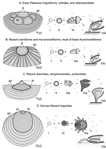
Among the life cycles that may have occurred in the evolution of brachiopods (Kuzmina et al. Reference Kuzmina, Malakhov and Temereva2019), those discussed in the following sections may be reconstructed based on available fossil material (Table 2; Fig. 7).
Structure of the first-formed shells of some extinct brachiopods with generalized schemes of their hypothetical life cycles. A, Large first-formed shell of the early Paleozoic brachiopods with assumed true planktotrophic larva in the ontogeny based on the paterinide dorsal valve (Ushatinskaya Reference Ushatinskaya1997); central mound possibly reflecting the position of the protegulum and anterolateral mounds reflecting the position of the larval setal sacks. B, First-formed shell of the brachiopods with true lecitotrophic larva based on the dorsal valve of the Ordovician orthides (Madison Reference Madison2009). C, First-formed shell of strophomenides and productides with assumed lecitotrophic larva transforming into the planktotrophic juvenile at the pelagic stage based on the strophomenide ventral valve (Madison and Kuzmina Reference Madison and Kuzmina2019b). D, First-formed shell of lingulides with protegulum formed inside the egg envelopes based on the dorsal valve of the Late Devonian “Lingula” (Baliński Reference Baliński, Brunton, Cocks and Long2001). The protegulum–brephic shell boundaries are indistinct in A–C. Abbreviations: ad, adult shell; br, brephic shell; ff, first-formed shell; pr, protegulum; ss, mounds of larval setal sacks; I, egg; II, blastula; III, true planktotrophic larva; IIIa, true lecitotrophic larva; IV, early planktotrophic juvenile; IVa, late planktotrophic juvenile; IVb, settled juvenile.

Life Cycle with True Planktotrophic Larvae That Form a Shell after Settlement
This type of ontogeny (Fig. 7A) possibly characterizes most of the early Paleozoic brachiopods (linguliforms, clitambonitids, and orthides) (Madison and Kuzmina Reference Madison and Kuzmina2019a). Because no sharp protegulum border has been reported for Cambrian brachiopods (e.g., Williams et al. Reference Williams, Popov, Holmer and Cusack1998; Skovsted et al. Reference Skovsted, Ushatinskaya, Holmer, Popov and Kouchinsky2015; Ushatinskaya Reference Ushatinskaya2016), the recent lingulide-like life cycle was evidently not present in the Cambrian. The early Paleozoic brachiopods are generally characterized by a large first-formed shell bearing one posteromedian mound and one to three pairs of anterolateral mounds that lack any sharp borders between the protegulum and brephic shell; in addition, the protegulum and brephic shell are composed of the same microstructural elements (e.g., Williams et al. Reference Williams, Popov, Holmer and Cusack1998; Madison Reference Madison2004; Balthasar Reference Balthasar2009; Zhang et al. Reference Zhang, Popov, Holmer and Zhang2018). Because the pelagic stage of the early Paleozoic brachiopods was possibly represented by true planktotrophic larvae and because the anterolateral mounds probably reflect the position of larval setal sacks, this shell region may be a first-formed region comprising both the protegulum and brephic shell. As such, it would be the largest of all known first-formed shells of brachiopods with true larvae. The large size of ancient first-formed shells may be explained by the competent (i.e., ready for settlement) planktotrophic larvae, which greatly increased in size during the pelagic stage. No extant brachiopods have planktotrophic larvae in their life cycles, and no large first-formed shells have been detected in extant forms. This life cycle is thought to be the plesiomorphic life cycle in brachiopod evolution (Kuzmina et al. Reference Kuzmina, Malakhov and Temereva2019).
Life Cycle with True Lecitotrophic Larvae
This life cycle (Fig. 7B) may be reconstructed based on the small first-formed shell, which may bear poorly pronounced relief corresponding to the larval setal sacks and indistinct boundaries between the protegulum and brephic shell. These features characterize extant and most fossil craniiforms and rhynchonellates (Freeman and Lundelius Reference Freeman and Lundelius2005).
Life Cycle with Lecitotrophic Larvae and Planktotrophic Juveniles That Settled after the Formation of the Protegulum
Although presently unknown for both modern and extinct brachiopods, this scenario seems to be possible and may be detected by future research (Fig. 8).
Hypothetical shell of the first-formed shell of the brachiopod with lecitotrophic larvae and planktotrophic juveniles that settled after the formation of the protegulum. Abbreviations: ad, adult shell; bba, border between brephic shell and adult shell; bpb, border between protegulum and brephic shell; br, brephic shell; cgl, concentric growth line; pr, protegulum.

Life Cycle with Lecitotrophic Larvae and Planktotrophic Juveniles That Settled after the Formation of the Brephic Shell
This life cycle (Fig. 7C) is characteristic of extant discinides and possibly of Paleozoic strophomenates (Madison and Kuzmina Reference Madison and Kuzmina2019b). Both groups have large brephic shells. Because there is no change in the mode of life, there is no distinct boundary between the protegulum and brephic shell, and that is the case for the orders Strophomenida and Productida (Madison and Kuzmina Reference Madison and Kuzmina2019b). Extant discinides, however, secrete siliceous tablets at the pelagic stage, and the prominent ledge-shaped boundary between the protegulum and brephic shell probably reflects the change in the mantle secretory mode (Williams et al. Reference Williams, Mackay and Cusack1992, Reference Williams, Lüter and Cusack2001).
Life Cycle Lacking Lecitotrophic Larvae and Including Only Planktotrophic Juveniles
This is the most advanced brachiopod life cycle, because its larval stage is completely embryonic and develops inside the egg envelope (Fig. 7D). It is known only for lingulides, both extinct and extant, whose embryonic protegulum–brephic shell boundary is sharp and narrow and whose brephic shell is large, bears faint growth lines, and lacks adult ornamentation. The protegula of Ordovician–Devonian lingulides (Baliński Reference Baliński, Brunton, Cocks and Long2001; Tapanila and Holmer Reference Tapanila and Holmer2006) fit this criterion, but their ornamentation still requires explanation. For brachiopods with this life cycle, the outer boundary of the embryonic protegulum forms a semicircle whose regularity may be explained by uniform growth of the mantle margins.
Conclusion
The life cycles of extinct and extant brachiopods of all three subphyla are highly diverse. The early Cambrian brachiopods had true planktotrophic larvae in their life cycles. This type of ontogeny is the most primitive brachiopod life cycle and resembles the life cycles of other Bilateria (Malakhov et al. Reference Malakhov, Bogomolova, Kuzmina and Temereva2019). Extant lingulides possess the most advanced life cycle; they have planktotrophic juveniles and do not have larvae.
Acknowledgments
The authors are very grateful to E. Mekhova (Moscow), who collected the animals in Vietnam. U. Toom from the Institute of Geology at Tallinn University of Technology is acknowledged for providing the shells of the Philippine Lingula anatina for study. The authors are also grateful to R. Rakitov (Paleontological Institute of RAS, Moscow, Russia) for assistance with the scanning electron microscope. This study was supported by funds from the Russian Science Foundation (no. 18-04-00082: the description of morphology and ornamentation of the shell) and the Russian Foundation for Basic Researches (no. 19-04-00449: material treatment and processing of the paper).