Introduction
Childhood trauma is a general term that encompasses unwanted physical or psychological harm (Morgan & Fisher, Reference Morgan and Fisher2006), and has been associated with susceptibility to psychopathology later in life, putatively by its effect on neural architecture (Heim & Nemeroff, Reference Heim and Nemeroff2001; Kessler et al., Reference Kessler, McLaughlin, Green, Gruber, Sampson, Zaslavsky and Williams2010). The sequelae of traumatic experiences may differ based on the type of trauma (Williams, Debattista, Duchemin, Schatzberg, & Nemeroff, Reference Williams, Debattista, Duchemin, Schatzberg and Nemeroff2016) and the period of individual's developmental stage at which the event occurred (Andersen et al., Reference Andersen, Tomada, Vincow, Valente, Polcari and Teicher2008; Gerritsen et al., Reference Gerritsen, Kalpouzos, Westman, Simmons, Wahlund, Bäckman and Wang2015; Lupien, McEwen, Gunnar, & Heim, Reference Lupien, McEwen, Gunnar and Heim2009). Physical abuse, for example, may increase risk of several illnesses, including schizophrenia, anxiety disorders, major depressive disorder and bipolar disorder [for review see (Carr, Martins, Stingel, Lemgruber, & Juruena, Reference Carr, Martins, Stingel, Lemgruber and Juruena2013)]. Physical abuse may also influence responsiveness to treatment in adulthood (Williams et al., Reference Williams, Debattista, Duchemin, Schatzberg and Nemeroff2016).
The effects of trauma or early life stress on the developing brain may alter the structure of certain brain regions in ways that are still detectable in adulthood (Davidson & McEwen, Reference Davidson and McEwen2012). Magnetic resonance imaging (MRI) studies have found that childhood trauma alters structures in limbic brain regions that are sensitive to the cumulative neurotoxic effects of stress hormones (Lupien, Juster, Raymond, & Marin, Reference Lupien, Juster, Raymond and Marin2018), including the hippocampus and amygdala (Davidson & McEwen, Reference Davidson and McEwen2012; Paquola, Bennett, & Lagopoulos, Reference Paquola, Bennett and Lagopoulos2016). Reduced grey matter volumes in the hippocampus are consistently reported in adults with a history of childhood trauma [for meta-analyses (Calem, Bromis, McGuire, Morgan, & Kempton, Reference Calem, Bromis, McGuire, Morgan and Kempton2017; Paquola et al., Reference Paquola, Bennett and Lagopoulos2016)].
Whether early life experiences of trauma affect the amygdala, is less clear, however (Paquola et al., Reference Paquola, Bennett and Lagopoulos2016). Some studies of asymptomatic participants report no relations between amygdala volumes and trauma history (Gerritsen et al., Reference Gerritsen, Kalpouzos, Westman, Simmons, Wahlund, Bäckman and Wang2015; Gorka, Hanson, Radtke, & Hariri, Reference Gorka, Hanson, Radtke and Hariri2014), while other studies show increased amygdala volumes in non-help-seeking individuals who reported early exposure to emotional and/or physical neglect (see review by Teicher and Samson, Reference Teicher and Samson2016). LoPilato and colleagues recently analyzed whole amygdala volumes of participants from the North American Prodrome Longitudinal Study (NAPLS), reporting no significant associations between childhood maltreatment and amygdala volumes in youth at clinical high risk for psychosis (LoPilato et al., Reference LoPilato, Goines, Addington, Bearden, Cadenhead, Cannon and Walker2019). Similarly, an earlier large-sample, longitudinal study of young participants found a nonsignificant increase in left amygdala volume associated with childhood maltreatment (Whittle et al., Reference Whittle, Dennison, Vijayakumar, Simmons, Yücel, Lubman and Allen2013). In this study, longitudinal mediation modelling revealed that although childhood maltreatment is associated with large amygdala size, the development of subsequent psychopathology is associated with small amygdala volumes (Whittle et al., Reference Whittle, Dennison, Vijayakumar, Simmons, Yücel, Lubman and Allen2013). Supporting the notion that the presence of full threshold psychopathology is associated with small amygdala volumes, there are studies of symptomatic participants with major depressive disorder (Malykhin, Carter, Hegadoren, Seres, & Coupland, Reference Malykhin, Carter, Hegadoren, Seres and Coupland2012), bipolar disorder (Hibar et al., Reference Hibar, Westlye, Van Erp, Rasmussen, Leonardo, Faskowitz and Andreassen2016) and psychotic-like disorders (Aas et al., Reference Aas, Navari, Gibbs, Mondelli, Fisher, Morgan and Dazzan2012; Hoy et al., Reference Hoy, Barrett, Shannon, Campbell, Watson, Rushe and Mulholland2012), which report smaller amygdala sizes in these groups. The left and right amygdala may have different sensitivity to trauma during certain developmental periods (Teicher & Parigger, Reference Teicher and Parigger2015), adding to the complexity of the relations between trauma, psychopathology and amygdala volumes. It appears, therefore, that there are complex relations between the timing of traumatic experiences, the subsequent evolution of psychopathology and the structure of the amygdala.
Another reason for the uncertainty regarding the impact of trauma on the amygdala is that most studies have examined the amygdala as a whole structure, while from an anatomical perspective the amygdala is comprised of at least 13 nuclei (Sah, Faber, Lopez De ArmentiaA, & Power, Reference Sah, Faber, Lopez De ArmentiaA and Power2003) with unique inputs and outputs that form physiologically distinct divisions. The major divisions within this construct include the basolateral amygdala (BLA), the central nucleus (Ce), the cortical nucleus (Co) and the medial nucleus (Me) (Sah et al., Reference Sah, Faber, Lopez De ArmentiaA and Power2003) with the latter three nuclei forming the evolutionarily primitive cortico-medial division (CeM) that is involved in controlling emotional reactions. In contrast, the BLA is an evolutionarily newer division associated with emotional actions and incorporates the lateral (La), basal (Ba), accessory basal (AB) and paralaminar (PL) nuclei (Sah et al., Reference Sah, Faber, Lopez De ArmentiaA and Power2003). Preclinical studies consistently report that the BLA is a storage site for fear memories and is involved in the regulation of fear extinction (Janak & Tye, Reference Janak and Tye2015; Sah et al., Reference Sah, Faber, Lopez De ArmentiaA and Power2003), suggesting that the BLA may be heavily involved in modulation of anxiety-like behaviours associated with past stressful events (Tripathi, Chakraborty, Srikumar, Raju, & Shankaranarayana Rao, Reference Tripathi, Chakraborty, Srikumar, Raju and Shankaranarayana Rao2019).
With the recent introduction of brain image segmentation algorithms that employ probabilistic atlases, it is now possible to look at the human amygdala structure partitioned into specific subregions using MRI T1 images (Saygin et al., Reference Saygin, Kliemann, Iglesias, van der Kouwe, Boyd and Reuter2017). Oshri and colleagues recently examined the subregion-specific presentation of the amygdala in youth at risk for general psychopathology and reported that higher scores on the Adverse Childhood Experience Scale (ACE) were associated with smaller amygdala volumes in key subregions of the amygdala, BLA and Centro-Medial segments (Oshri et al., Reference Oshri, Gray, Owens, Liu, Duprey, Sweet and MacKillop2019). While this report shows a selective association between ACE severity and reduced volumes in specific subregions of the amygdala (Oshri et al., Reference Oshri, Gray, Owens, Liu, Duprey, Sweet and MacKillop2019), it is unclear whether different types of traumatic experiences can instigate differential effects on the structure of the amygdala. This is important because trauma-associated brain changes can be dependent on the type of the experience, with sexual and physical abuse exerting the most pronounced negative effects on the limbic brain (Teicher, Samson, Anderson, & Ohashi, Reference Teicher, Samson, Anderson and Ohashi2016). In a study with a sample of depressed patients, Grant and colleagues showed that aberrant amygdala hyperactivation in response to negative stimuli was more strongly associated with reported physical abuse than other forms of abuse or neglect (Grant, Cannistraci, Hollon, Gore, & Shelton, Reference Grant, Cannistraci, Hollon, Gore and Shelton2011). Extant clinical studies also support the notion that physical and sexual abuse is associated with a wider spectrum of later psychopathology in life [for systematic review see (Carr et al., Reference Carr, Martins, Stingel, Lemgruber and Juruena2013)].
The primary aim of this work was, therefore, to examine the impact of childhood trauma on the subregion-specific structure of the amygdala in young individuals at risk for mental illness. We hypothesized that youth who experienced physical or sexual abuse would have structural changes within the BLA, due to its important role in fear memory regulation (Tripathi et al., Reference Tripathi, Chakraborty, Srikumar, Raju and Shankaranarayana Rao2019; Yang et al., Reference Yang, Mozhui, Karlsson, Cameron, Williams and Holmes2008). A secondary aim was to understand whether trauma-related changes in amygdala volumes mediate symptoms of anxiety and depression, hypothesizing that trauma-associated amygdala alterations would predict severity of depression and anxiety symptoms in youth who did not meet criteria for a mood or anxiety disorder diagnosis. To test these hypotheses, we applied a novel subfield-specific amygdala characterization (Saygin et al., Reference Saygin, Kliemann, Iglesias, van der Kouwe, Boyd and Reuter2017) to MRI scans obtained from participants in Canadian Psychiatric Risk and Outcome (PROCAN) study (Addington et al., Reference Addington, Goldstein, Wang, Kennedy, Bray, Lebel and MacQueen2018). The PROCAN study was designed to identify risk factors for youth at risk for SMI and the baseline characteristics of the sample have been previously described (27–29).
Methods
Participants
Participants were recruited from two university-based clinical sites in Canada through public advertisement and mental-health services (Sunnybrook Health Sciences Centre in Toronto and the University of Calgary) (Addington et al., Reference Addington, Goldstein, Wang, Kennedy, Bray, Lebel and MacQueen2018). This report included 182 PROCAN youth aged 12–25 years (mean = 18.1; s.d. = 3.6) who completed clinical and MRI assessments and whose MRI scans passed quality inspection. Young individuals within the PROCAN sample were stratified into risk-stages (Addington et al., Reference Addington, Liu, Goldstein, Wang, Kennedy, Bray and MacQueen2019) following transdiagnostic staging model for youth at risk for SMI [proposed by (McGorry, Hickie, Yung, Pantelis, & Jackson, Reference McGorry, Hickie, Yung, Pantelis and Jackson2006)]. The present sample included low-risk healthy controls (HC, n = 37) and participants that encompassed a range of risk levels for SMI; (a) asymptomatic young individuals who have a first-degree relative with a SMI or a history of obstetric complications (Stage 0, n = 32); (b) help-seeking youth who are experiencing distress and may have unspecific symptoms of anxiety and depression (Stage 1a, n = 41); (c) youth who are already experiencing attenuated symptoms of SMI [Stage 1b (n = 72)]. Participants had no current or lifetime Axis I psychotic or bipolar disorder at the time of scanning, determined using The Structured Clinical Interview for DSM – 5 [SCID – 5 (Kessler et al., Reference Kessler, Andrews, Colpe, Hiripi, Mroczek, Normand and Zaslavsky2002)].
As previously described (Addington et al., Reference Addington, Goldstein, Wang, Kennedy, Bray, Lebel and MacQueen2018), participants were recruited according to the risk stage stratification, but not based on past history of childhood traumatic experiences (Stowkowy et al., Reference Stowkowy, Goldstein, MacQueen, Wang, Kennedy, Bray and Addington2019). All participants signed informed consent (for minors, signed by parents/guardians).
Clinical measures
Presence and severity of attenuated psychotic symptoms, depression and anxiety were evaluated via clinical interviews and self-administered questionnaires. The Structured Interview for Psychosis-Risk Syndromes (SIPS) was used to evaluate whether participants met risk criteria for psychosis (McGlashan, Walsh, & Woods, Reference McGlashan, Walsh and Woods2010). Depressive symptoms were assessed with the Quick Inventory of Depressive Symptomatology (QIDS SR-16) (Rush et al., Reference Rush, Trivedi, Ibrahim, Carmody, Arnow, Klein and Keller2003) and the Beck Depression Scale (Beck, Ward, Mendelson, Mock, & Erbaugh, Reference Beck, Ward, Mendelson, Mock and Erbaugh1961). Anxiety symptoms were scored with the Generalized Anxiety Disorder Scale-7 (GAD-7) (Dear et al., Reference Dear, Titov, Sunderland, McMillan, Anderson, Lorian and Robinson2011) and the Social Interaction Anxiety Scale (SIAS) (Olivares, García-López, & Hidalgo, Reference Olivares, García-López and Hidalgo2001).
History of childhood trauma
Participants completed an adapted version of the Childhood Trauma and Abuse Scale (Janssen et al., Reference Janssen, Krabbendam, Bak, Hanssen, Vollebergh, de Graaf and van Os2004). The version used in this study included ‘yes’ or ‘no’ question about the experience of emotional neglect, or psychological, physical or sexual abuse prior to the age of 18 years; in our adapted version there were also additional questions about the experience of psychological or physical bullying. A total trauma score represented participant's scores ranging from 0 to 6 based on the additive occurrence of all six types of traumatic experiences that occurred before 18 years of age. If the history of trauma was present, participants were asked at which age the traumatic event had occurred (years: 0–6, 7–12, 13–17) and whether the traumatic event continued for more or less than a year. To account for possible cumulative effects of childhood abuse that may have occurred at each stage of development we calculated total abuse score. This score represented a sum of sexual and/or physical abuse that may have occurred at any of the three developmental time windows (score range 0–6).
Imaging
MRI Data were acquired on a GE 3.0 Tesla Discovery scanner (in Calgary) and a Phillips 3.0 Tesla Achieva scanner (in Toronto), equipped with eight channel head coils. At each study site, there were predetermined imaging protocols for GE 3.0 Tesla Discovery/Phillips 3.0 Tesla Achieva scanner. T1-weighted images were collected with the following scanner parameters: turbo gradient echo sequence 3:33/9:53 min at 1 mm3 resolution; repetition time = 6.4/6.5 (ms); echo time = 2.8/3 (ms); flip angle = 15 /8; inversion time = 450/950 (ms); field of view = 256 mm; matrix = 240 × 240 [for further details see (Macqueen et al., Reference Macqueen, Hassel, Arnott, Addington, Bowie, Bray and Kennedy2019)].
All morphometric measures were obtained via an automated segmentation algorithm that is freely available as a part of the FreeSurfer 6.0 (FS6.0) software package (http://surfer.nmr.mgh.harvard.edu/). Global brain measurements were extracted from the recon-all workflow [e.g. total brain volume (TBV), total grey volume (TGV), 3-rd Ventricle etc.]. Images were processed through an amygdala subfield-specific segmentation module that employs Bayesian probabilistic brain atlases (Saygin et al., Reference Saygin, Kliemann, Iglesias, van der Kouwe, Boyd and Reuter2017). We obtained 10 volumetric measurements of the amygdala for each hemisphere, with the following regions: lateral nucleus (La), basal nucleus (Ba), accessory basal nucleus (AB), anterior amygdaloid area (AAA), central nucleus (Ce), medial nucleus (Me), cortical nucleus (Co), cortico-amygdaloid-transition-area (CAT) and paralaminar nucleus (PL). Volumes of the whole amygdala were calculated as a sum of all available subregions. For further analyses, we computed BLA (Ba + La + AB + PL) and CeM (Ce + Me) volumes; Co was evaluated separately.
All output was manually inspected. Out of 188 MRI images available, the present analysis included 182 images that passed quality assurance and segmentation quality control procedures (for details of quality control procedures see Supplementary Information).
Statistical analysis
SPSS version 25.0 (IBM SPSS Statistics for iMac, Armonk, NY, USA) was used for all statistical analyses. To address our first aim, to examine possible effects of childhood trauma(s) on amygdala volumes, we used automatic linear modelling (ALM). ALM is a regression procedure with two principal advantages over traditional regression analyses – ALM allows for automatic data preparation (e.g. transform variables; trim outliers; remove missing variables) and performs automatic subset selection to construct variable importance measures for a chosen target predictor (Yang, Reference Yang2013). Variable importance measures were used to identify which of the subregions of the amygdala were the most meaningful in the association with childhood trauma and which types of trauma show a stronger relationship with predetermined subregions.
To confirm the significance and strength of the identified relationships between specific subregions of the amygdala and selected types of trauma, the resultant subset of predictors selected with ALM were then subjected to the curve estimation procedure allowing for statistical diagnosis of possible linear or non-linear relations between the target variable and the predicted variable. Additionally, the primary correlates of the amygdala volumes were evaluated using hierarchical stepwise multiple linear regressions.
To address our second aim, that is to examine possible direct/indirect effects of amygdala volumes between childhood trauma and clinical presentation, the PROCESS version 3.3 macro (Hayes, Reference Hayes2015) for SPSS was used. To improve the numerical stability of linear models we used Principal Component Analysis (PCA) as a data reduction method for multivariate analyses (Abdi & Williams, Reference Abdi and Williams2010). Descriptive statistics employed a general linear model (GLM), analyses of covariance (ANCOVA) and Pearson correlations (r) by variable characteristics. All analyses were two-tailed and significance was set at the 0.05 alpha level.
Results
Clinical And demographic characteristics of the sample
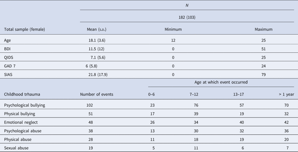
Table 1 summarizes clinical and demographic information of the whole sample. One hundred and eighty-two (103 female) participants with a mean age of 18.3 years had MRI T1-weighted brain scans that were considered evaluable.
Demographic characteristics, measures and reported childhood traumas of PROCAN study participants

BDI, Beck Depression Inventory; QIDS, Quick Inventory of Depressive Symptomatology; SIAS, Social Interaction Anxiety Scale; GAD7, Generalized Anxiety Disorder 7-item' scale.
Approximately 56% of the participants reported an occurrence of psychological bullying and 28% reported physical bullying, highlighting bullying as the predominant type of trauma in our sample. Emotional neglect and psychological abuse were reported by 26% and 20% of the participants, respectively. A history of physical abuse was present in 15% of the participants, out of which the majority (70%) indicated that physical abuse lasted in duration for more than a year. Sexual abuse was the least common type of trauma reported among all participants (10%) and the least frequent in duration as only 7 out of 20 participants indicated that history of sex abuse lasted for over a year (See Table 1).
Identifying covariate terms
To identify covariate terms for whole amygdala volumes, we used hierarchical stepwise linear regression modelling. The model comprised of the independent variables: age, sex, left/right inferior lateral ventricle, 3rd and 4th ventricle and TBV; and the dependent variable of interest – whole amygdala volumes. For left whole amygdala, nearly 60% of the variance was explained by TBV, left-Inf-lat-ventricle, age and sex (R 2 = 0.57, F = 58.5, df = 4181, p = 0.000). Covariate terms for right whole amygdala were TBV, right-inf-vent and age (R 2 = 0.59, F = 88.6, df = 3181, p = 0.000).
Effects of childhood trauma on the amygdala
A preliminary analysis of the data did not show significant associations between whole amygdala volumes and total trauma or total abuse scores. Next, we aimed to identify whether childhood trauma was associated with any structural differences in specific subregions of the amygdala. For the probative analysis, the target variable was total trauma score and 20 volumetric parameters of the amygdala (with additional age, sex and TBV) were loaded into the ALM. Following ALM, predictor importance chart identified right Ba (r = −0.217, p = 0.004), right AB (r = −0.219, p = 0.004), left PL (r = −0.21, p = 0.005) and right PL (r = −0.19, p = 0.009) and left Ba (r = −0.2, p = 0.007) as the first five important terms explaining total trauma scores (ensemble mean accuracy 17%). Next, using the curve estimation procedure we regressed total trauma scores with subfields of the amygdala selected by ALM procedure. When the resultant predicted variables were regressed, both linear [R 2 = 0.17, F(1177) = 36, p = 0.000] and non-linear, cubic functions [R 2 = 0.18, F(3175) = 12.8, p = 0.000] confirmed the significance of the ALM predictors.
When we controlled for TBV, sex and age, GLM further detailed that nearly 50% of the variance in total trauma scores were explained by components of the BLA complex; that is, participants with greater trauma scores were more likely to have small left Ba [R 2 = 0.46, F(9178) = 2.7, p = 0.014] and right Ba [R 2 = 0.48, F(9178) = 2.7, p = 0.013] and left aBa [R 2 = 0.48, F(9178) = 2.3, p = 0.034] right aBA [R 2 = 0.51, F(9178) = 3.6, p = 0.002].
Next, we focused on which of the types of traumatic experience appeared to influence the volume of the amygdala. An ALM procedure with the whole amygdala volume as a target variable and occurrence of psychological, physical bullying, emotional neglect, psychological, physical and sexual abuse were used as inputs (including age, sex, TBV and inf-lat-vent volumes). ALM showed that for whole amygdala volumes, the occurrence of sexual abuse (r = −0.21, p = 0.004) and physical abuse (r = −0.15, p = 0.044) were more important features than the occurrence of psychological (r = −0.11, p = 0.13) or physical (r = −0.08, p = 0.24) bullying, emotional neglect (r = −0.09, p = 0.22) or psychological abuse (r = −0.09, p = 0.2); overall accuracy 57.2%. Therefore, further analyses were focused only on investigating the effects of sexual and physical abuse on the structural presentation of the amygdala volumes.
Sexual abuse:
On the subregion-specific level, right Co [R 2 = 0.46, F(3, 179) = 5.6, p = 0.035] and right Ba [R 2 = 0.25, F(3, 179) = 4.4, p = 0.037] right aBa [R 2 = 0.52, F(4, 179) = 4.8, p = 0.029] were more likely to be smaller in those individuals who reported a history of sexual abuse as compared to those who did not. Notably, those who reported sexual abuse that lasted for over a year were more likely to exhibit smaller left La [R 2 = 0.28, F(5, 18) = 8, p = 0.014] and right La [R 2 = 0.17, F(5, 18) = 4.8, p = 0.046] compared to those who reported sexual abuse for less than a year.
Physical abuse:
There was a significant reduction of the BLA complex in both left and right hemispheres (R2 = 0.44, F = 4.8, df = 3179, p = 0.028)/(R 2 = 0.45, F = 4.7, df = 3179, p = 0.03).
We also conducted additional analysis to test whether the identified subregions of amygdala are significantly different depending on the developmental period at which sexual or physical abuse had occurred (For details see Section 1.1. of Supplementary Results and online Supplementary Figure 1.).
Cumulative effects of sexual and physical abuse on the BLA complex:
Linear regression analysis confirmed a strong and significant association between right and left subregions of BLA and total abuse score, where participants with higher total abuse scores were more likely to have smaller BLA volumes (R 2 = 0.80, F = 155.7, df = 1,33, p = 0.000).
Associations among childhood trauma, amygdala volume, and clinical presentation:
To determine whether amygdala volumes mediate the association between childhood trauma and the severity of clinical symptoms, a bootstrap estimation approach was used (Hayes, Reference Hayes2015; Mackinnon, Warsi, & Dwyer, Reference Mackinnon, Warsi and Dwyer1995). For the present analysis, 95% confidence intervals of the indirect effects were obtained with 5000 bootstrap samples. As the occurrence of sexual and physical abuse were associated with volume decrease in the right Ba/AB and right Co, only these regions of the amygdala were considered as potential mediators. Prior to the mediation analysis, we extracted a single principal component as a regression factor score (Kuhn & Johnson, Reference Kuhn and Johnson2013) derived from right Ba, right AB and right Co (sum of squared loadings 91.5%, direct oblimin rotation). The resultant principal component was tested as a mediating variable (M).
Sexual abuse:
There was a significant direct effect of sexual abuse on QIDS scores [B = 3.7, s.e. = 1.5, t(1164) = 2.4, p = 0.014, CI = 0.7–6.7] and a significant indirect effect mediated by the principal component of volumes of the right subregions of the amygdala (B = 0.9, s.e. = 0.35, CI = 0.31–1.6); total effect [B = 4.6, s.e. = 1.4, t(1164) = 3.2, p = 0.0016, CI = 1.7–7.5]. More pronounced indirect (B = 2.4, s.e. = 0.84, CI = 0.94–4.2) and direct effects were detected in the relation between BDI and sexual trauma [B = 7.5, s.e. = 3.2, t(1176) = 2.3, p = 0.022, CI = 1.08–14]; total effect [B = 9.9, s.e. = 3.1, t(1176) = 3.2, p = 0.0016, CI = 3.8–16.1].
There was no direct effect between sexual trauma and GAD-7 total scores [B = 2.6, s.e. = 1.6, t(1176) = 1.6, p = 0.098, CI = −0.5 to 5.8], but a significant indirect effect was detected (B = .8, s.e. = 0.34, CI = 0.16–1.51]; total effect [B = 3.4, s.e. = 1.5, t(1176) = 2.2, p = 0.027 CI = 0.38–6.5]. Similarly, no direct effects were apparent between sexual trauma and SIAS scores [B = 7.8, s.e. = 5.3, t(1177) = 1.4, p = 0.13, CI = −2.5 to 18.3], but there was a significant indirect effect [B = 2.6, s.e. = 1.5, CI = 0.63–5.2]; total effect [B = 10.5, s.e. = 5, t(1177) = 2.1, p = 0.036, CI = 0.66–20.5]. Across all four clinical scores, the sum of total effects of sexual abuse across was 28.4; and 6.7 of the totals was accounted by indirect effects.
Physical abuse:
Physical abuse had direct [B = 6.9, s.e. = 1, t(1164) = 6.6, p = 0.000, CI = 4.9–9] and indirect effects on QIDS scores (B = .5, s.e. = 0.24, CI = 0.09–1); total effect [B = 7.4, s.e. = 1, t(1164) = 7, p < 0.001, CI = 5.4–9.5]. Similar direct [B = 12.6, s.e. = 2, t(1176) = 5.3, p < 0.001, CI = 8–17.2] and indirect effects (B = 1.3, s.e. = 0.57, CI = 0.39–2.6) were detected between physical trauma and BDI scores; total effect [B = 13.9, s.e. = 2.3, t(1164) = 5.9, p < 0.001, CI = 9.3–18.5]. Physical abuse had weaker but significant direct effects on GAD7 scores [B = 6.7, s.e. = 1.1, t(1163) = 5.7, p < 0.001, CI = 4.3–8.9], but there was no significant mediation effect detected (B = .3, s.e. = 0.23, CI = −0.02 to 0.9); total effect [B = 7, s.e. = 1.1, t(1177) = 2.1, p < 0.001, CI = 4.8–9.2]. There was a significant direct effect between physical abuse and SIAS scores [B = 8.3, s.e. = 3.6, t(1177) = 2.2, p = 0.023, CI = 1.1–15.6], and a significant indirect effect (B = 1.7, s.e. = 0.82, CI = 0.4–3.5); total effect [B = 10.1, s.e. = 3.6, t(1177) = 2.7, p = 0.006, CI = 2.8–17.4].
Discussion
We examined associations between types of childhood trauma and subregion-specific characteristics of the amygdala in youth at various stages of risk for SMI. Participants with higher total trauma scores were more likely to have smaller amygdala volumes. Specifically, the cumulative effects of childhood trauma had strong linear relations with reduced volumes in the basal and accessory basal nuclei. This relationship was detected exclusively at the subregion-specific level because we did not find significant associations between trauma exposure and whole amygdala volumes.
Our total trauma score incorporated a range of experiences such as psychological or physical bullying, emotional neglect and psychological, physical or sexual abuse. Sexual and physical abuse were reported less frequently but appeared to have the most deleterious effects on the amygdala subregional volumes. Smaller BLA volumes were apparent in adolescents who reported a history of sexual and/or physical abuse. However, participants with a prolonged history of sexual abuse (that is, duration of over 1 year), demonstrated changes not only in the basal region but also in the cortical and lateral nuclei. We additionally explored whether sexual or physical abuse during different developmental age periods had different effects on volumes of the amygdala. Although the age-specific analysis was underpowered we observed, generally, that participants with a history of abuse during 0–6 years and 0–17 years tended to have observably smaller volumes of amygdala (see online Supplementary Fig. 1).
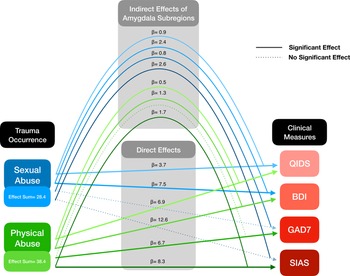
Notably, we did not find the same pattern of volumetric deficits in those participants with a prolonged history of physical abuse. However, physical abuse did have strong direct effects with clinical symptoms of anxiety and especially depression. Abuse-associated changes in the right BLA and cortical region mediated the relations between most symptomatic features of anxiety and depression. Notably, the reduced volumes of these two amygdalae regions strongly mediated the association between sexual abuse and higher scores on clinical scales. While less pronounced mediation effects (9%) were observed between physical abuse and clinical symptoms, there were stronger direct effects (See Fig. 1).
Statistical mediational model reveals the associations between right Basal and Cortical amygdala complex, childhood abuse and clinical features of anxiety and depression. Sexual and physical abuse demonstrate differential patterns of direct and indirect associations with clinical presentation. Physical abuse shows pronounced direct effects on the clinical scores of depression and anxiety. Sexual abuse had a specific indirect effect on anxiety scores. Effect sum = represents the sum of all beta values from total effects. BDI = Beck Depression Inventory, QIDS = Quick Inventory of Depressive Symptomatology; SIAS = Social Interaction Anxiety Scale; GAD7 = Generalized Anxiety Disorder 7-item' scale. Only solid line represents significant associations (p < 0.05).

To compare our present results with other clinical work, we found only one recent report by Oshri and colleagues who used the same amygdala subregion-specific segmentation workflow (Saygin et al., Reference Saygin, Kliemann, Iglesias, van der Kouwe, Boyd and Reuter2017) on a large sample of youth at risk for general psychopathology (Oshri et al., Reference Oshri, Gray, Owens, Liu, Duprey, Sweet and MacKillop2019). Consistent with their report, we also found lateral effects, that is reductions in the right BLA were associated with high scores on adverse childhood experience scale and high levels of anxiety and depression (Oshri et al., Reference Oshri, Gray, Owens, Liu, Duprey, Sweet and MacKillop2019).
While the role of the amygdala in the pathophysiology of mood disorders is well-established, it remains unclear why dysregulation in the BLA, in particular, is associated with increased risk for mood disorders and can be more sensitive to the effects of childhood abuse. From the physiological prospective, the BLA is a key site of neural plasticity and thought as a sensory gateway for the amygdala whereas the central region is an important output structure relaying emotional signals to the upper cortical levels (Phelps & LeDoux, Reference Phelps and LeDoux2005). As evidenced from preclinical animal models, chronic stress exposure can irreversibly change the structure of BLA, but not CeM neurons (Vyas, Bernal, & Chattarji, Reference Vyas, Bernal and Chattarji2003; Vyas, Mitra, Shankaranarayana Rao, & Chattarji, Reference Vyas, Mitra, Shankaranarayana Rao and Chattarji2002). In contrast, experimental inactivation of BLA can abolish the anxiety-like behaviours and even prevent chronically stressed rats from volumetric reductions within the limbic system (Tripathi et al., Reference Tripathi, Chakraborty, Srikumar, Raju and Shankaranarayana Rao2019). It is important to mention that mice with naturally smaller BLA volume can exhibit significantly greater levels of conditioned freezing compared to strains with larger BLA reflecting a high anxiety-like phenotype (Yang et al., Reference Yang, Mozhui, Karlsson, Cameron, Williams and Holmes2008). Therefore, there exists a possibility that a significant history of early life stress exposure can induce BLA atrophy as a result of aberrant neural hyperexcitability and the latter process is linked with enhanced anxiety-like behaviours (Guadagno, Wong, & Walker, Reference Guadagno, Wong and Walker2018; Rau, Chappell, Butler, Ariwodola, & Weiner, Reference Rau, Chappell, Butler, Ariwodola and Weiner2015).
Taken together, these findings suggest that a history of physical and sexual abuse may leave detectable structural deficits in the amygdala, mediating the risk for SMI. In addition, we observed an intricate differentiating pattern of structural alterations within the amygdala that may depend on the type of abuse experienced. These, and other results (Hoy et al., Reference Hoy, Barrett, Shannon, Campbell, Watson, Rushe and Mulholland2012; Oshri et al., Reference Oshri, Gray, Owens, Liu, Duprey, Sweet and MacKillop2019), indicate that morphologic changes in the amygdala might be detectable even before clinical manifestation of SMI in youth with a history of trauma.
The results should be interpreted with caution for the following reasons. Participants were not recruited on the basis of the presence of childhood trauma and information was gathered retrospectively introducing report bias. We observed a stronger association between amygdala volume reductions and specific types of abuse; however, trauma exposures tend to co-occur thus limiting inferences with respect to causative links between these associations. Although we reported significant associations between childhood trauma and amygdala changes and clinical status, our present analysis was cross-sectional in nature and these findings should not be interpreted as a developmental sequelae. Furthermore, the impact of neurodevelopmental and cognitive differences on the neuroimaging results remains unclear and warrants research interest for future works.
Another limitation is that we used T1-images as input for amygdala segmentation workflow, thus the volumes of internal boundaries of the amygdala should be interpreted with caution (Iglesias et al., Reference Iglesias, Augustinack, Nguyen, Player, Player, Wright and Van Leemput2015; Saygin et al., Reference Saygin, Kliemann, Iglesias, van der Kouwe, Boyd and Reuter2017).
Nonetheless, these results support the concept that early adverse experiences can induce structural changes in specific nuclei of the amygdala. These trauma-associated alterations are apparent before the onset of psychiatric illness and may mediate the degree of pre-morbid symptomatology. Future studies should aim to confirm the subregion specificity of these findings and to track the development of amygdala abnormalities over time.
Supplementary material
The supplementary material for this article can be found at https://doi.org/10.1017/S0033291720003177.
Acknowledgements
This study was supported by the Brain Canada Foundation and the Mathison Centre for Mental Health Research & Education at the University of Calgary through a research grant to Dr J Addington. Dr J Addington, Mr. N Nogovitsyn, Dr R Souza, Dr S Bray, Dr B Goldstein, Dr JL Wang, Ms. T Placsko and Ms. J Stowkowy have no financial disclosures or other potential conflicts of interest to declare.
Declaration of Interest
Dr Kennedy has been an advisor/consultant for Abbott, Alkermes, Allergan, BMS, Janssen, Lundbeck, Lundbeck Institute, Otsuka, Pfizer, Servier, Sunovion; and participated in clinical trials/studies for Abbott, BMS, Janssen, Pfizer, Servier; speaking engagements with the following organizations BMS, Lundbeck, Pfizer, Servier, Xian-Janssen; and has received research support from OBI, CIHR, BMS, Brain Canada, Janssen, Lundbeck, ORF, Pfizer, Servier. Dr MacQueen has received honoraria from Lundbeck, Janssen and AllerGen in the past 36 months. Dr Taylor received Honoria from Sunovion in the last 36 months.