Introduction
A subset of three-dimensional (3D) biomaterial printing is 3D printing of biomaterials containing cells (cell-laden), called bioinks. Bioprinting is an approach to on-demand 3D placement of cells that provides the means to create complex structures. Although there are many challenges, the most pressing is maintaining cell viability pre-, during, and post-3D printing. This article discusses those challenges along with what has been reported thus far with regard to preliminary relationships between bioink material properties and cells.
Current 3D bioprinting strategies
This article is entirely focused on extrusion (filament)-based 3D printing, which is the most widely used and versatile additive manufacturing platform for printing a variety of inks, including those with higher viscosities. Reference Malda, Visser, Melchels, Jüngst, Hennink, Dhert, Groll and Hutmacher1 For extrusion-based 3D printing, ink “printability” can have different definitions depending on the approach and end goals. Liquid bioinks of varying viscosities can be printed onto a stage and subsequently exposed to a stimulus that induces gelation ( Figure 1 ). This stimulus can be in the form of light (i.e., ultraviolet [UV] cross-linking), heat (thermal physical cross-linking), or cross-linker baths or mists (i.e., ionic cross-linking). In the case of liquid bioinks with gelation on-stage, the kinetics of cross-linking and the transition from sol to gel must be rapid enough to prevent substantial spreading and collapse of the extruded bioink. The stimulus must be presented either immediately when the ink exits the nozzle or after completion of a single printed layer (layer-by-layer cross-linking).
Bioinks can be prepared and three-dimensionally printed in liquid phase or gel phase. (a) Liquid-phase inks can have varying viscosities and are cross-linked post-printing. Once extruded, liquid bioink filaments can flatten and spread causing an increase in filament diameter. Cell settling in liquid bioinks can result in a heterogeneous printed object. (b) Gel-phase inks can be prepared with varying robustness (e.g., G′ storage modulus). Since gel inks are already in solid form, extruded filaments can better maintain shape. Furthermore, gel bioinks can maintain cell dispersion and therefore, homogeneity in the object.

Alternatively, viscous (honey-like) liquid-phase bioinks or even weak gel-phase bioinks can be printed. The gel-phase inks provide enhanced structure fidelity over less viscous (viscosity close to that of water) liquid-phase inks. If cross-linking is not conducted layer-by-layer, it can be introduced all at once to the printed structure as a post-printing processing step. The viscosity of liquid-phase bioinks can be tuned by adding an inherently viscous substance, such as hyaluronic acid, or increasing the polymer fraction. For gel-phase inks, the mechanical properties can be tuned by changing the polymer fraction, polymer properties, or degree of cross-linking (Figure 1). One disadvantage of liquid-phase bioinks is cell sedimentation in the ink during 3D printing and the resulting inhomogeneity in the 3D printed structures. Gel-phase inks have been shown to overcome this challenge by “locking” cells in place by the rapid increase in pre-gel viscosity that occurs upon synthesis and the quick gelation that follows. Reference Rutz, Hyland, Jakus, Burghardt and Shah2,Reference Dubbin, Hori, Lewis and Heilshorn3
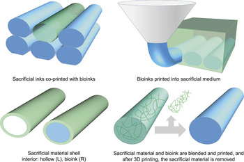
The use of sacrificial materials in the 3D bioprinting process has emerged in several strategies: (1) co-printing a bioink with a sacrificial ink; (2) a sacrificial material shell printed around the bioink filament; (3) a sacrificial material additive within the bioink to temporarily improve printability; and (4) a sacrificial medium into which bioinks are printed ( Figure 2 ). Co-printing self-supporting inks has been presented as a strategy to mold and hold in place inks with less than ideal printability. Reference Schuurman, Khristov, Pot, van Weeren, Dhert and Malda4–Reference Pati, Jang, Ha, Won Kim, Rhie, Shim, Kim and Cho6 Printed filaments of these inks have also been used to create empty microchannels once the sacrificial material is removed. Reference Bertassoni, Cecconi, Manoharan, Nikkhah, Hjortnaes, Cristino, Barabaschi, Demarchi, Dokmeci, Yang and Khademhosseini7–Reference Miller, Stevens, Yang, Baker, Nguyen, Cohen, Toro, Chen, Galie, Yu, Chaturvedi, Bhatia and Chen9 Another strategy is to use a sacrificial material within the bioink. This sacrificial material provides a self-supporting structure during printing, and is leached out after cross-linking the matrix to be left behind. Reference Tamayol, Najafabadi, Aliakbarian, Arab-Tehrany, Akbari, Annabi, Juncker and Khademhosseini10,Reference Armstrong, Burke, Carter, Davis and Perriman11 Alternatively, a sacrificial material “shell” can be provided by coaxial extrusion where the prepolymer shell is in the outer diameter and the cross-linker is in the inner diameter. Reference Onoe, Okitsu, Itou, Kato-Negishi, Gojo, Kiriya, Sato, Miura, Iwanaga, Kuribayashi-Shigetomi, Matsunaga, Shimoyama and Takeuchi12–Reference Colosi, Shin, Manoharan, Massa, Costantini, Barbetta, Dokmeci, Dentini and Khademhosseini14
To overcome limitations of poor printability in bioinks (blue), several strategies utilizing sacrificial materials (green) have been presented. Co-printing of bioinks and sacrificial material inks; sacrificial medium; coaxial extrusion of sacrificial material “shells” that are hollow or filled with bioink; mixing of sacrificial materials and bioink polymers into a single ink and subsequent sacrificial material removal after 3D printing and cross-linking.

To simplify this approach, others have demonstrated the use of support media into which inks can be deposited. Reference Wu, DeConinck and Lewis15–Reference Highley, Rodell and Burdick17 These media are sensitive to shear, allowing the nozzle of the 3D printer to pass through. In the case of support inks or media, the ink printed into the medium will usually require post-printing cross-linking; therefore, support inks and media must be compatible with such conditions and permit adequate diffusion of cross-linkers. Furthermore, most applications will require support inks or media to be removed, and methods to do so must be cell friendly.
Considerations when printing cells: A brief guide to the preparation of bioinks and their 3D printing and post-processing
Materials
Present bioink work often utilizes traditional biomaterials, including natural proteins (collagen, gelatin, fibrin/fibrinogen, tissue-specific extracellular matrix) and polysaccharides (chitosan, hyaluronic acid, alginate), recombinant proteins, engineered peptides, and synthetic polymers (poly(ethylene glycol)[PEG], poly(N-isopropylacrylamide), polaxamers [triblock copolymers of poly(propylene oxide) and poly(ethylene oxide)]). Both natural and synthetic materials are important to the future of the field. Natural-synthetic composite inks, in particular, can leverage the advantageous properties of both—natural polymers tend to be inherently bioactive while synthetics can offer superior and controllable mechanical and degradation properties, as well as greater reproducibility. The development of new polymers Reference Peppas and Khademhosseini18 with unique properties, such as stimuli-responsiveness to induce shape transformation, Reference Li, Zhang, Akpek, Shin and Khademhosseini19 is a new concept that may be leveraged in next-generation bioinks.
Physical and chemical cross-linking in the presence of cells
Before, during, or after 3D printing, precursor polymer solutions must be cross-linked to form a hydrogel. In nearly all cases, encapsulated cells must be subjected to this step and therefore, cytocompatibility of the cross-linking reaction must be thoroughly investigated.
The most commonly used cross-linking reactions are physical, including ionic, thermal, and enzymatic (i.e., thrombin with fibrinogen). These reactions are “gentle” in that the cross-linking typically cannot cross-react with cells, and gelation can be reversible. Although mild, physical cross-linking tends to be unstable in large media reservoirs and for in vivo studies. Reference Shoichet, Li, White and Winn20,Reference Shikanov, Zhang, Xu, Smith, Rajan, Woodruff and Shea21 For example, the cross-linker calcium will leach out of alginate when the extra-gel concentration (i.e., interstitial tissue fluid) is below the intra-gel concentration used for cross-linking. In another example, gels cross-linked via hydrogen bonding can be disrupted by competing in vivo milieu, such as plasma proteins and proteases.
Alternatively, chemical cross-linking can be performed, but in this case, bio-orthogonality is a necessity. UV cross-linking, thiol Michael type addition (reaction between thiol and alkene), Reference Phelps, Enemchukwu, Fiore, Sy, Murthy, Sulchek, Barker and García22–Reference Jin, Teixeira, Krouwels, Dijkstra, van Blitterswijk, Karperien and Feijen26 and emerging “click” reactions Reference Madl, Katz and Heilshorn27–Reference Alge, Azagarsamy, Donohue and Anseth32 (e.g., copper-free reaction between azides and activated alkynes) have all been used with success for cell encapsulation. UV cross-linking remains the most frequently used method for bioinks, while the thiol Michael type has found limited use and to the best of our knowledge, click reactions have yet to be utilized with bioinks. In addition to enzymatic cross-linkers that initiate physical cross-linking, there are also several enzymatic, covalent cross-linkers, Reference Heck, Faccio, Richter and Thöny-Meyer33 such as genipin Reference Chen, Chang, Cheng, Tsai, Yao and Liu34,Reference Macaya, Ng and Spector35 and transglutaminase, Reference Schense and Hubbell36,Reference Homan, Kolesky, Skylar-Scott, Herrmann, Obuobi, Moisan and Lewis37 which have been used to manipulate hydrogel degradation and mechanical properties.
An interesting subset of cross-linking for 3D printing includes dynamic cross-linking or “adaptable linkages”—guest–host interaction, biorecognition, hydrophobic interactions, hydrogen bonding, ionic cross-linking, and dynamic covalent reactions. Reference Wang and Heilshorn38,Reference Burdick and Murphy39 This special set of reactions yields shear-thinning and self-healing properties ideal for material extrusion through fine nozzles and the ability to maintain extruded filament shape, respectively. Researchers have long used these types of reactions for injectable hydrogels and have recently used them with success in 3D printing. Reference Wang and Heilshorn38,Reference Foster, Marquardt and Heilshorn40 These include the development of gel-phase hydrogel bioinks Reference Dubbin, Hori, Lewis and Heilshorn3,Reference Highley, Rodell and Burdick17,Reference Ouyang, Highley, Rodell, Sun and Burdick41 and support medium Reference Highley, Rodell and Burdick17 as well as post-processing methods. Reference Rutz, Hyland, Jakus, Burghardt and Shah2
Preparation of cells
The choice of cells is greatly dependent on each tissue engineering application and can range in type (primary, immortalized, stem cells), age (embryonic, fetal, or adult derived), and source (animal, human). Dependent on each cell type, cell passage conditions such as passage number and protocols for dissociating cells from culture substrates should not be overlooked since it is required that cells be highly viable before being exposed to the stress of 3D printing. The environment of the cells during the process should also be carefully examined. Temperature, pH, and ionic strength of the bioink and post-printing cross-linking solutions must be adjusted accordingly. Oxygen and CO2 conditions, which can affect media pH, are often not controlled during bioink synthesis and 3D printing, but could easily be incorporated. After passage, cells must be homogeneously mixed into bioinks or precursor bioink solutions. This mixing should be gentle, avoiding bubbles and turbulent flow, to ensure cell viability.
Sterility
A seldom-discussed challenge in bioprinting research is sterility. Starting with sterility of bioink polymers, filtration and autoclaving of polymer solutions or gamma irradiation of a polymer solid (frequently used for proteins) can be used; each of these has its advantages and disadvantages. Many researchers use 70% ethanol to sterilize their materials; however, it is not a sterilization process that can eliminate all types of contamination and therefore, has limited use in eventual clinical translation. Anything that comes into contact with the bioink, such as cartridges, nozzles and substrates, manipulators (forceps, spatulas), and fluids, must also be sterilized. Ethylene oxide, plasma irradiation, and 10% bleach are additional choices for sterilization of equipment, but their use with bioinks is likely limited.
Three-dimensional printing and post-processing
During 3D printing, characterizing the flow properties of inks is good practice. Flow properties are usually determined by extruding ink into a tared tube for a chosen amount of time to determine the mass flow rate. Characterization performed simultaneous to the experiment can give the most accurate reflection of material properties, as opposed to a separate analysis conducted on a rheometer. Particularly, for bioinks sensitive to certain conditions, such as time or temperature, an instrument separate from the 3D printer may not fully recapitulate printing conditions. Separate analysis by other instruments, however, is still required and will complement such data acquired on the 3D printer. Characterizing material properties helps build relationships between bioink flow properties and printability, and help with reproducibility within the research community.
The build time of 3D printed structures is another variable for encapsulated cells. The conditions during 3D printing are likely different from those used for long-term cell culture (controlled CO2, media, humidity), and prolonged absences from ideal culture conditions during the printing process can lead to drops in cell viability. Related to build times, hydration of the printed structures must be carefully monitored. If cells dehydrate, cell viability cannot be recovered. Humid chambers, mists, and drops of sterile buffers onto printed structures can be employed. Alternatively, inks can be 3D printed into media baths, where the structure is either completely submerged or gradually lowered into the bath with the top layer being actively printed on the top. This approach, however, may compromise adhesion between printed layers.
After 3D printing, the bioinks and printed structures may not have the final desired material properties. For example, additional cross-linking and application of bioactive components (e.g., tethering peptides or absorption of growth factors) can be performed. Additional cross-linking is particularly important for achieving appropriate stiffness and delaying hydrogel degradation. The components of the incubating media can also have an effect on bioink degradation. In our studies, we found that the addition of serum proteins (fetal bovine serum) greatly quickened the degradation of lightly PEG cross-linked gelatin. Reference Rutz, Hyland, Jakus, Burghardt and Shah2 Media components could also be carefully selected to enhance viability and recovery post-3D printing. For example, methods for fluorescence-activated cell sorting, in which cells are exposed to shear stress—not unlike 3D printing—may be useful to apply post-extrusion (i.e., supplementing the media with additional fetal bovine serum after shearing the cells).
Finally, 3D printing studies report several types of characterization and assessments, such as bioink rheology, “printability,” preciseness of the printed structure, and mechanical properties of the printed structure. Methods and types of assessments are still being developed, but cell viability is arguably the most critical measure. Different methods for measuring cell viability have been used, but this should be developed and standardized in a rigorous effort.
Characterization of cell viability should include quantitative analysis of live, stressed, and dead or dying (apoptotic, necrotic) cell populations, Reference Nair, Gandhi, Khalil, Yan, Marcolongo, Barbee and Sun42 as we have observed that only quantifying the live population can seriously overestimate the percentage of healthy cells. Ideally, characterization should be conducted with 3D methods. For example, confocal fluorescence microscopy gives the enormous advantage of not degrading the bioink for evaluation, unlike methods such as flow cytometry. With optically transparent hydrogels, an accurate picture of the extruded sample is obtained. While many current bioink studies are publishing new synthetic methods and associated cell viabilities, next-generation bioink studies will begin to incorporate investigations of cell phenotype and function as well as evaluation toward targeted applications.
The relationship among printing parameters, bioink material properties, and cell viability
Past studies show that the act of extrusion and 3D printing is a stress to cells. Typical cell viability after extrusion reported in bioink literature is usually, at best, 70–90% in optimized conditions and with lower reported in suboptimal conditions. Higher cell viabilities are also often reported in cast controls compared to extruded samples. The responsible factors of cell stress (i.e., mechanically induced) and their severity are still not completely understood. Here, we review findings that show how bioink material properties influence cells ( Figure 3 ) as well as how cells influence bioink material properties.
Bioink design, printing parameters, and post-printing processing impact cell viability. The viability of the cells, in turn, impacts subsequent cellular events, such as proliferation, differentiation, and tissue formation, and therefore, is a key challenge in bioink synthesis.

Printing parameters
Printing parameters, such as extrusion pressure and dispensing speed, are usually varied for optimal printed structure features—filament diameter matching the nozzle diameter, filament uniformity, and precise filament placement. However, variations in these parameters influence extruded cell viability. Researchers have reported, in both liquid-phase Reference Nair, Gandhi, Khalil, Yan, Marcolongo, Barbee and Sun42–Reference Fedorovich, Schuurman, Wijnberg, Prins, van Weeren, Malda, Alblas and Dhert44 and gel-phase Reference Billiet, Gevaert, De Schryver, Cornelissen and Dubruel45 bioinks, that increasing applied pressure decreased extruded cell viability. Additional care must also be given to the nozzle shape and diameter. Billiet et al. found that conical nozzles yielded better cell viability than cylindrical nozzles when using gel-phase bioinks. Reference Billiet, Gevaert, De Schryver, Cornelissen and Dubruel45 However, as nozzle length increases, shear stress increases, and therefore, drawing firm conclusions between tapered conical or cylindrical nozzles requires analysis of varying nozzle lengths. Decreasing nozzle diameter in cylindrical nozzles also causes a decrease in extruded cell viability in both liquid-phase Reference Nair, Gandhi, Khalil, Yan, Marcolongo, Barbee and Sun42,Reference Chang, Nam and Sun43 and gel-phase bioinks. Reference Billiet, Gevaert, De Schryver, Cornelissen and Dubruel45
Nair et al. compared cell viability with varying pressure and nozzle diameter and concluded that the extrusion pressure more significantly affects cell viability than nozzle diameter. Reference Nair, Gandhi, Khalil, Yan, Marcolongo, Barbee and Sun42 Because extrusion pressure influences flow rate, this finding may have implications in the total build time of cell-laden structures. Under severely stressing conditions (high pressures and small diameters), the majority of cells were necrotic rather than apoptotic and cell nuclei suffered morphological damage, which indicated irreparable damage. Reference Nair, Gandhi, Khalil, Yan, Marcolongo, Barbee and Sun42
It is worth noting that increasing the printing speed at a given pressure can result in strand stretching and thinning of the filament diameter. Reference Khalil and Sun46 This may help achieve cell alignment along the printed strand; however, this likely introduces potentially undesirable tensile and compressive forces from the polymer matrix to the cells. Strand diameter is a significant factor in cell alignment as others have reported in micromolding techniques. Reference Aubin, Nichol, Hutson, Bae, Sieminski, Cropek, Akhyari and Khademhosseini47,Reference Baranski, Chaturvedi, Stevens, Eyckmans, Carvalho, Solorzano, Yang, Miller, Bhatia and Chen48 Choi et al. and Kang et al. observed alignment of myotubes (fused muscle cells) in the direction of extrusion from bioprinting. Reference Choi, Kim, Jeong, Yi, Park, Hwang and Cho49,Reference Kang, Lee, Ko, Kengla, Yoo and Atala50 Choi et al. observed increasing myotube alignment with decreasing nozzle diameter after prolonged culture.
Cell viability and morphology may depend on the location of the cells within a given filament. Cell viability, for example, might be lower at the periphery of the strand due to increased shear stress at the nozzle wall. Others have reported a greater degree of cell spreading and cellular network formation on the periphery filaments, likely because these cells are close to the surface of the hydrogel and are not fully encapsulated. Reference Colosi, Shin, Manoharan, Massa, Costantini, Barbetta, Dokmeci, Dentini and Khademhosseini14 Morphological differences between cells in the exterior and interior locations of filaments may also result in functional differences between cells. An additional variable is the diffusion of oxygen and nutrients, and how that may differ in the interior of large (>500 µm) filaments. To the best of our knowledge, there have been no reported studies on the difference in cell viability on the periphery and interior portions of extruded filaments.
The impact of cells on bioinks
The presence of cells and their density change the rheological properties of inks and the final mechanical properties of the hydrogel. Most often, researchers present the corresponding ink formulation without cells, but a few studies have reported cell-laden bioink rheology. In physically cross-linked gel-phase bioinks, the inclusion of cells to a cell-barren ink, as well as increasing cell densities, decreased the degree of cross-linking, decreased viscosity in the liquid-phase, and decreased final mechanical properties. Reference Billiet, Gevaert, De Schryver, Cornelissen and Dubruel45,Reference Kesti, Müller, Becher, Schnabelrauch, D’Este, Eglin and Zenobi-Wong51,Reference Zhao, Li, Mao, Sun and Yao52
Zhao et al. reported a decrease (∼30%) in the storage modulus of a gelatin-alginate gel when 1 million cells/mL were incorporated. Reference Zhao, Li, Mao, Sun and Yao52 Billiet et al. found bioink viscosity in gelatin methacrylamide bioinks decreased twofold for 0.5 and 1.5 million cells/mL and fourfold when prepared with 2.5 million cells/mL. Reference Billiet, Gevaert, De Schryver, Cornelissen and Dubruel45 Physical gelation temperatures of 4°C less were used for cell-laden inks in order to compensate for decreased mechanical properties relative to ink without cells. Furthermore, the moduli of thermally cross-linked gelatin methacrylamide decreased by as much as several thousand Pa when cells were added at the densities previously described. Therefore, studies comparing inks of varying cell densities will likely require alteration of the bioink formulation to achieve constant rheological properties. When cell-laden inks were UV-cross-linked post-printing, an increase in the modulus of one polymer ink and a decrease in a different polymer ink were found when compared to the respective cell-barren inks. Reference Kesti, Müller, Becher, Schnabelrauch, D’Este, Eglin and Zenobi-Wong51 These results highlight the importance of conducting cell-laden bioink rheology.
Bioink design: Gel-phase bioink rheological properties
It is established that cells are mechanoresponsive, and there are many studies reporting the influence of substrate stiffness. Reference Vogel and Sheetz53,Reference Discher, Mooney and Zandstra54 However, how bioink materials respond to the shear stress of extrusion and subsequently, how the material deformation then influences cell viability and cellular functions (i.e., gene expression, differentiation, proliferation) are largely unknown. Printability of gel-phase inks is connected to the storage modulus of the gel (better structure fidelity at higher G′), but a few studies have shown this variable can have significant consequences on cell viability.
Heilshorn and colleagues investigated injectable cell therapies and found that alginate gels (calcium cross-linked) provided a protective effect to extruded cells and showed higher cell viability than saline solution and the corresponding alginate solution (not cross-linked). Reference Aguado, Mulyasasmita, Su, Lampe and Heilshorn55 They further examined the effects of the mechanical properties (G′ = 0.33–58.1 Pa) of the gels by changing the degree of cross-linking and the average molecular weight of the alginate. Cell viability was highest at 29.6 Pa and decreased with G′ higher or lower than this critical point of highest cell viabilty. Reference Aguado, Mulyasasmita, Su, Lampe and Heilshorn55 Interestingly, exposing cell-laden hydrogels to shear rates equivalent to that of extrusion in a rheometer did not decrease cell viability. Based on these findings, the Heilshorn lab chose to use hydrogels with G′ between ∼10–50 Pa for injectable cell therapies Reference Cai, Dewi, Goldstone, Cohen, Steele, Woo and Heilshorn56,Reference Cai, Dewi and Heilshorn57 and 3D bioprinting. Reference Dubbin, Hori, Lewis and Heilshorn3
Sun and colleagues examined cell viability in gelatin/alginate blend gel-phase bioinks of varying mechanical properties that were manipulated by altering the gelatin concentration as well as the bioink incubating temperature to induce physical cross-linking of the gelatin. Cell viability decreased from ∼97.5% at G′ ∼ 50–125 Pa to ∼72.5% at G′ ∼ 2000 Pa with cell viability dropping below 90% at 382 Pa, although it was unclear which bioink compositions and conditions were studied to obtain 16 different storage moduli. Reference Zhao, Li, Mao, Sun and Yao52
Lewis and colleagues decreased the mechanical properties of gelatin-based gel-phase bioinks by decreasing the average molecular weight of gelatin with processing gelatin at increasing temperatures. Examining several processing temperatures and therefore moduli, the researchers reported ∼50% cell viability at G′ ∼ 1200 Pa (70°C) to 95% cell viability at G′ ∼ 150 Pa (95°C). The rheology conditions used to determine bioink moduli, however, were different from printing conditions. First, the inks were not cell-laden, and second, moduli reported were the plateau moduli from cooled temperatures (<4°C) whereas printing was conducted at room temperature. Reference Kolesky, Homan, Skylar-Scott and Lewis58 Therefore, these reported moduli do not reflect the true moduli of the tested bioinks. Nevertheless, these three studies demonstrate that the modulus of printed gel-phase inks is a critical variable for cell viability and requires more attention in future studies.
Bioink design: Composition
The type of polymer(s) used in the bioink composition can influence cell viability as well as long-term behavior and function. As previously discussed, rheological properties influence cell viability and these properties change with varying bioink compositions. The incorporation of alginate into a gelatin methacrylate bioink prevented thermal gelation at room temperature, however, how this affected cell viability was not examined. Reference Colosi, Shin, Manoharan, Massa, Costantini, Barbetta, Dokmeci, Dentini and Khademhosseini14 Although several multimaterial strategies to prepare bioinks have been presented, Reference Rutz, Hyland, Jakus, Burghardt and Shah2,Reference Tamayol, Najafabadi, Aliakbarian, Arab-Tehrany, Akbari, Annabi, Juncker and Khademhosseini10,Reference Hinton, Jallerat, Palchesko, Park, Grodzicki, Shue, Ramadan, Hudson and Feinberg16 comparing cell viability across polymer types has not been undertaken. This is a complex endeavor in that many material properties will also likely be simultaneously changed and therefore, careful characterization, analysis, and bioink manipulation is required.
Post-printing: Cross-linking
A theme central to current bioink printing work is the use of post-printing cross-linking, most frequently UV-based, to increase final degradation and mechanical properties. Increasing the post-printing degree of cross-linking by varying UV exposure times decreases cell viability. Reference Colosi, Shin, Manoharan, Massa, Costantini, Barbetta, Dokmeci, Dentini and Khademhosseini14,Reference Billiet, Gevaert, De Schryver, Cornelissen and Dubruel45,Reference Zhang, Davoudi, Walch, Manbachi, Luo, Dell’Erba, Miri, Albadawi, Arneri, Li, Wang, Dokmeci, Khademhosseini and Oklu59 Khademhosseini and colleagues found this to be different from past studies in UV cross-linked cast hydrogels, Reference Fedorovich, Oudshoorn, van Geemen, Hennink, Alblas and Dhert60 where cells could tolerate a few minutes of UV exposure, compared to extruded cells that could only tolerate 30 s of UV exposure before cell viability diminished. Reference Colosi, Shin, Manoharan, Massa, Costantini, Barbetta, Dokmeci, Dentini and Khademhosseini14 These results highlight the fact that cells are already in a stressed state from extrusion prior to the stress of further cross-linking.
In addition to cell viability, the degree of total cross-linking (primary and secondary combined) can lead to different degrees of cell spreading and cellular network formation. Reference Colosi, Shin, Manoharan, Massa, Costantini, Barbetta, Dokmeci, Dentini and Khademhosseini14,Reference Jia, Gungor-Ozkerim, Zhang, Yue, Zhu, Liu, Pi, Byambaa, Dokmeci, Shin and Khademhosseini61 In cast hydrogels, increasing the degree of cross-linking Reference Chen, Lin, Qi, Yang, Bae, Melero-Martin and Khademhosseini62 or polymer fraction Reference Billiet, Van Gasse, Gevaert, Cornelissen, Martins and Dubruel63,Reference Nichol, Koshy, Bae, Hwang, Yamanlar and Khademhosseini64 can interfere with cell viability and cellular projections and network formation. Concentrations less than 10%, and ideally less than 5%, are almost always required, but this is polymer-dependent. This is critical when the bioink polymer concentration is increased to improve printability. In addition to providing cells with appropriate stiffness, higher modulus hydrogels achieved through post-printing cross-linking may also be required for handling and surgical implantation, as well as longer degradation rates both in vitro and in vivo. Strategies for synthesizing bioinks that simultaneously achieve high post-extrusion cell viability, excellent printability, and optimal mechanical properties post-printing will remain a key challenge in future work.
Post-printing: Recovery
Many factors, such as printing parameters, bioink gel mechanical properties, and post-printing cross-linking, can greatly decrease cell viability, as discussed in Figure 3. It is possible, however, for cells to recover from stress or proliferate to recuperate lost numbers. Chang et al. demonstrated marginal recovery with liquid-phase bioinks (no more than 5% increase in viability) at 6 h post-printing, but substantial recovery (up to ∼35% increase) after 24 h. Reference Chang, Nam and Sun43 Little to no additional recovery was found at three and seven days, when compared to viability at 24 h. Kolesky et al. also reported ∼10–20% increases in viability of different cell types in gel-phase bioinks over seven days. Reference Kolesky, Truby, Gladman, Busbee, Homan and Lewis8 It is important to note that increases in cell viability over several days could also be attributed to proliferation of healthy cells. Whether or not this is a universal trend across varying bioinks will require further study. Furthermore, it is difficult to imagine all cells will be able to recover when others have reported necrotic cells and nuclear damage. Reference Nair, Gandhi, Khalil, Yan, Marcolongo, Barbee and Sun42
Conclusion
We have discussed several considerations when synthesizing new bioinks. Preparation of bioinks and their 3D printing is significantly complicated by the presence of cells, which require strict conditions for viability. We also described the relationships between bioink material properties and cell viability and behavior ( Table I ).
Relationships between bioink material properties and cell viability and behavior.

Bioink properties impact cells and cells can impact bioink properties. Printing variables and bioink mechanical properties impact cell viability, while the presence of cells can impact bioink mechanical properties. After post-processing, final bioink mechanical properties can impact cell elongation and migration as well as connections with neighboring cells.
There are a few findings that will prove useful to future bioink development. First, bioinks should be synthesized to minimize the required pressure for extrusion. Second, there exists a circular relationship between cells and bioink rheology—cells impact bioink rheology and rheology impacts cell viability. Therefore, it is necessary to characterize rheological properties in the presence of cells. Third, the modulus of gel-phase bioinks is a variable that impacts cell viability, as others have reported decreasing cell viability with increasing bioink moduli. Comparing results in different hydrogels generated from different labs, we note that modulus alone may not effectively determine extruded cell viability. Another influencing variable that could be investigated is the polymer itself, either by changing type (i.e., gelatin versus alginate) or its properties (i.e., average molecular weight, polydispersity). Finally, even if extruded cell viability is optimized, post-printing cross-linking can further complicate and affect cell viability. In future work, although we must design bioinks and printing methods to be as cell-friendly as possible, investigating strategies to recover stressed cells would also be a valuable addition to the bioprinting community. These important relationships and developments are integral to accelerate progress in the bioprinting field, but at present, remain poorly understood.
Present bioprinting work is limited by a narrow window of printability in order to achieve high cell viability; however, manipulation of materials and their properties is at the core of biomaterials science and engineering research in order to both study and optimize the biological response. Toward developing next-generation bioinks with unique and tunable materials properties, the impact of bioink manipulation on cells and vice versa need to be more thoroughly investigated. Promising approaches include utilizing innovative chemistries for bioink synthesis. Click chemistries, in particular, provide bio-orthogonality that could allow safe manipulation of material properties in the presence of cells. Bioinks engineered to have unique rheological properties that minimize mechanical damage to cells are also of high value to the field. Furthermore, developing models that describe the relationships between printing parameters, bioink properties, printability, and cell viability will be useful for effectively predicting the success of new bioink designs. From whichever strategies prove to be fruitful, next-generation bioinks must simultaneously achieve excellent printability, high cell viability, and a wide range of material properties in order to make the greatest impact in the field of regenerative engineering.

Alexandra L. Rutz is a postdoctoral researcher at École Nationale Supérieure des Mines, France. She received her MS degree in 2014 and her PhD degree in 2016, both in biomedical engineering from Northwestern University. Her doctoral thesis focused on engineering gel-phase hydrogel bioinks through partial cross-linking, including the development of a customizable, multimaterial bioink method. Rutz can be reached by phone at 773-217-0730 or by email at arutz13@gmail.com.

Phillip L. Lewis is a doctoral candidate at the Simpson Querrey Institute at Northwestern University. He received his MS degree in biomedical engineering from Northwestern University in 2015. His current research focuses on liver tissue engineering through three-dimensional (3D) printing using novel bioactive 3D printable inks to control liver developmental morphogenic processes. Lewis can be reached by phone at 312-503-3052 or by email at philliplewis2015@u.northwestern.edu.

Ramille Shah holds a joint appointment in the Departments of Materials Science and Engineering and Surgery at Northwestern University (NU). She earned her BS degree in materials science and engineering at NU and her PhD degree in materials science and engineering at the Massachusetts Institute of Technology with a research focus on musculoskeletal tissue engineering. Her research focuses on the development, characterization, and translation of new functional material three-dimensional (3D) inks that are compatible with room-temperature extrusion-based 3D printing for both biomedical (e.g., complex tissue and organ engineering) and nonbiomedical (e.g., energy and advanced structural) applications. Shah can be reached by phone at 312-503-3931 or by email at ramille-shah@northwestern.edu.