Introduction
Nematodes of the dracunculoid family Philometridae represent a group of about 200 species parasitic in the body cavity and various body tissues of fish (Moravec & Barton, Reference Moravec and Barton2016). Due to morphological and biological peculiarities (Moravec, Reference Moravec2006; Moravec & de Buron, Reference Moravec and de Buron2013), most of them are known only by their large females. Rasheed (Reference Rasheed1963) created a taxonomic system for the Philometridae based principally on female morphology, which was followed in the monographs by Ivashkin et al. (Reference Ivashkin, Sobolev and Khromova1971) and Chabaud (Reference Chabaud, Anderson, Chabaud and Willmott1975) and became a basis for the present classification system of dracunculoids proposed by Moravec (Reference Moravec2006).
Data on philometrid nematodes parasitizing Neotropical freshwater fish are scarce, including 12 species belonging to the genera Alinema Rasheed, 1963, Neophilometroides Moravec, Salgado-Maldonado et Aguilar-Aguilar, 2002, Nilonema Khalil, 1960, Philometra Costa, 1845, Philometroides Yamaguti, 1935 and Rumai Travassos 1960, parasites of characiform, perciform, osteoglossiform, siluriform and synbranchiform fish (Moravec & de Buron, Reference Moravec and de Buron2013; Moravec & Diggles, Reference Moravec and Diggles2015). Nine of these species, belonging to all of the above-mentioned genera, were described in South America and, except for Neophilometroides paraguyanensis (Petter, 1995) from the bryconid Salminus brasiliensis (Cuvier) in Paraguay (Petter, Reference Petter1995), were also reported from Brazil. These are: Alinema amazonicum (Travassos, 1960) and Philometra baylisi Vaz & Pereira, 1934 from pimelodid catfish (Vaz & Pereira, Reference Vaz and Pereira1934; Travassos, Reference Travassos1960; Inglis & Ogden, Reference Inglis and Ogden1964), P. nattereri Cárdenas, Moravec, Fernandes & Morais, 2012 from the serrasalmid Pygocentrus nattereri Kner (see Cárdenas et al., Reference Cárdenas, Moravec, Fernandes and Morais2012), P. fariaslimai Fortes, 1981 [species inquirenda] from ariid catfish (Fortes, Reference Fortes1981; Moravec, Reference Moravec2006), Philometroides maplestoni (Travassos, Artigas & Pereira, 1928) from the bryconid Salminus hilarii Valenciennes (Travassos et al., Reference Travassos, Artigas and Pereira1928), Nilonema senticosum (Baylis, 1927) and Rumai rumai Travassos, 1960 from the arapaimid Arapaima gigas (Schinz) (see, for example, Baylis, Reference Baylis1927; Travassos, Reference Travassos1960; Santos & Moravec, Reference Santos and Moravec2009) and Philometra mirabilis Moravec & Diggles, 2015 from the cichlid Cichla mirianae Kullander et Ferreira (see Moravec & Diggles, Reference Moravec and Diggles2015).
During recent parasitological investigations of fish in the Acre River (Amazon River basin), Acre State, north-western Brazil, philometrid nematodes were collected from the stomach of the Bloch’s catfish Pimelodus blochii Valenciennes (Pimelodidae). A close examination revealed that they represent a new species of Philometroides, which is described herein.
Pimelodus blochii (maximum body length 35 cm) is a tropical freshwater and brackish-water fish commonly found in the lower reaches of large rivers in South America in the Gulf of Paria, Amazon, Corantijn, Essequibo and Orinoco basins (Froese & Pauly, Reference Froese and Pauly2016). Bloch's catfish is one of eight species of Pimelodus Lacépède occurring in the Amazon River drainage system. This is a highly appreciated and the most consumed species in the region, considered by locals to be a tasty and cheap fish (Padovani et al., Reference Padovani, Forsberg and Pimentel1996; Boujard et al., Reference Boujard, Pascal, Meunier and Le Bail1997; Ribeiro & Lucena, Reference Ribeiro and Lucena2006).
Materials and methods
A total of 120 specimens of the catfish P. blochii were caught between November 2013 and April 2014 by a local fisherman in the Acre River (10°39′40.33″S, 68°30′19.51″W).
Following post-mortem examination, one fish was found to be infected with two gravid nematode specimens, one of which was fixed in 70% ethanol and the other in 4% formalin. They were cleared in glycerine. Measurements are given in micrometres unless otherwise stated. Drawings were made with the aid of a drawing tube. Subsequently, the anterior and posterior parts of body were postfixed for 24 h at room temperature in 1% osmium tetroxide in cacodilate buffer, dehydrated through a graded alcohol series, critical-point dried and sputter-coated with gold for analysis by scanning electron microscopy (SEM). Samples were examined using a JEOL JSM-6390 LV scanning electron microscope (JEOL Ltd, Tokyo, Japan) at the Plataforma de Microscopia Eletrônica do Instituto Oswaldo Cruz, with an accelerating voltage of 15 kV. Specimens were deposited in the Helminthological Collection of the Instituto Oswaldo Cruz (CHIOC), Rio de Janeiro, Brazil. The fish nomenclature followed FishBase (Froese & Pauly, Reference Froese and Pauly2016).
Results
Philometroides acreanensis n. sp.
Family Philometridae Baylis et Daubney, 1926.
Description
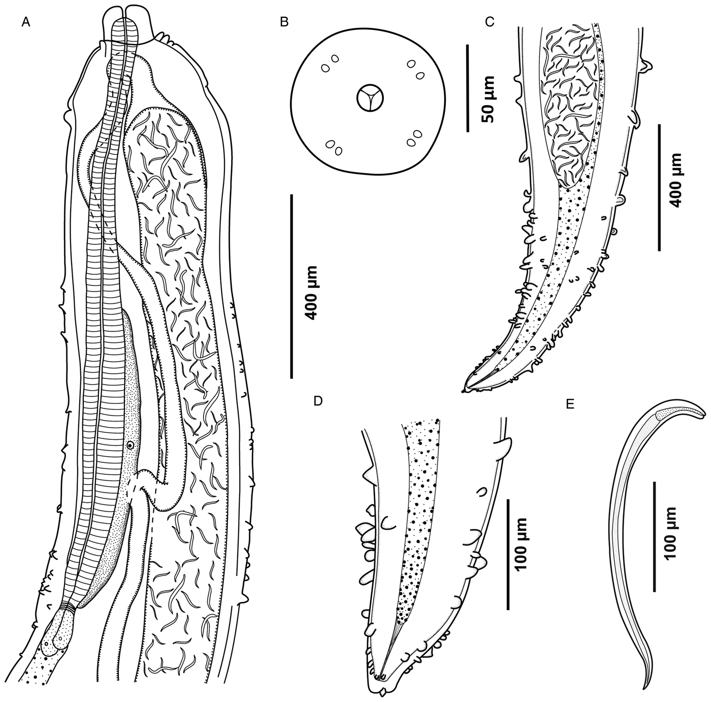
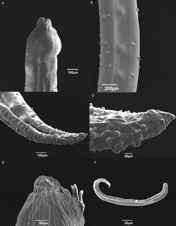
Measurements of the holotype of the gravid female and those of the paratype (in parentheses) are given. Large-sized whitish nematodes; posterior end markedly narrowed, cone-shaped (figs 1C, D and 2C, D). Cuticle with bosses irregularly distributed along body; bosses circular or transversely oval in apical view (figs 1A and 2A–D). Embossment starts anteriorly at level of mid-oesophagus and finishes close to posterior extremity of body (figs 1A, C, D and 2A–D). Body length 240 (280) mm long and 750 (875) wide. Oral aperture circular, surrounded by four submedian pairs of small cephalic papillae (fig. 1B). Oesophagus 1.25 (−) mm long and 550 (−) wide, without usual inflation at anterior end (fig. 1A). Oesophageal gland 330 long, starting at mid-length of oesophagus and extending posteriorly to oesophageal end. Small ventriculus present, opening into intestine through valve. Nerve ring not located. Intestine narrow at its anterior end; posterior end of intestine atrophied, attached by short ligament to body wall near posterior extremity (fig. 1C, D). Ovaries narrow, long, reflexed, situated near anterior and posterior extremities of body (fig. 1A). Vulva and vagina absent; instead, spot of ruptured cuticle with emerging larvae observed close to anterior end on one side of body (fig. 2E). Uterus occupies major part of body, filled with numerous first-stage larvae 300–330 long and 15–20 wide with oesophagus 50–55 long (figs 1A, E and 2E, F).
Gravid female of Philometroides acreanensis n. sp., showing: (A) anterior end, dorsal view; (B) schematic reconstruction of cephalic end, apical view; (C) posterior end, ventral view; (D) caudal region, lateral view; and (E) larva from the uterus, lateral view.

Scanning electron microscopy of the gravid female of Philometroides acreanensis n. sp., showing: (A) anterior end with with cuticular bosses; (B) mid-region with cuticular bosses; (C) caudal region, ventro-lateral view; (D) cuticular bosses on caudal end, lateral view; (E) apical region with emerging larvae; and (F) larval stage from uterus.

Male: not known.
Taxonomic summary
Type host
Pimelodus blochii Valenciennes (Siluriformes: Pimelodidae).
Site in host
Stomach wall.
Type locality
Acre River (Amazon River basin), Acre State, Brazil (10°39′40.33″S, 68°30′19.51″W).
Prevalence and intensity
0.8% (1 fish infected/120 fish examined); two nematodes.
Deposition of type specimens
Holotype and paratype in the Helminthological Collection, Oswaldo Cruz Institute, Rio de Janeiro (CHIOC numbers 38325a–b).
Etymology
The specific name acreanesis relates to the type locality.
Discussion
The general morphology, especially the occurrence of irregularly scattered cuticular bosses on the body surface, suggest that the present specimens belong to the genus Philometroides (see Rasheed, Reference Rasheed1963; Moravec, Reference Moravec2006). At the present time, this genus includes 35 species (Moravec & de Buron, Reference Moravec and de Buron2013; Moravec et al., Reference Moravec, Halajian, Tavakol, Nyagura and Luus-Powell2015; Montes et al., Reference Montes, Plaul and Martorelli2016; Moravec & Barton, Reference Moravec and Barton2016), which are tissue-dwelling parasites of marine (12 species), brackish-water (3 species) and freshwater fish (20 species). Most of them were described only from females, whereas conspecific males are known for eight species. Like other philometrids, Philometroides spp. are known to exhibit a relatively high degree of host specificity, and individual species are characterized, in addition to morphological features and molecular sequence data, by the location of gravid females in the host (Moravec, Reference Moravec2006; Moravec & de Buron, Reference Moravec and de Buron2013).
Of the 20 species of Philometroides parasitizing freshwater fish, P. maplestoni from the abdominal cavity of the characiform fish S. hilarii in the Paraná River basin is the only one reported from the Neotropical Region (Brazil) (Travassos et al., Reference Travassos, Artigas and Pereira1928; Luque et al., Reference Luque, Aguiar, Vieira, Gibson and Santos2011). Another species of Philometroides, P. tahieli Montes, Plaul et Martorelli, 2016, has recently been described from the operculum of juvenile Micropogonias furnieri Desmarest (Sciaenidae, Perciformes) in estuarine waters of Argentina (Montes et al., Reference Montes, Plaul and Martorelli2016).
The original description of P. maplestoni is inadequate, but it can be distinguished from P. acreanensis n. sp. by the body length of gravid female (144 mm vs. 240 mm), absence of cuticular bosses (as apparent from drawings) at the oesophageal region of body and on the caudal end (vs. bosses present both in the oesophageal region and on the caudal end), by a distinctly shorter oesophagus (710 μm vs. 1250 μm) and the anterior ovary reaching anteriorly the posterior end of oesophagus (vs. reaching almost the anterior end of oesophagus). The gravid female of P. tahieli is much shorter than that of the new species (29.8–31.4 mm vs. 240–280 mm), the length of its oesophagus represents 38–42% (vs. 0.5%) of the body length and its caudal end is not conically narrowed as in P. maplestoni and P. acreanensis. The new species also differs from P. maplestoni and P. tahieli in the site of infection in the host (stomach wall vs. abdominal cavity or operculum), the order of the fish host (Siluriformes vs. Characiformes and Perciformes) and the geographical distribution (Amazon River basin vs. Paraná River basin and estuaries in Argentina). Philometroides acreanensis can be distinguished from the remaining Philometroides spp. parasitizing freshwater or brackish-water fish in other continents, or in marine fish, by a combination of morphological and biometrical features.
As mentioned by Moravec & Barton (Reference Moravec and Barton2016), the stomach wall as the site of infection of gravid females, as found in P. acreanensis, is quite unusual for philometrids. Of about 200 recognized philometrid species, this site in the host was previously reported only for Philometra ivaschkini Parukhin, 1976, Philometroides atropi (Parukhin, 1966) [probably misidentified] and P. stomachicus Moravec et Barton, 2016, all parasites of marine fish (Parukhin, Reference Parukhin1971, Reference Parukhin1976; Moravec & Barton, Reference Moravec and Barton2016). Philometroides acreanensis n. sp. is the first known philometrid species with this location in freshwater fish. Moreover, the body length of the gravid female of P. acreanensis is conspicuously longer than that of both P. atropi and P. stomachicus (240–280 mm vs. 19.5–36.0 mm and 85–90 mm, respectively), the cuticular embossment of the new species starts at the mid-length of the oesophagus (vs. the oesophageal portion of body without cuticular bosses in P. atropi and P. stomachicus) and the bosses near the caudal end are large, mostly transversely oval in apical view (vs. bosses near the caudal end being fairly small and circular in apical view in P. atropi and P. stomachicus).
It should be remarked that the larvae of P. acreanensis n. sp. were observed to leave the body of the gravid female only via a small spot of the ruptured body cuticle located at a short distance posterior to the anterior extremity on one side of the body (fig. 2E). The same was observed in the recently examined gravid females of Philometra gracilis Moravec et Barton, 2016, a parasite of tissues behind the gills of the marine fish Lutjanus johnii (Bloch) off Australia (Moravec & Barton, Reference Moravec and Barton2016), in which the small spot of ruptured cuticle was always found to be at the level of the mid-length of the oesophagus on one side of the body (unpublished). The same finding was illustrated previously by Thomas (Reference Thomas1929) for Philometroides nodulosus (Thomas, 1929) from subcutaneous tissues of catostomids in North America, and it was also observed in Philometroides paralichthydis Moravec et de Buron, 2006 from the subcutaneous tissues of the estuarine flatfish Paralichthys lethostigma (Jordan et Gilbert) in the USA (I. de Buron, pers. comm.). Since, generally, gravid females of philometrids have no vulva, apparently some adaptations associated with the parasite location in the host developed in some species, in that their larvae are released via the spot situated near the anterior extremity of the nematode, where the cuticle ruptures.
To date, the following ten nominal species of Philometroides have been reported from freshwater, brackish-water and marine fish in waters of North and South America: P. aphanonaris Moravec, Crosby, de Buron, González-Solís et Roumillat, 2008, P. grandipapillatus Moravec et Bakenhaster, 2010, P. huronensis Uhazy, 1976, P. maplestoni (Travassos, Artigas et Pereira, 1928), P. marinus Moravec et de Buron, 2009, P. nodulosus (Thomas, 1929), P. paralichthydis Moravec et de Buron, 2006, P. sanguineus (Rudolphi, 1819), P. tahieli Montes, Plaul et Martorelli, 2016 and P. wellborni Moravec, Crosby, de Buron, González-Solís et Roumillat, 2008 (see Hoffman, Reference Hoffman1999; Moravec, Reference Moravec2006; Moravec et al., Reference Moravec, Crosby, de Buron, González-Solís and Roumillat2008; Moravec & de Buron, Reference Moravec and de Buron2009; Montes et al., Reference Montes, Plaul and Martorelli2016).
Note: Philometroides caudata Moravec, Scholz et Vivas-Rodríguez, 1995 was subsequently transferred to the newly established genus Neophilometroides Moravec, Salgado-Maldonado et Aguilar-Aguilar, 2002 as N. caudatus (Moravec, Scholz et Vivas-Rodríguez, 1995) by Moravec et al. (Reference Moravec, Salgado-Maldonado and Aguilar-Aguilar2002).
Key to species of Philometroides occurring in fish species of America
-
1 Parasites of marine fish………2
-
– Parasites of freshwater or estuarine fish………3
-
-
2 Female with eight markedly large outer cephalic papillae. Cuticular bosses transversely oval, distributed throughout body length, but more frequent on its posterior half. Caudal projections absent. Female body 20–45 mm long. Maximum body width/length ratio 1:38–52. Parasitic in subcutaneous tissue and muscles of Carangidae (Caranx) in Gulf of Mexico………P. grandipapillatus
-
– Outer cephalic papillae small, located in pairs on four dome-shaped cephalic elevations. Cuticular bosses small, circular, restricted to anterior part of body. Caudal projections large. Gravid female 550 mm long. Parasitic in abdominal cavity of Rachycentridae (Rachycentron) in northern West Atlantic………P. marinus
-
-
3 Parasites of estuarine fish………4
-
– Parasites of freshwater fish………5
-
-
4 Eight outer cephalic papillae large, dome-shaped. Cuticular ornamentations in form of circular bosses, circular or transverse mounds distributed throughout body. Caudal projections absent. Anterior oesophageal inflation large. Body of gravid female rather plump, 10–29 mm long; maximum width/length ratio 1:11–22. Parasitic in subcutaneous tissues of mouth, head and fins of Paralichthyidae (Paralichthys) in estuarine systems of northern West Atlantic (USA)………P. paralichthydis
-
– Eight outer cephalic papillae minute. Cuticular ornamentations in form of circular bosses and transverse mounds distributed throughout body. Caudal projections absent. Anterior oesophageal inflation moderately developed. Body of gravid female elongate, 30–31 mm long. Parasitic in inner opercular muscle of Sciaenidae (Micropogonias) in estuarine waters in Argentina………P. tahieli
-
-
5 Parasites of fish in North America. Body length of gravid female at most 122 mm. Anterior oesophageal inflation present. Parasitic in host's subcutaneous tissues………6
-
– Parasites of fish in South America. Body length of gravid female at least 144 mm. Anterior oesophageal inflation absent. Parasitic in host's abdominal cavity or digestive tract………10
-
-
6 Parasitic in Catostomidae………7
-
– Parasitic in Centrarchidae or Cyprinidae………8
-
-
7 Gravid female 35–101 mm long. Cuticular bosses cover also posterior end of body, forming two ventrolateral clusters; bosses present also in space between caudal projections. Spicules 122–140 μm and 110–134 μm long, length of gubernaculum 70–82 μm. Females parasitic in fins of Catostomidae (Catostomus, Moxostoma) in Canada (Great Lakes)………P. huronensis
-
– Gravid female 37–45 mm long; cuticular bosses sparse anteriorly and absent on posterior body end. Spicules 143 μm and 152 μm long, length of gubernaculum 52 μm. Females parasitic in subcutaneous tissues of head of Catostomidae (Catostomus, Carpioides) in Canada and USA………P. nodulosus
-
-
8 Female with three small, sharp projections (oesophageal teeth) protruding out from mouth opening and with numerous cuticular bosses densely distributed almost throughout body. Caudal projections large. Length of almost equal spicules 66–84 μm; gubernaculum 63–70 μm long, its distal tip with small dorsal barb. Females parasitic in fins of Palaearctic cyprinids (Carassius); recorded also from introduced Carassius auratus in USA………P. sanguineus
-
– Female without oesophageal teeth. Parasites of Centrarchidae………9
-
-
9 Female body 21–41 mm long; maximum width/length ratio 1:29–75. Entire body closely covered by fairly large, circular cuticular bosses, not forming clusters on caudal end; bosses absent from space between large caudal projections. Parasitic under epidermis in nares, upper jaw near vomer or inner side of operculum of some Centrarchidae (Micropterus) in USA (Alabama, Georgia, S. Carolina)………P. aphanonaris
-
– Female body 78–122 mm long; maximum width/length ratio 1:78–121. Cuticular bosses small, less numerous, restricted to mid-body. Caudal projections very small. Parasitic mainly in oculo-orbits of some Centrarchidae (Lepomis) in USA (Alabama, Georgia, S. Carolina)………P. wellborni
-
-
10 Gravid female 144 mm long. Cuticular bosses circular; embossment absent from oesophageal region of body and on caudal end. Parasitic in abdominal cavity of Characidae (Salminus) in Paraná River basin, Brazil………P. maplestoni
-
– Gravid female 240 mm long. Cuticular ornamentations in form of circular to transversely oval bosses densely distributed throughout body. Parasitic in stomach wall of Pimelodidae (Pimelodus) in Amazon River basin, Brazil………P. acreanensis n. sp.
-
Acknowledgements
We thank Ana Cristina Ferreira and Pedro Moraes for technical support.
Financial support
This work was carried out as a part of P.H.O.C.'s PhD thesis. The study was supported financially by Conselho Nacional de Desenvolvimento Científico e Tecnológico (CNPq-Universal nos. 449658/2014-7 and 440410/2015-0), Coordenação de Aperfeiçoamento de Pessoal de Ensino Superior (CAPES-Parasitologia 247/2012), Fundação Oswaldo Cruz (PAEF no. IOC-008-FIO-04), Instituto Federal do Acre (IFAC), Fundação de Amparo à Pesquisa do Acre (FAPAC no. 025/2013) and, partly, by the institutional support (RVO:60077344) of the Institute of Parasitology, BC CAS. C.P.S. was sponsored by a CNPq fellowship.
Conflict of interest
None.
Ethical standards
This research was licensed by the Brazilian Institute Chico Mendes for Conservation of Biodiversity (ICMBio No. 43450-1) in accordance with the guidelines of the Brazilian College for Animal Experiments (COBEA).