Sinus node dysfunction affects approximately 10–15% of patients with single-ventricle physiology who have undergone staged surgical palliation culminating with the Fontan procedure.Reference Petko, Myung and Wernovsky1 Pacemaker implantation is frequently indicated for symptomatic sinus node dysfunction and is a common reason for late reoperation after a Fontan palliation.Reference Pundi, Johnson, Dearani, Pundi, Li and Hinck2 Device implantation may be attempted via an epicardial or a transvenous approach, with permanent epicardial pacing being the norm. Transvenous pacing in this patient group is generally avoided as having a pacing lead in the low-flow pulmonary circuit potentially can create an increase in pulmonary vascular resistance due to micro-emboli from the pacing lead, vascular obstruction in an already-fragile venous circuit, and the ability to obtain tissue suitable for pacing may be compromised due to limited access to the atrial myocardium resulting from the prior surgeries. However, studies have demonstrated that epicardial leads may be associated with higher pacing thresholds and lower sensing thresholds, particularly following multiple surgeries with the associated residual epicardial scarring.Reference Heinemann, Gass, Breuer and Ziemer3 The potential for sub-optimal lead characteristics is coupled with the associated risks of cardiac surgery and the challenge of having to dissect through significant scar tissue in a patient with a history of multiple prior surgical procedures. On the other hand, the Fontan palliation inherently limits the access to atrial tissue with adequate characteristics for pacing from a transvenous approach, and consideration must be given to the need for long-term anticoagulation with lead implantation via the transvenous approach.Reference Alberto Lopez4 Use of 3-dimensional electro-anatomic mapping of the endocardial surface of the heart has been described as an adjunct in placing transvenous pacing leads in an effort to decrease fluoroscopy exposure.Reference Ruiz-Granell, Ferrero, Morell-Cabedo, Martinez-Brotons, Bertomeu and Llacer5 We hypothesised that this technology may facilitate the identification of atrial tissue with satisfactory sensing and pacing thresholds within a patient’s Fontan baffle where limited mobility and diffuse scar is present.Reference Leary6 Here we report a case of a patient with a Fontan palliation for complex cyanotic CHD in whom epicardial pacing was thought to carry an excessive risk due to exuberant scarring around the heart noted during prior surgeries who underwent electro-anatomic voltage mapping and real-time 3-dimensional anatomic mapping to guide a transvenous single chamber lead placement within their Fontan baffle.
Case report
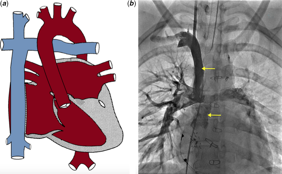
A 19-year-old female presented for pacemaker placement secondary to symptomatic sinus node dysfunction. Her underlying anatomy was congenitally corrected-transposition of the great arteries with tricuspid atresia, and she had undergone initial palliation with a pulmonary artery band followed by a bidirectional Glenn/DKS anastomosis and finally a lateral tunnel Fontan surgery at 4 years of age (Fig 1a and b). She subsequently started complaining of significant exertional fatigue and orthostatic dizziness. She underwent a 24-hour Holter monitor recording that demonstrated marked sinus bradycardia with an average heart rate of 55 bpm and a minimum heart rate of 34 bpm. A dual chamber epicardial pacemaker was placed when she was 16 years old. This surgery was complicated by a long and difficult dissection and the ability to obtain suitable myocardium for pacing. At the end of the procedure, the capture thresholds for the atrium and the ventricle were 1.0 and 0.4 mV, respectively, at a pulse width of 0.40 ms. She was followed closely by the electrophysiology service and was noted to have progressively increasing atrial lead impedances (Supplementary Fig S1). A chest radiograph confirmed fracture of her atrial lead. Her echocardiogram demonstrated normal single ventricle function with only trace mitral valve regurgitation. Review of her Holter monitor did not reveal any concern for atrioventricular node dysfunction, and it was felt that a single chamber atrial lead would be sufficient. After discussion with the family and after weighing the risks and benefits of the epicardial versus transvenous approach, the decision was made to proceed with the transvenous approach.
( a ) Diagrammatic representation of congenitally corrected transposition of the great arteries/tricuspid atresia status post Fontan palliation. ( b ) Contrast angiography demonstrating the Glenn and Fontan anastomoses (yellow arrows).

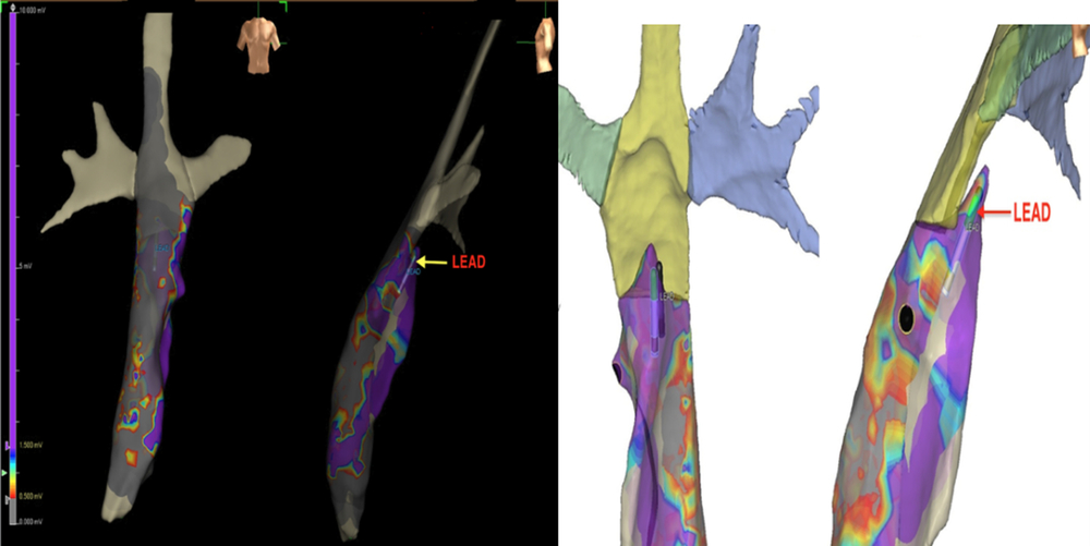
After creation of a left pre-pectoral pocket, venous access was obtained via the left subclavian vein and a 7Fr tear-away sheath was placed. A 6 French decapolar electrophysiology catheter was advanced through the sheath into Fontan pathway. The catheter was connected to the St. Jude Precision Mapping System, and an anatomic shell of the Fontan baffle from the inferior caval vein to the proximal pulmonary arteries was created. A voltage map of the Fontan baffle with the voltage scale set between 0.5 and 1.5 mV to be marked as non-viable tissue or patch material and above 1.5 mV marked as viable atrial tissue. The decapolar catheter was then exchanged for a 69 cm Medtronic 3830 transvenous pacing lead (serial # LFF150494V). The back end of the lead was connected to the St. Jude Precision system using alligator clips for real-time lead guidance which was overlaid on our previously created voltage map. Using this map as a guide, numerous sites were sampled within the Fontan baffle. A site with excellent pacing and sensing thresholds was identified in the postero-superior aspect of the baffle in an area of viable atrial tissue based on the voltage map (Fig 2). The lead was subsequently attached to the myocardium with active fixation, and stable lead position was confirmed fluoroscopically. The sensed P wave was 2.625 mV at this location, and the capture threshold was 0.50 V at 0.40 ms. The lead was attached to a generator which was placed in the previously created pre-pectoral pocket which was then closed in the usual fashion. The fluoroscopy time was 14:02 minutes, and the procedural time was 2 hours and 47 minutes. A chest radiograph the following morning demonstrated stable lead position (Supplementary Fig S2). Pacemaker analysis revealed stable capture and sensing thresholds. She was discharged home the day after the procedure. At most recent follow-up at 2.5 months post-procedure, her sensed P wave was 3.6 mV, and her capture threshold was 0.625 V at 0.4 ms. The lead impedance was also stable, and the patient had no significant complications from the procedure.
Voltage mapping used to guide lead placement in the postero-superior aspect of the Fontan baffle.
Discussion
Patients with Fontan palliation for single ventricle physiology are at significant risk of sinus node dysfunction. This may be secondary to scarring in the right atrium, direct injury to the sinus node while making the surgical incision, cannulating the heart for bypass or interruption of the sinus node artery.Reference Singh and Webster7 The literature is divided on the type of Fontan palliation associated more frequently with sinus node dysfunction (lateral tunnel versus the extracardiac Fontan).Reference Azakie, McCrindle and Van Arsdell8 The second-stage palliative surgery may have an important bearing on the incidence of subsequent sinus node dysfunction, as data demonstrate that a hemi-Fontan followed by lateral tunnel completion may have an advantage over bidirectional Glenn followed by lateral tunnel completion. This may be secondary to the lesser need for redo dissection near the sinus node and its blood supply.Reference Epstein, DiMarco and Ellenbogen9 In either case, symptomatic sinus node dysfunction is a class I indication for a permanent pacemaker placement.Reference Epstein, DiMarco and Ellenbogen9
The decision regarding approach to placement of a pacemaker in a patient with CHD and history of cardiac surgery is guided by age, complexity of the underlying anatomy, chambers which need to be paced, number of prior cardiac surgeries and shared decision making with the family. However, the nature of the Fontan palliation limits access to atrial tissue with adequate characteristics for pacing from a transvenous approach. Depending on how the Fontan baffles are created and the material of which they are made, isolated islands of “paceable” tissue may be available within the baffle itself. A prior heart catheterisation or cardiac CT can be considered to better understand the anatomical relationship of the Fontan pathway with the common atrium and determine whether transvenous pacing is potentially feasible. Consideration may be given to mapping the Fontan baffle prior to lead insertion in patients with a Fontan palliation who present for transvenous placement of pacemaker leads. This can be done at the time of the pacemaker insertion itself with an electrophysiology catheter or at a prior cardiac catheterisation. The goal of such voltage mapping is to locate areas of “paceable” tissue with low-capture thresholds and high sensing thresholds, thus avoiding placement of a permanent lead in an area of scar tissue and to also reduce the trial and error of lead implantation. This technique was first described by Kloosterman et al in a patient with Ebstein’s anomaly.Reference Kloosterman, Yamamura, Alba, Mitrani, Myerburg and Interian10 They used the Biosense CARTO system to create a voltage map to guide atrial lead position. Using the technique of voltage mapping allowed accurate identification of a location for placement of a transvenous pacemaker lead, significantly shortened procedural time, potentially reduced fluoroscopy time and reduced the risk the patient encounters from the trial-and-error method of lead placement. All of these are advantages for patients with complex CHD set against standard methods of pacemaker implantation. Others have applied this technique to children or those needing cardiac resynchronisation in an effort to minimise fluoroscopy.Reference Ruiz-Granell, Ferrero, Morell-Cabedo, Martinez-Brotons, Bertomeu and Llacer5 This case suggests that indications for this technique could be expanded to include lead implantation in patients with complex CHD in whom an epicardial pacemaker implantation is not deemed feasible.
As a counterbalance to the transvenous approach, the Fontan operation is known to increase the risk of thromboembolic complications, particularly systemic venous thrombosis secondary to increased venous pressures, a generalised low-flow state and the presence of prosthetic material.Reference Rosenthal, Friedman, Kleinman, Kopf, Rosenfeld and Hellenbrand11 The risk of thrombosis is believed to be higher in the presence of pacing leads within the baffle requiring initiation of anticoagulation therapy.Reference Leary6 This becomes particularly important when the transvenous pacing approach is utilised for a fenestrated Fontan which has the additional risk of an embolic stroke as the baffle “pops off” into the pulmonary venous atrium. Hence, it is paramount to weigh these risks against the potential benefit of avoiding a repeat sternotomy. Discussions with the family and patient are critical to enable shared decision making in this complex group of patients.
Our patient demonstrated appropriate capture and sensing thresholds in the postero-superior aspect of the Fontan baffle. We used the St. Jude Precision system to generate a voltage map to identify a suitable target for the transvenous pacemaker lead with limited fluoroscopic exposure. This was placed without any complications, and she tolerated the procedure well. Though some previous reports have reported difficulty in securing a lead in the appropriate location,Reference Alberto Lopez4 some of this is dictated by the technique used to create the Fontan baffle which affects the manoeuvrability of the lead and access to satisfactory tissue. The rate of lead dislocation may be exacerbated by the complex course the lead has to take as it courses through the innominate vein, superior caval vein, a branch of pulmonary artery and finally the Fontan baffle which is often narrowed. This tortuous course also adds to the difficulty of securing the lead in position and limits the amount of slack that is able to be left on the lead in the Fontan pathway. In spite of these challenges, transvenous pacemaker leads are an appealing alternative to epicardial pacemaker placement. In our patient, many of these potential problems were overcome with the use of a pre-formed sheath and an active fixation lead with an exposed screw. The transvenous approach spares the patient a repeat sternotomy with all its associated co-morbidities and can potentially significantly shorten the hospital stay while providing excellent pacing characteristics.
Conclusion
Electro-anatomic voltage mapping is an attractive method to guide accurate placement of transvenous pacemaker leads in patients with complicated atrial anatomy such as following the Fontan palliation. Appropriate use of this technique reduces the difficulty in locating myocardial tissue suitable for pacing and may significantly reduce procedural time. It also prevents the need for an epicardial pacemaker placement, thus avoiding a redo sternotomy in a patient with multiple prior cardiac surgeries. However, the potential benefit of this approach must be weighed against the risk of increased thromboembolic complications and the need for long-term anticoagulation.
Acknowledgements
None.
Financial support
This research received no specific grant from any funding agency, commercial or not-for-profit sectors.
Conflicts of interest
None.
Ethical standards
The authors assert that all procedures contributing to this work comply with the ethical standards of the relevant national guidelines on human experimentation and with the Helsinki Declaration of 1975, as revised in 2008. Since this is a case report, no institutional review board approval was necessary.
Supplementary material
To view supplementary material for this article, please visit https://doi.org/10.1017/S1047951119002166