Introduction
Magnetic nanoparticles (NPs), particularly superparamagnetic iron oxide nanoparticles (SPIONPs) with appropriate surface chemistry have emerged as a promising new platform in biomedical and bioengineering applications, such as protein and enzyme immobilization, tissue repair, immunoassay, magnetic resonance imaging, hyperthermia, hybridization and detection of DNA, drug delivery, etc.[Reference Gupta and Gupta1–Reference Hou, Zhou, Zhu, Wang, Hu, Zeng, Wang and Yin3] Modern applications require that these NPs have high magnetization values and size in nano regime with overall narrow particle size distribution so that the particles have identical physicochemical properties.[Reference Gupta and Gupta1] Magnetite is the most stable among iron oxide (IO; Fe3O4) with a high resistance to corrosion, superparamagnetic, high coercivity, high magnetic susceptibility, low cost, and low Curie temperature.[Reference Wu, He and Jiang4]
Currently, biomolecules-mediated synthesis of IONPs has been considered a more environmentally sound, non-toxic, biocompatible, and more cost-effective alternative to chemical and physical methods of synthesis.[Reference Nagajyothi, Pandurangan, Kim, Sreekanth and Shim5] With this view, the present study focuses the actinobacterial metabolites-mediated synthesis of SPIONPs for extended in vitro anticancer applications.
Materials and methods
Biosynthesis of magnetite-NPs
IONPs were synthesized using the co-precipitation method defined by Qu et al.[Reference Qu, Yang, Ren, Kan, Zou, Li and Li6] with minor modifications; the basic reaction involved in the reduction process is shown below:
 $${\rm 2Fe}^{{\rm 3} +} + {\rm Fe}^{{\rm 2} +} + {\rm 8NH}_{\rm 3} + {\rm 4H}_{\rm 2} {\rm O} = {\rm Fe}_{\rm 3} {\rm O}_{\rm 4} ({\rm s}) + {\rm 8NH}_{\rm 4} ^{+}.$$
For reaction, FeCl3 (0.1 mol/L) solution was added to the actinobacterial crude extract in an equal (1:1 ratio) volume. IONPs were immediately synthesized by the bioreduction process. The mixture was stirred for 120 min and then allowed to stand at room temperature for another 30 min. The reaction mixture was then centrifuged and washed several times with ethanol and then dried at 50°C under vacuum to purify the magnetite-NPs.
Characterization of NPs
Biosynthesized NPs were instrumentally characterized for determination of physicochemical properties. Fourier-transform infrared spectroscopy (FTIR) spectrum was recorded on a FTIR model EXI on KBr pellets. Phase purity and particle size were determined by x-ray diffraction (XRD) (Model Shimadzu XRD 6000) measurement with Cu-Kα x-ray source (λ-1.54056 Å) at a generator voltage 40 kV. Measurements of magnetic moment and oxidation state of paramagnetic ions of the NPs were determined using Lakeshore VSM 7410 model. Weight loss during the continuous heating and heat flux differences were studied using NetZSch TGA instrument. Energy dispersive x-ray spectroscopy (EDX) (ZWISS, Germany) was used to record the elemental composition of the sample. The shape and size of the IONPs were characterized by using electron microscopes such as field-emission transmission electron microscopy (FE-SEM; ZWISS, Germany), high-resolution transmission electron microscopy (HR-TEM; TEM JEOL JEM 2100), and selected area (electron) diffraction (SEAD) pattern analysis.
In vitro anticancer studies
HeLa (Cervical cancer cells taken from Henrietta Lacks) cell line was obtained from National Centre for Cell Sciences, Pune (NCCS). The cell line was grown in RPMI-1640 medium with optimal parameters under aseptic condition. The cytotoxicity of biosynthesized IONPs was assessed by the MTT method[Reference Mosmann7] and nuclear condensation in cells was visualized by Hoechst nuclear staining.[Reference Crowley, Marfell and Waterhouse8] The probable pathway of cell death was confirmed by acridine orange/ethidium bromide (AO/EB) dual staining method.[Reference Ho, Yazan, Ismail and Ismail9] Flow cytometric analysis was used to determine the DNA content and peaks of the apoptotic cells.[Reference Qi, Yoshida, Wang and Ueda10]
Results and discussion
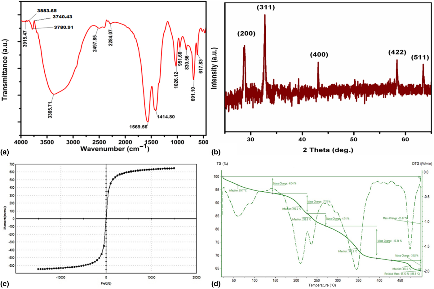
In the present study, Streptomyces sp. MA30 was isolated from magnesite mine soil samples collected from Salem, India and which is successfully used for the synthesis of IONPs with superparamagnetic properties. Conclusive properties of the synthesized magnetite-NPs could be confirmed by IR, x-ray-based spectroscopic, thermal gravimetric analysis (TGA), vibrating sample magnetometer (VSM) and electron microscopic analysis. The interactions between the Fe3+ precursor and biomolecules for capping and effective stabilization of the IONPs were confirmed by FTIR [Fig. 1(a)] analysis. The spectra clearly showed strong bands at 3365.71 (OH/Primary NH2 stretch), 2284.07 (R–C≡C–CH3–C≡C stretch), 1569.56 (C=C), 1414.80 (C=O stretch), 1026.12 (C–C stretch), 951.66 (C–C stretch), 830.56 (C–O–C stretch), 691.10 (alkyl halide), and 617.83 cm−1 (alkyl halide). Subsequently, bands at 691.10 and 617.83 cm−1 were assigned to alkyl halide functional groups, which are raised due to the metal–oxygen stretching vibration modes (Fe–O bonds) in the crystalline lattice of magnetite-NPs.[Reference Makarov, Makarova, Love, Sinitsyna, Dudnik, Yaminsky, Taliansky and Kalinina11] The phase structure and crystallite sizes of IONPs synthesized by Streptomyces sp. MA30 has been characterized using XRD [Fig. 1(b)]. All the peaks of XRD patterns were evaluated and indexed using JCPDS database, matching with IONPs standards.[Reference Prabhu, Venkateswara Rao, Siva Kumari, Kumar and Pavani12] In this study, the diffraction peaks obtained at 28.74°, 32.72°, 43.04°, 51.84°, 58.34°, and 63.43° (2θ) correspond to (220), (311), (400), (422), (511), and (440) planes of Fe3O4, respectively. The XRD peaks reveal the formation of a cubic spinel structure and were in good agreement with the JCPDS pattern 85-1436.[Reference Su, Fang, Liao, Lin, Li and Hu13]
Analysis of physicochemical properties of Fe3O4-NPs, (a – FTIR, b – XRD, c – VSM, and d – TGA).
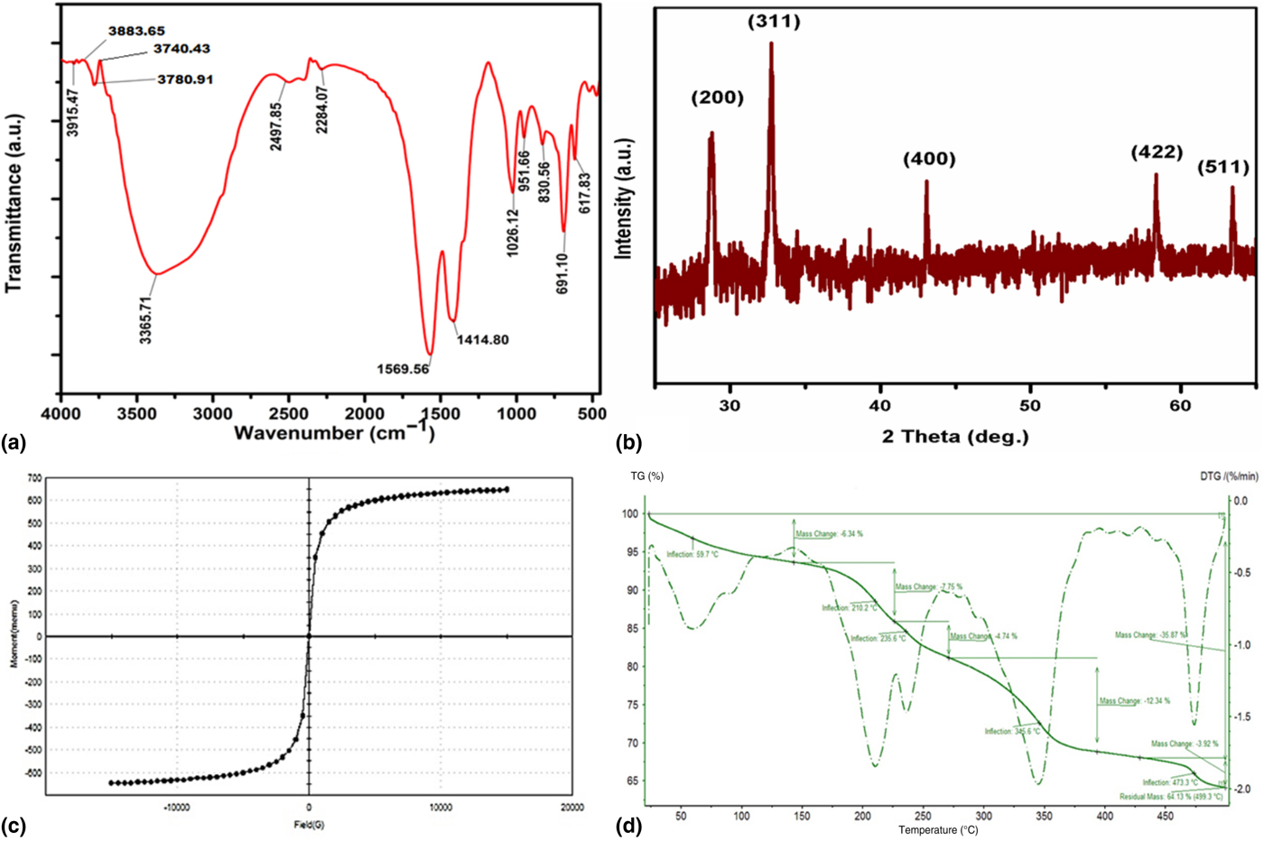
The magnetic properties of the synthesized NPs were examined by VSM analysis [Fig. 1(c)]. The hysteresis loops clearly specified that synthesized magnetite-NPs were superparamagnetic in nature.[Reference Rahman, Mohapatra and Ahmad14] Additionally, coercivity (Hci), saturation magnetization (Ms) and retentivity (Mr) values were 0 G, 64.871 and 2.0258 × 10⁻³ emu g−1, respectively. The results attained in this study were similar to those previously reported for magnetic NPs.[Reference Mahdavi, Ahmad, Haron, Namvar, Nadi, Rahman and Amin15] The TGA/derivative thermo-gravimetry (DTG) analysis of the synthesized IONPs showed in Fig. 1(d) revealed the weight loss steps from room temperature to 500°C. The maximum weight loss was obtained as 35.87% and residual mass found 64.13% in the IONPs. The weight loss obviously indicating that actinobacterial metabolites attached to the surface of the NPs were degraded at higher temperatures.[Reference Prabhu, Venkateswara Rao, Siva Kumari, Kumar and Pavani12]
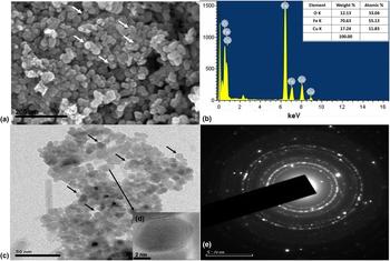
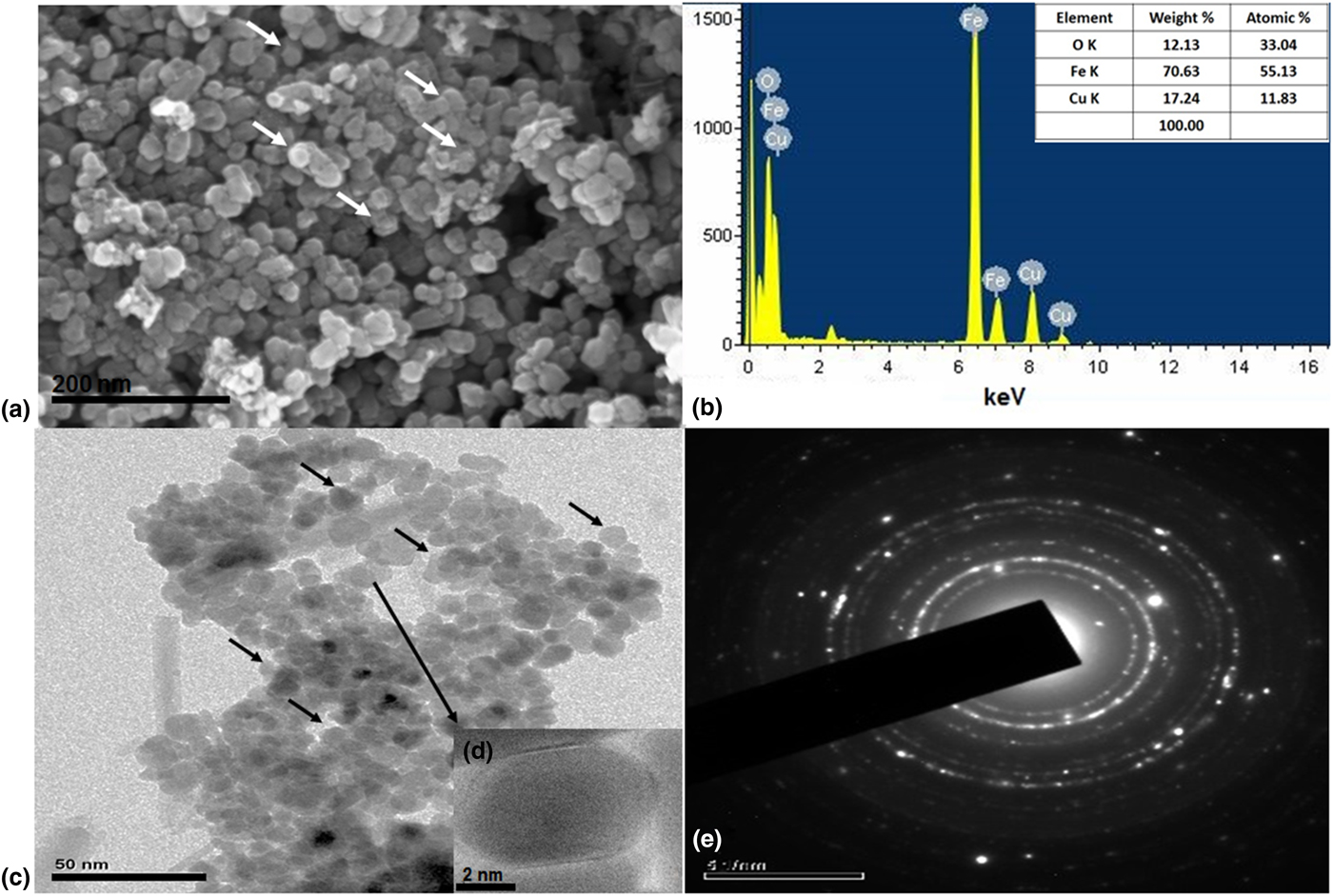
The morphology and distribution of the synthesized IONPs were confirmed by FE-SEM and HR-TEM analyses [Fig. 2(a), 2(c) and 2(d)]. The electron microscopic images established that porous nature of the NPs with size ranges from 15 to 30 nm. The EDX analysis confirmed that NPs sample composed of 70.63% Fe, 12.13% O, and 17.24% Cu, respectively [Fig. 2(b)]. The two typical absorption peaks at 6.5 and 0.6 keV were observed in EDX spectra is due to surface plasmon resonance, which confirmed that the synthesized NPs were composed of Fe and O.[Reference Prabhu, Venkateswara Rao, Siva Kumari, Kumar and Pavani12] The presence of Cu in the spectra might be raised from copper grid used in the TEM analysis. SEAD pattern of the sample reveals the well-established crystalline property of the NPs [Fig. 2(e)]. Various research groups previously established similar results.[Reference Prabhu, Venkateswara Rao, Siva Kumari, Kumar and Pavani12, Reference Mahdavi, Ahmad, Haron, Namvar, Nadi, Rahman and Amin15]
Morphology and elemental analysis of synthesized Fe3O4-NPs (a – FE-SEM image, b – EDX, c & d – HR-TEM image, and e – SAED pattern).
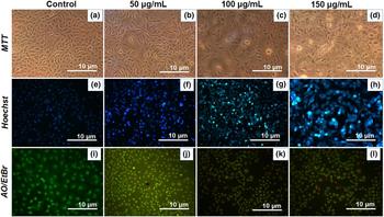
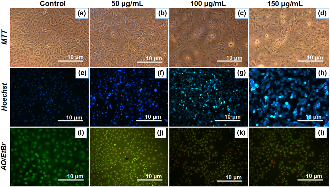
The biosynthesized IONPs are tested against the HeLa cell line. The tested NPs showed potent anticancer activity in the tested cell line. The anti-proliferative study (MTT assay) clearly showed about 50–60% of growth inhibition at 150 µg/mL concentration of NPs on the HeLa cell line [Fig. 3(a)–3(d)]. Cells with Hoechst staining is an ideal assay for differentiating apoptotic cells from viable or necrotic cells[Reference Zhivotosky, Orrenius, Bonifacino, Dasso, Harford, Schwartz and Yamada16] because apoptotic cells will generally display condensed DNA and fragmented nuclei,[Reference Errami, Naura, Kim, Ju, Suzuki, El-Bahrawy, Ghonim, Hemeida, Mansy and Zhang17] whereas live and necrotic cells do not. In the present study, Hoechst nuclear staining visualized about 55–60% of the cells in apoptotic phase, DNA of the apoptotic cells keep interacting with tested nuclear dye, such as Hoechst, whereas live cells do not [Fig. 3(e)–3(h)]. Based on the results, the IC50 value is calculated as 78.42 µg/mL. AO/EtBr dual staining technique showed that the normal viable cells appeared with green fluorescence, apoptotic cells with orange and necrotic cells with red fluorescence color. The orange and red color indicates that cells undergo nuclear shrinkage, blebbing, and loss of membrane integrity by NPs induced cytotoxicity [Fig 3(i)–3(l)].
Cytotoxicity test of Fe3O4-NPs against HeLa cell line (MTT assay – a to d, Hoechst nuclear staining – e to h, and AO/EtBr dual staining – i to l) at various concentrations.
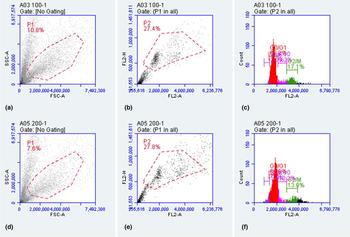
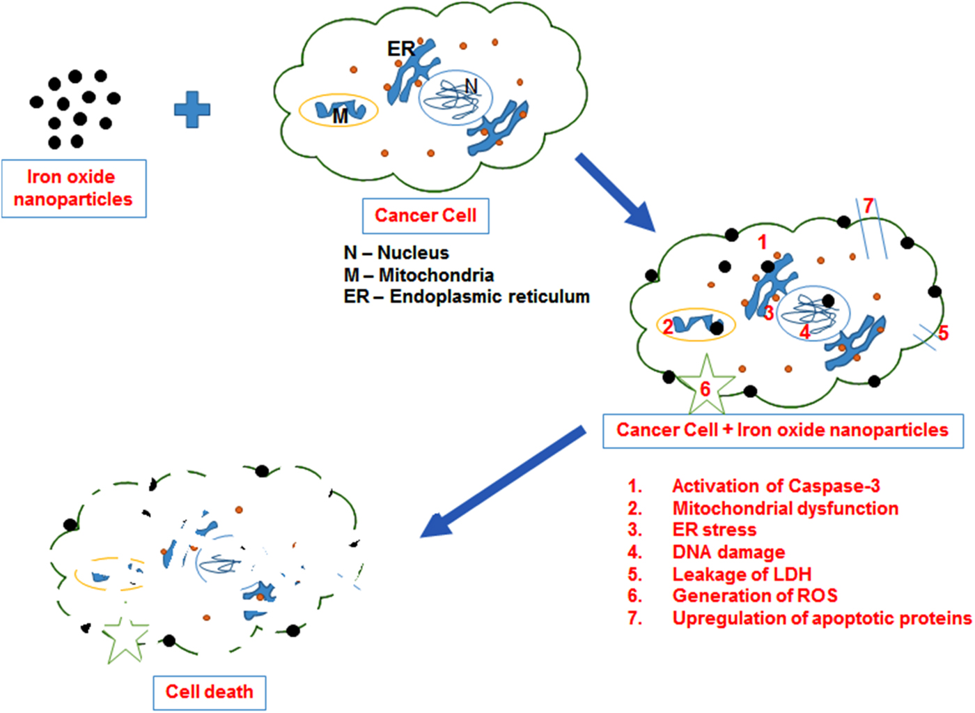
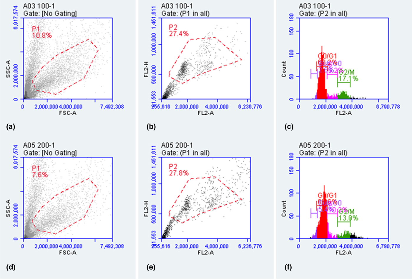
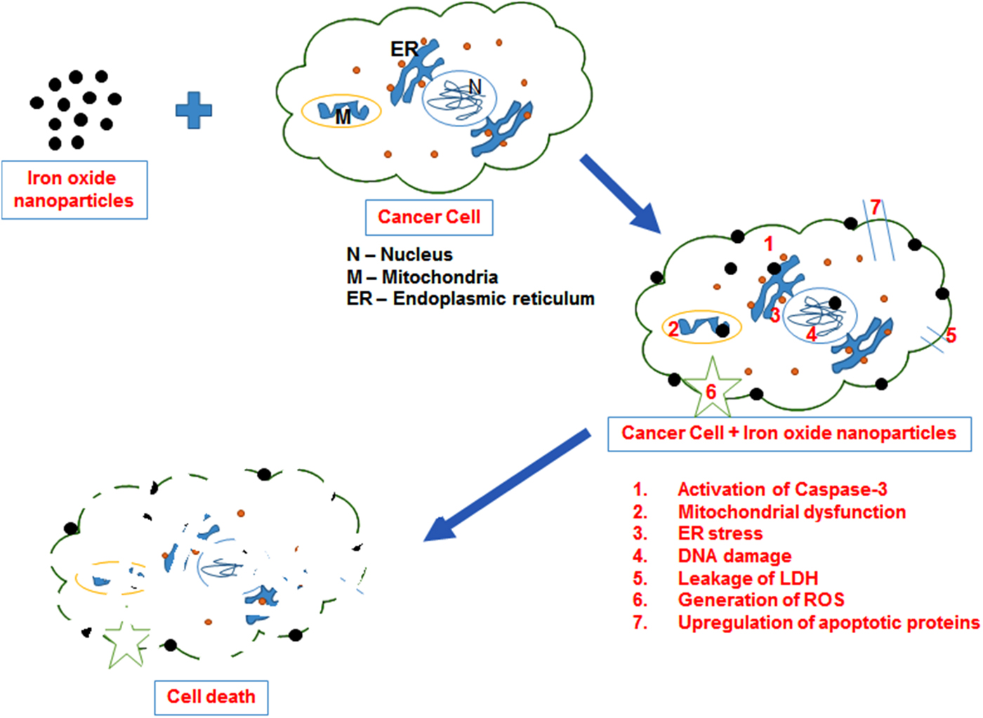
Finally, flow cytometric analysis confirmed the presence of apoptotic cells in growth phases. The control sample consists 2.64% cells in sub G0 (apoptotic) phase, 10.13% in G0/G1 phase, 9.49% in S phase and 17.14% in G2/M phase and whereas, the IONPs treated sample consist 5.43% cells in sub G0 (apoptotic) phase, 11.20% in G0/G1 phase, 9.77% in S phase and 12.95% in G2/M phase [Fig. 4(a)–4(f)]. Very few research groups published the anticancer effect of the Fe3O4 and iron NPs.[Reference Nagajyothi, Pandurangan, Kim, Sreekanth and Shim5, Reference Mahdavi, Ahmad, Haron, Namvar, Nadi, Rahman and Amin15] The possible mechanism behind the anticancer potential of IONPs on the cancer cells are represented in Fig. 5. These findings visibly indicate that synthesized IONPs in the present study may be used as a potent anticancer drug for treating human cervical cancer and as well as used for drug delivery, cancer diagnostics, etc. effectively without any side effects.
Flow cytometric analysis of control and NPs treated HeLa cells where, a to c – Control cells and d to f – Fe3O4-NPs treated HeLa cells.
Schematic diagram represent the mechanistic aspect of the iron oxide nanoparticles on cancer cells.
Conclusion
In this study, the influence of actinobacterial metabolites in the formation of superparamagnetic IONPs with effective anticancer activity was investigated. Nature of crystalline phase, magnetic property, thermal behavior and morphological features of the NPs were analyzed in detail. In vitro anticancer activity was tested on HeLa cell line and IC50 value was calculated as 78.42 µg/mL. The knowledge of present study will be helpful for further evaluation and management of actinobacterial sources for the large-scale production of IONPs for different kind of biomedical applications, particularly in advanced approaches like cancer theranostics.
Acknowledgments
One of the authors, T. Shanmugasundaram wishes to thank the SERB, India for financial support in the form of National Post-Doctoral Fellowship (PDF/2016/003119).