Introduction
Folliculogenesis is coordinated by various hormones and growth factors that are responsible for ensuring the success of follicular development (Bristol-Gould & Woodruff, Reference Bristol-Gould and Woodruff2006), but the mechanisms that control primordial follicle activation are not yet completely elucidated. Knowledge of this event is essential to understand fully female gamete development. In this respect, considerable research has been done to study the factors involved in early folliculogenesis by in vitro culture of ovarian cortical tissue from different species [bovine (Wandji et al., Reference Wandji, Srsen, Voss, Eppig and Fortune1996); goat (Silva et al., Reference Silva, Van den Hurk, Matos, Santos, Pessoa, Moraes and Figueiredo2004; Magalhães-Padilha et al., Reference Magalhães-Padilha, Fonseca, Haag, Wischral, Gastal, Jones, Geisler-Lee, Figueiredo and Gastal2012) and ovine (Peng et al., Reference Peng, Yang, Wang, Tong and Guo2010)]. Follicular activation is characterized by transformation of the pre-granulosa cells from a flattened to a cuboidal shape, followed by their proliferation. Cuboidal granulosa cells from growing follicles express proliferating nuclear antigen (PCNA), which is a nuclear protein essential for follicular growth and thus is considered to be a useful marker of proliferating granulosa cells (Wandji et al., Reference Wandji, Srsen, Voss, Eppig and Fortune1996).
Several growth factors are involved in follicular activation, survival and proliferation of granulosa cells. Studies on the expression patterns and biological functions of growth differentiation factor 9 (GDF-9) and bone morphogenetic protein 15 (BMP15) have demonstrated that they play a critical role in early follicular development (Dong et al., Reference Dong, Albertini, Nishimori, Kumar, Lu and Matzuk1996; Knight & Glister, Reference Knight and Glister2006). Moreover, various studies have shown that the interaction between Kit ligand (KL) and its receptor c-kit is important for primordial follicle activation (Parrott & Skinner, Reference Parrott and Skinner1999; Hutt et al., Reference Hutt, McLaughlin and Holland2006; Kim, Reference Kim2012), oocyte growth and survival (Jin et al., Reference Jin, Han, Yu, Wei, Hu and Liu2005), and granulosa cell proliferation (Oktay et al., Reference Oktay, Schenken and Nelson1995; Kalich-Philosoph et al., Reference Kalich-Philosoph, Roness, Carmely, Fishel-Bartal, Ligumsky, Paglin, Wolf, Kanety, Sredni and Meirow2013). However, the results achieved until now, especially on primordial follicle activation, are limited and inconclusive. To improve our knowledge about the factors that control ovarian follicle development in caprine species and to explore possible physiologic activities, it is important to conduct studies with chemical substances other than hormones and growth factors. Lectins, for example, could play an important role in folliculogenesis. These compounds have specific antigen binding to carbohydrates and are capable of interaction with several molecules of biological fluids and cell surface receptors, acting as decoders of information exchanged between molecules and cells (Misquith et al., Reference Misquith, Rani and G. Surolia1994).
Jacalin is one of the two lectins present in jackfruit (Artocarpus integrifolia) seeds. This lectin has specific residues of d-galactose and is characterized as a tetrameric molecule with a molecular mass of 39.5 kDa, consisting of α and β chains (Young et al., Reference Young, Johnston and Watson1991). Jacalin has received considerable attention because of its interesting biological properties. More importantly, it was shown to possess a potent and selective mitogenic effect on distinct T-cell and B-cell functions (Bunn-Moreno & Campos-Neto, Reference Bunn-Moreno and Campos-Neto1981). Due to its ability to stimulate subpopulations of T-cells, jacalin has been used as a mitogen factor to increase in vitro proliferation of lymphocytes (Saxon et al., Reference Saxon, Tsui and Martinez-Maza1987). Because of the ability of lectins to promote cell–cell and cell–matrix interactions, they may play a key role in proliferation, differentiation and cell migration (Stulnig et al., Reference Stulnig, Schuweiger and Hirsch-Kauffmann1993). This fact is strengthened by the demonstrated binding of jacalin to the zona pellucida and the oolemma of ovarian follicles (canine: Blackmore et al., Reference Blackmore, Baillie, Holt, Dierkx, Aitken and Mclaughlin2004; human: Jiménez-Movilla et al., Reference Jiménez-Movilla, Aviles, Gomez-Torres, Fernandez-Colom, Castells, Juan, Romeu and Ballestal2004).
The aim of the present study was to evaluate the effect of different concentrations of jacalin on the in vitro activation, survival and development of primordial follicles, and to investigate the interaction of jacalin and FSH on growth and expression of mRNA for BMP-15, KL (Kit ligand), c-kit, GDF-9 and PCNA in cultured goat ovarian cortical tissue.
Materials and methods
Chemicals
Unless mentioned otherwise, the culture media, jacalin and other chemicals used in the present study were purchased from Sigma Chemical Co. (St Louis, MO, USA).
Experiment 1: Effect of different concentrations of jacalin on activation and survival of goat primordial follicles
Ovaries (n = 12) were obtained from six cross-breed goats (Capra hircus) collected at a local slaughterhouse. Immediately postmortem, the ovaries were washed in 70% alcohol for 10 s following twice in saline solution (0.9% NaCl) that contained antibiotics (100 IU/ml penicillin and 100 mg/ml streptomycin). The ovaries were transported within 1 h to the laboratory in saline solution that contained antibiotics at 4°C.
The culture system used was described in detail earlier by Silva et al. (Reference Silva, Van den Hurk, Matos, Santos, Pessoa, Moraes and Figueiredo2004). Ovarian cortical tissue from the same ovarian pair was cut in 22 slices (3 mm × 3 mm × 1 mm) using a scissor and scalpel under sterile conditions. The tissue pieces were then either directly fixed for histology (uncultured control) or placed in culture for 1 or 6 days. Cortical tissues were transferred to 24-well culture dishes that contained 700 μl of culture media. Culture was performed at 39°C in 5% CO2 in a humidified incubator. The basic culture medium consisted of α-MEM (pH 7.2–7.4) supplemented with ITS (10 μg/ml insulin, 5.5 μg/ml transferrin, and 5 ng/ml selenium), 2 mM glutamine, 2 mM hypoxanthine, antibiotics (100 IU/ml penicillin and 100 mg/ml streptomycin), 50 μg/ml ascorbic acid, 3.0 mg/ml of bovine serum albumin (α-MEM+). The ovarian cortical fragments were cultured in control medium (α-MEM+) alone or supplemented with different concentrations of jacalin (10, 25, 50, or 100 μg/ml), in accordance with Sell & Costa (Reference Sell and Costa2002). Every 2 days, the culture medium was replaced with fresh medium. After 1 and 6 days of culture, the pieces of ovarian tissue were fixed overnight at room temperature in 4% paraformaldehyde in phosphate-buffered saline (PBS; pH 7.4) for histological studies. After fixation, the ovarian fragments were dehydrated in a graded series of ethanol, clarified with xylene, and embedded in paraffin wax. For each piece of ovarian cortex, 7-μm sections were mounted on slides and stained with eosin and hematoxylin. Coded anonymized slides were examined under a microscope (Nikon, Tokyo, Japan) at ×100 and ×400 magnification. The developmental stages of follicles were classified as primordial (one layer of flattened or flattened and cuboidal granulosa cells around the oocyte) or growing follicles (primary: one layer of cuboidal granulosa cells, and secondary: two or more layers of cuboidal granulosa cells around the oocyte). These follicles were classified further individually as histologically normal when an intact oocyte was present, surrounded by granulosa cells that are well organized in one or more layers, and have no pyknotic nucleus. Degenerated follicles were defined as those with a retracted oocyte, that has a pyknotic nucleus and/or is surrounded by disorganized granulosa cells, which are detached from the basement membrane. Overall, from 141–212 follicles were evaluated for each treatment. The percentages of healthy primordial and developing follicles were calculated before (fresh control) and after culture in a particular medium. The follicular diameter was determined as the mean of two perpendicular measures of each follicle, using an ocular micrometer. This experiment was repeated six times.
The percentages of primordial and developing follicles, as well as of those classified as morphologically normal after 1 or 6 days of culture in medium supplemented with different concentrations of jacalin were compared by Fisher's exact test (GraphPad Instat). The differences were considered significant when P < 0.05.
Experiment 2: Effects of jacalin and FSH on follicle growth and gene expression in cultured goat cortical tissue
Ovaries (n = 12) from six adult mixed-breed goats were collected as described in experiment 1. After fragmentation, for uncultured controls, some pieces of goat ovarian cortex were directly fixed for histological and ultrastructural analysis or stored at –80°C for extraction of total RNA. The remaining fragments were cultured in vitro for 6 days in 24-well culture dishes that contained 700 μl of culture medium. The basic culture medium consisted of α-MEM (pH 7.2–7.4) supplemented with ITS (10 μg/ml insulin, 5.5 μg/ml transferrin and 5 ng/ml selenium), 2 mM glutamine, 2 mM hypoxanthine, antibiotics (100 IU/ml penicillin and 100 mg/ml streptomycin), 50 μg/ml ascorbic acid, 3.0 mg/ml of bovine serum albumin (α-MEM+). Ovarian fragments were cultured at 39°C in 5% CO2 in a humidified incubator in MEM+ alone or α-MEM+ supplemented with jacalin (50 μg/ml), FSH (50 ng/ml) or both. Every 2 days, the culture medium was replaced with fresh medium. To evaluate caprine follicular morphology after 6 days of culture, the ovarian fragments were fixed overnight at room temperature in 4% paraformaldehyde in PBS (pH 7.4) for histological studies and evaluated as described in experiment 1. The follicular diameter was determined as the mean of two perpendicular measures of each follicle, using an ocular micrometer. This experiment was repeated six times.
To better examine follicular morphology, transmission electron microscopy (TEM) was performed to analyze the ultrastructure of pre-antral follicles from control, as well as from treatments that showed the best results during the histological analysis after 6 days of culture. A portion with a maximum dimension of 1 mm3 was cut from each fragment of ovarian tissue and fixed in Karnovsky solution (4% paraformaldehyde and 2.5% glutaraldehyde in 0.1 mol/l sodium cacodylate buffer, pH 7.2) for 3 h at room temperature (RT). After three washes in sodium cacodylate buffer, specimens were postfixed in 1% osmium tetroxide, 0.8% potassium ferricyanide, and 5 mmol/l calcium chloride in 0.1 mol/l sodium cacodylate buffer for 1 h at room temperature (RT). The samples were then dehydrated through a gradient of acetone solutions and embedded in SPINPON resin (Structure Probe Inc., West Chester, Pennsylvania, USA). Afterward, semi-thin sections (3 mm) were cut on an ultramicrotome (Reichert-Jung Ultra-cut 701701, Leica Microsystem, Wetzlar, Germany), stained with toluidine blue, and analyzed by light microscopy under ×400 magnification. The ultra-thin sections (60–70 nm) were contrasted with uranyl acetate and lead citrate, and examined under a Morgani-FEI TEM (Oregon). The density and integrity of ooplasmic and granulosa cell organelles, as well as vacuolization, and basement membrane integrity, were evaluated.
To evaluate the effects of jacalin, FSH and their combination on mRNA expression for BMP-15, KL, c-kit, GDF-9 and PCNA, 6-day cultured fragments from each treatment were collected and then stored at ‒80°C until extraction of total RNA.
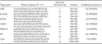
Total RNA extraction was performed using a Trizol® purification kit (Invitrogen, São Paulo, Brazil). In accordance with the manufacturer's instructions, 800 μl of Trizol solution was added to each frozen samples and the lysate was aspirated through a 20-gauge needle before centrifugation at 10,000 g for 3 min at room temperature. Thereafter, all lysates were diluted 1:1 with 70% ethanol and subjected to a mini-column. After binding of the RNA to the column, DNA digestion was performed using RNase-free DNase (340 Kunitz units/ml) for 15 min at room temperature. After washing the column three times, the RNA was eluted with 30 μl RNase-free water. The RNA concentration was estimated by reading the absorbance at 260 nm and was checked for purity at 280 nm in a spectrophotometer (Amersham Biosciences, Cambridge, UK) and 2 μg of total RNA was used for reverse transcription. Before the reverse transcription reaction, samples of RNA were incubated for 5 min at 70°C and then cooled in ice. Reverse transcription was performed in a total volume of 20 μl, which was comprised of 10 μL of sample RNA, 4 μl 5× reverse transcriptase buffer (Invitrogen, São Paulo, Brazil), 8 units RNase out, 150 units Superscript III reverse transcriptase, 0.036 U random primers (Invitrogen, São Paulo, Brazil), 10 mM DTT, and 0.5 mM of each dNTP. The mixture was incubated for 1 h at 42°C, for 5 min at 80°C, and then stored at –20°C. Negative controls were prepared under the same conditions, but without the inclusion of the reverse transcriptase. Quantification of mRNA was performed using SYBR Green. PCR reactions were composed of 1 μl cDNA as a template in 7.5 μl of SYBR Green Master Mix (PE Applied Biosystems, Foster City, CA, USA), 5.5 μl of ultra-pure water, and 0.5 μM of each primer. The primers were designed by using the PrimerQuestSM program (http://www.idtdna.com) to perform amplification of BMP-15, KL, c-kit, GDF-9, PCNA, and housekeeping genes ubiquitin-C (UBC) and β-actin (Table 1). These housekeeping genes have shown highest stability in caprine pre-antral follicles (Frota et al., Reference Frota, Leitão, Costa, van den Hurk, Brito, Saraiva, Figueiredo and Silva2011) and, thus, were used to normalize expression of target genes. The specificity of each primer pair was confirmed by melting curve analysis of PCR products. The efficiency amplification for all genes was verified according to Pfaffl et al., Reference Pfaffl2001. The thermal cycling profile for the first round of PCR was: initial denaturation and activation of the polymerase for 10 min at 95°C, followed by 50 cycles of 15 s at 95°C, 30 s at 58°C, and 30 s at 72°C. The final extension was for 10 min at 72°C. All reactions were performed in a real-time PCR Realplex (Eppendorf, Germany). The ΔΔCt method was used to transform the Ct values into normalized relative expression levels (Livak & Schmittgen, Reference Livak and Schmittgen2001).
Primer pairs used in real-time PCR

The percentages of morphologically normal follicles, as well as those of primordial and developing follicles after 6 days of culture were subjected to Fisher's exact test (P < 0.05). Levels of mRNA for BMP-15, KL, c-kit, GDF-9 and PCNA in cultured fragments were analyzed by using the non-parametric Kruskal–Wallis test (P < 0.05). Data were expressed as mean ± standard error of the mean (sem).
Results
Experiment 1: Effect of different concentration of jacalin on activation and survival of goat primordial follicles
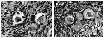
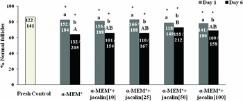
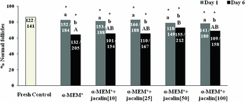
Histological analysis showed the presence of degenerated (Fig. 1A) and normal (Fig. 1B) follicles in cultured ovarian cortical fragments. After 6 days, the percentage of normal follicles after culture of ovarian tissue in different concentrations of jacalin (0, 10, 25, 50, or 100 μg/ml) had decreased significantly when compared with uncultured control or with tissues cultured for 1 day in the same condition. After 6 days, the percentage of normal follicles after culture in medium with 50 μg/ml of jacalin was significantly higher than those in tissues culture in α-MEM+ alone (Fig. 2).
Histological section of non-cultured tissue after staining with hematoxylin and eosin (H&E), showing a degenerated follicle (A) and a normal follicle (B). O, oocyte; Nu, nucleus; GC, granulosa cells (original magnification ×400).

Percentage of morphologically normal follicles in uncultured control tissue and after culture for 1 or 6 days in α-MEM+ supplemented with different concentrations of jacalin (0, 10, 25, 50 or 100 μg/ml). *Differs significantly from fresh control; A,BDifferences between treatments after 6 days. a,bDifference between days cultured (P < 0.05).
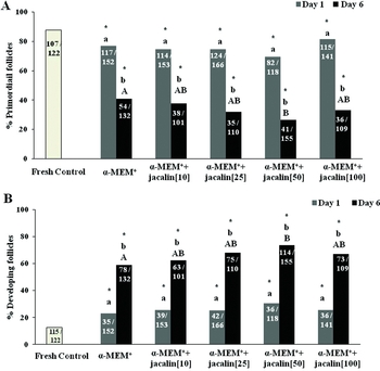
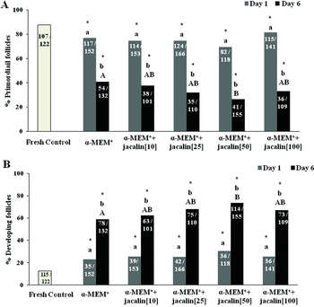
After 1 or 6 days, culture of ovarian fragments in the different media significantly reduced the percentage of primordial follicles (Fig. 3A) compared with that in fresh control tissue (P < 0.05), but increased the percentage of developing follicles (Fig. 3B). Additionally, a progressive reduction of primordial follicles and increase of developing follicles (P < 0.05) was observed with the increase of culture period from 1 to 6 days (Fig. 3A, B). Moreover, when compared to culture in α-MEM+, supplementation of 50 μg/ml of jacalin to this medium even further reduced the percentage of primordial follicles and increased the percentage of developing follicles (Fig. 3A, B). After in vitro culture, the diameter of primordial (28.1 ± 0.82 to 29.8 ± 0.77) and developing follicles (38.2 ± 0.52 to 40.0 ± 0.6) were not influenced by tested treatments.
Percentage of primordial (A) and development (B) follicles in uncultured control tissue and after culture for 1 or 6 days in α-MEM+ supplemented with different concentrations of jacalin (0, 10, 25, 50 or 100 μg/ml). *Differs significantly from fresh control. A,BDifferences between treatments after 6 days. a,bDifference between days cultured (P < 0.05).
Experiment 2: Effects of jacalin and FSH on follicles growth, gene expression in cultured ovarian cortical tissue
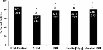
Ovarian fragments cultured in all tested media reduced the percentage of normal follicles, when compared with uncultured control (day 0) (P < 0.05). After 6 days, higher percentages of normal follicles were observed in ovarian tissue cultured in presence of FSH (50 ng/ml), jacalin (50 μg/ml) or both, when compared with control medium (α-MEM+), but no synergistic interaction between FSH and jacalin was observed (Fig. 4).
Percentage of morphologically normal follicles in uncultured control tissue (day 0), and in tissue cultured for 6 days in medium containing FSH, jacalin or both. *Differs significantly from fresh control follicles. A,BDifferences between treatments after 6 days (P < 0.05).

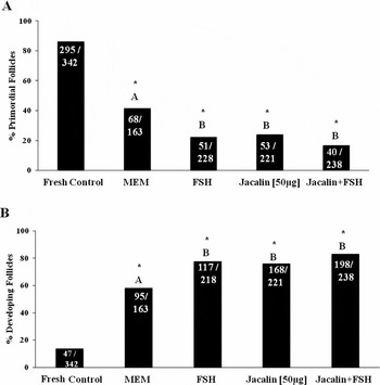
Compared to uncultured control group, a significant reduction (P < 0.05) in the percentage of primordial follicles and increase of developing follicles was observed in all other tested media after 6 days of culture. When compared with tissue cultured in control medium (α-MEM+), a significant reduction (P < 0.05) in the percentage of primordial follicles and increase of developing follicles was observed in tissues cultured in medium supplemented with FSH (50 ng/ml), jacalin (50 μg/ml) or both, but no synergistic interaction was observed between FSH and jacalin (Fig. 5A, B). After in vitro culture, the diameter of primordial (28.9 ± 0.76 to 29.8 ± 0.77) and developing follicles (38.4 ± 0.46 to 40.1 ± 0.55) were not influenced by tested treatments.
Percentage of primordial (A) and development (B) follicles in uncultured control tissue (day 0) and in tissue cultured for 6 days in medium that contained follicle stimulating hormone (FSH), jacalin or both. *Differs significantly from control fresh follicles. A,BDifferences between treatments after 6 days (P < 0.05).

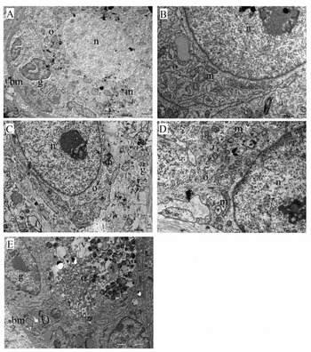
Ultrastructural analysis showed that non-cultured follicles (Fig. 6A) had rounded oocytes, well preserved organelles and nucleus with apparent and well defined nucleolus. Follicles cultured in α-MEM+ (Fig. 6B) showed oocytes with regular size and the presence of normal mitochondria and endoplasmic reticulum. Follicles cultured in α-MEM+ supplemented with FSH (Fig. 6C) presented a detachment of the oocyte from granulosa cells. In addition, follicles cultured in the presence of the jacalin alone (Fig. 6D) showed the same detachment observed in the FSH group. Organelles had well preserved ultrastructure, and it was possible to observe mitochondrial cristae, as well as endoplasmic reticulum and ribosomes. Follicles cultured in the presence of both FSH and jacalin (Fig. 6E) showed vesicular bodies and poorly preserved organelles.
Ultrastructural characteristics of ovarian follicles. (A) Follicle from uncultured control. (B) Follicle cultured in α-MEM+ presenting normal mitochondria and endoplasmic reticulum. (C) Follicle cultured in α-MEM+ supplemented with FSH, presenting a oocyte detached from granulosa cells. (D) Follicles cultured in α-MEM+ supplemented with jacalin. (E) Follicles cultured in α-MEM+ in the presence of FSH and JAC showing vesicular bodies. Legends: bm: basal membrane, g: granulosa cells, m: mitochondria, n: nucleus, o: oocyte, v: vacuole.

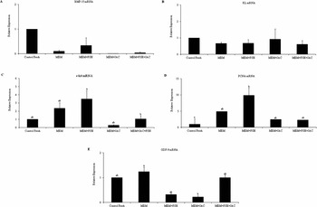
Efficiency values obtained after amplification of the primers varied from 1.00 to 1.08, showing that the values were near the theoretical optimum level. Uncultured control tissue (day 0) and tissue cultured for 6 days in α-MEM+ alone or supplemented with FSH, jacalin or both had similar levels of mRNA for BMP-15 and KL (Fig. 7A, B). In contrast, the presence of FSH in culture medium significantly increased the levels of mRNA for c-kit, when compared to tissues cultured with FSH and jacalin (Fig. 7C). In addition, the presence of FSH increased the levels of mRNA for PCNA when compared with uncultured control (P < 0.05, Fig. 7D). When compared with control medium, the level of GDF-9 mRNA was significantly reduced in ovarian tissue cultured in medium containing jacalin (Fig. 7E).
Levels of mRNA for BMP-15 (A), KL (B), c-kit (C), PCNA (D), and GDF-9 (E) in uncultured control tissue (day 0) and in tissue cultured for 6 days in medium containing FSH, jacalin or both. a,bSignificant difference between treatments (P < 0.05).

Discussion
The present study demonstrated the importance of jacalin and FSH on in vitro survival and follicular activation of caprine primordial follicles in a 6-day culture system. The percentage of primordial follicles was dramatically reduced, with a concomitant increase in the number of developing follicles after in vitro culture of ovarian tissue. Supplementation of culture medium with 50 μg/ml of jacalin or FSH caused further decrease of primordial follicles and increase of developing follicles, and contributed in maintaining follicle survival after 6 culture days. As jacalin has the property to bind carbohydrates and to improve cell to cell adhesion (Yagi et al., Reference Yagi, Campos-Neto and Gollahon1995; Sharma & Surolia, Reference Sharma and Surolia1997), this lectin may have recognized and mediated adhesion between carbohydrates, that are present in granulosa cells and oocyte. The communication between granulosa cells during the pre-antral and early antral follicle stages is necessary to ensure survival and subsequent oocyte developmental competence. The interaction between oocytes and granulosa cells during follicular development is mutually beneficial, since the oocytes regulate granulosa cell functions and inhibit apoptosis in the layers of cells immediately around the oocyte (Hussein et al., Reference Hussein, Froiland, Amato, Thompson and Gilchrist2005). On the other hand, granulosa cells support oocyte competence (Fatehi et al., Reference Fatehi, Van den Hurk, Colenbrander, Daemen, Van Tol, Monteiro, Roelen and Bevers2005).
Apart from its favorable role in cellular adhesion, jacalin may have a mitogenic function on granulosa cells from activated primordial follicles, since lectins are considered to be mitogenic factors for different kinds of cells (Bunn-Moreno & Campos-Neto, Reference Bunn-Moreno and Campos-Neto1981; Misquith et al., Reference Misquith, Rani and G. Surolia1994; Sell & Costa Reference Sell and Costa2002). It has been suggested that mitogenic lectins interact with components of cell membranes to stimulate cell proliferation (Lis & Sharon, Reference Lis and Sharon1986). Unfortunately, the exact mechanism of lectin-stimulated mitosis is unknown. Because this is the first study dealing with the effects of jacalin on the survival and growth of primordial follicles cultured in vitro follicles, our findings cannot be compared with those of other studies.
The addition of FSH and jacalin was effective in promoting follicular viability and activation, but there was no synergistic interaction between FSH and jacalin. The effect of FSH on follicular growth and survival in vitro is well described and many studies have emphasized the important role of FSH in maintaining viability (Matos et al., Reference Matos, Lima-Verde, Luque, Maia-Jr, Silva, Celestino, Martins, Báo, Lucci and Figueiredo2007a) and stimulation of in vitro follicle growth (Cecconi et al., Reference Cecconi, Barboni, Coccia and Mattioli1999; Mao et al., Reference Mao, Wu, Smith, McCauley, Cantley, Prather, Didion and Day2002; Adriaens et al., Reference Adriaens, Cortvrindt and Smitz2004; Andrade et al., Reference Andrade, Seneda and Alfieri2005; Saraiva et al., Reference Saraiva, Celestino and Chaves2008; Magalhães et al., Reference Magalhães, Araújo and Verde2009; Lima et al., Reference Lima, Rocha, Alves, Saraiva, Araújo, Lima, Lopes, Báo, Campello, Rodrigues and Figueiredo2013).
It has been demonstrated the presence of FSHR (follicle-stimulating hormone receptor) protein in primordial, primary, secondary and antral follicles in caprine ovaries (Barros et al., Reference Barros, Cavalcante, Macedo, Barberino, Lins, Gouveia, Menezes, Queiroz, Araújo, Palheta, Leite and Matos2013; Saraiva et al., Reference Saraiva, Celestino, Araújo, Chaves, Almeida, Lima-Verde, Duarte, Silva, Martins, Bruno, Matos, Campello, Silva and Figueiredo2011), which shows that FSH can act directly on very early follicle, or indirectly via factors released by larger follicles or ovarian stromal cells (Silva et al., Reference Silva, Van den Hurk, Matos, Santos, Pessoa, Moraes and Figueiredo2004). The developing follicles may produce growth factors that act in a paracrine manner on surrounding primordial follicles. Among the local factors that may be involved in early folliculogenesis are fibroblast growth factor (FGF) (Matos et al., Reference Matos, van den Hurk, Lima-Verde, Luque, Santos, Martins, Báo, Lucci and Figueiredo2007d; Songsasen et al., Reference Songsasen, Fickes, Pukazhenthi and Wildt2009; Bruno et al., Reference Bruno, Matos, Chaves, Celestino, Saraiva, Lima-Verde, Araújo and Figueiredo2009; Markholt et al., Reference Markholt, Grøndahl, Ernst, Andersen, Ernst and Lykke-Hartmann2012), epidermal growth factor (EGF) (Silva et al., Reference Silva, Van den Hurk, Matos, Santos, Pessoa, Moraes and Figueiredo2004; Tse & Ge, Reference Tse and Ge2010; Markholt et al., Reference Markholt, Grøndahl, Ernst, Andersen, Ernst and Lykke-Hartmann2012), BMP-15 (McNatty et al., Reference McNatty, Juengel, Reader, Lun, Myllymaa, Lawrence, Western, Meerasahib, Mottershead and Groome2005a; McMahon et al., Reference McMahon, Hashimoto, Mellon and Shimasaki2008; Otsuka et al., Reference Otsuka, McTavish and Shimasaki2011), KL (Parrott & Skinner, Reference Parrott and Skinner1999; Hutt et al., Reference Hutt, McLaughlin and Holland2006; Celestino et al., Reference Celestino, Matos, Saraiva and Figueiredo2009) and GDF-9 (Dong et al., Reference Dong, Albertini, Nishimori, Kumar, Lu and Matzuk1996; Orisaka et al., Reference Orisaka, Tajima, Tsang and Kotsuji2009; Myllymaa et al., Reference Myllymaa, Pasternack, Mottershead, Poutanen, Pulkki, Pelliniemi, Ritvos and Laitinen2010; Kristensen et al., Reference Kristensen, Andersen, Clement, Franks, Hardy and Andersen2013).
The ultrastructural analysis of the ovarian fragments cultured with α-MEM+ or supplemented with FSH or jacalin, demonstrated well preserved organelles such as mitochondria and endoplasmic reticulum. It is known that the lectins are able to promote cell-matrix adhesion, which can play a key role in cell morphology (Stulnig et al., Reference Stulnig, Schuweiger and Hirsch-Kauffmann1993). Jeyaprakash et al. (Reference Jeyaprakash, Jayashree, Mahanta, Swaminathan, Sekar and Vijayan2005) reported that jacalin is able to bind to d-galactose residues and has the ability to bind carbohydrates and improve cell to cell adhesion (Yagi et al., Reference Yagi, Campos-Neto and Gollahon1995; Sharma & Surolia, Reference Sharma and Surolia1997). Thus, jacalin may have recognized and mediated adhesion between carbohydrates presents in granulosa cells and oocyte and contributed to maintain the follicular ultrastructure. In addition, previous study demonstrated that jacalin increases the expression of integrin beta 3 in monocytic lineage (Yagi et al., Reference Yagi, Campos-Neto and Gollahon1995). The communication between granulosa cells during the pre-antral and early antral stages is necessary to ensure subsequent oocyte developmental competence (Albertini et al., Reference Albertini, Combelles, Benecchi and Carabatsos2001). However, in presence of both FSH and jacalin, the oocytes had vesicular bodies and poorly preserved organelles, which according to Silva et al. (Reference Silva, Báo, Lucci, Carvalho, Andrade, Ferreira and Figueiredo2001) are signs of follicular degeneration. Recently, several lectins have been speculated to possess pro-apoptotic, pro-autophagic, and anti-angiogenic properties in cancer cell lines (Fu et al., Reference Fu, Zhou, Yao, Yu, Liu and Bao2011; Li et al., Reference Li, Yu, Xu and Bao2011). In this context, Velasquez et al., (Reference Velasquez, Ríos, Ortiz, Lizama, Nuñez, Abramovich, Orge, Oliva, Orellana, Villalon, Moreno, Tesone, Rokka, Corthals, Croxatto, Parborell and Owen2013) demonstrated that the lectin concanavalin A (Con A) inhibited the stimulatory effect of FSH and induces oocyte degeneration and death of rat granulosa cells. On the other hand, addition of 10 mg/ml phytohemagglutinin (PHA) during in vitro culture caprine secondary follicles stimulated antrum formation and expression of FSH-R and PCNA mRNAs, which helped to keep ultrastructural integrity of cultured follicles (Cunha et al., Reference Cunha, Costa, Rossi, Silva, Passos, Portela, Pereira, Donato, Campello, Saraiva, Peixotos, Silva and Santos2013).
The presence of FSH in culture medium stimulated the expression of mRNA for PCNA in cultured ovarian tissue. PCNA performs the essential function of providing replicative polymerases with the high processivity required to duplicate an entire genome (Maga & Hubscher, Reference Maga and Hubscher2003) and has been used as a marker of granulosa cell proliferation in various species [bovine: (Wandji et al., Reference Wandji, Srsen, Voss, Eppig and Fortune1996); caprine: (Silva et al., Reference Silva, Van den Hurk, Matos, Santos, Pessoa, Moraes and Figueiredo2004); rodents: (Muskhelishvili et al., Reference Muskhelishvili, Wingard and Latendresse2005)]. Furthermore, the expression of PCNA by granulosa cells is closely associated with the initiation of growth primordial follicle (Figueiredo et al., Reference Figueiredo, Rodrigues, Santos, Lopes and Silva2006). Several authors have demonstrated that FSH promotes an increase in follicular diameter and proliferation of granulosa cells (rats: McGee et al., Reference McGee, Spears, Minami, Hsu, Chun, Billig and Hsueh1997; bovine: Wandji et al., Reference Wandji, Srsen, Voss, Eppig and Fortune1996; caprine: Silva et al., Reference Silva, Van den Hurk, Matos, Santos, Pessoa, Moraes and Figueiredo2004; ovine: Cecconi et al., Reference Cecconi, Barboni, Coccia and Mattioli1999). FSH-induced proliferation of granulosa cells is mediated by various paracrine factors (Van den Hurk & Zhao, Reference van den Hurk and Zhao2005).
Apart from PCNA mRNA expression, FSH also increased the expression of c-kit mRNA. The presence of jacalin, however, blocked this effect. Previous studies indicate that the KL/c-kit system is considered a key regulator of follicular growth, acting on the activation of primordial follicles and in its development (Lima et al., Reference Lima, Celestino, Figueiredo and Rodrigues2010). Lima et al. (Reference Lima, Brito, Rodrigues, Silva, Magalhães-Padilha, Lima, Celestino, Faustino, Campello, Silva, Figueiredo and Rodrigues2011) quantified the expression of mRNA for c-kit and demonstrated a decrease in the levels of c-kit mRNA during the transition from the primary to the secondary follicle stage. Because lectins, as jacalin, are able to bind to glycoproteins (Roque-Barreira & Campos-Neto, Reference Roque-Barreira and Campos-Neto1985), it is possible that binding of jacalin either to FSH or its receptors, since both are glycoproteins, can have reduced the effects FSH during culture. Previous study showed that FSH is able to control the expression of GDF-9 (Wang & Roy, Reference Wang and Roy2006). The currently found jacalin-induced reduction of GDF-9 mRNA expression can also be due to a jacalin-evoked reduced ability of FSH to bind to its receptors.
In conclusion, jacalin (50 μg/ml) and FSH (50 μg/ml) are able to improve activation and survival of caprine primordial follicles within 6-days in vitro-cultured ovarian cortical fragments. The presence of FSH increases the ovarian cortical tissue expression of mRNA for PCNA and c-kit, but jacalin blocks its effect on c-kit expression. Jacalin furthermore reduced the GDF-9 mRNA expression in cultured caprine ovarian cortical tissue. This study opens new perspectives to use jacalin as supplement in culture medium of pre-antral follicles.
Acknowledgements
This research was supported by National Council for Scientific and Technological Development (CNPq, Brazil, Grant number: 562686/2010–0).