Introduction
Lichen is a symbiotic organism usually comprised of a fungus (mycobiont) and a green algae or cyanobacterial partner (photobiont), although multiple algal genotypes in a thallus have been reported from several lichen species (Guzow-Krzeminska Reference Guzow-Krzeminska2006, Ohmura et al. Reference Ohmura, Kawachi, Kasai, Watanabe and Takeshita2006, Piercey-Normore Reference Piercey-Normore2006, Grube & Muggia Reference Grube and Muggia2010, Casano et al. Reference Casano, del Campo, García-Breijo, Reig-Armiñana, Gasulla, del Hoyo, Guéra and Barreno2011, Park et al. Reference Park, Kim, Elvebakk, Kim, Jeong and Hong2015). They are abundant and often dominant life form in Antarctic terrestrial ecosystems (Øvstedal & Lewis Smith Reference Øvstedal and Lewis Smith2001).
Lichens contain many bacterial inhabitants as well as lichenized fungi and algae. Bacterial communities in the lichen thalli have been researched by cultural approaches, fluorescence in situ hybridization (FISH) staining, molecular fingerprinting methods and recently by next-generation sequencing approaches (Scott Reference Scott1956, González et al. Reference González, Ayuso-Sacido, Anderson and Genilloud2005, Cardinale et al. Reference Cardinale, Puglia and Grube2006, Reference Cardinale, de Castro, Müller, Berg and Grube2008, Liba et al. Reference Liba, Ferrara, Manfio, Fantinatti-Garboggini, Albuquerque, Pavan, Ramos, Moreira-Filho and Barbosa2006, Grube et al. Reference Grube, Cardinale, de Castro, Muller and Berg2009, Bates et al. Reference Bates, Cropsey, Caporaso, Knight and Fierer2011, Hodkinson et al. Reference Hodkinson, Gottel, Schadt and Lutzoni2012, Lee et al. Reference Lee, Kim, Lee and Hong2014). Predominance of Alphaproteobacteria has been observed in many of the bacterial community studies (Cardinale et al. Reference Cardinale, de Castro, Müller, Berg and Grube2008, Grube et al. Reference Grube, Cardinale, de Castro, Muller and Berg2009, Hodkinson et al. Reference Hodkinson, Gottel, Schadt and Lutzoni2012) and it has also been shown that lichen-associated bacteria form highly structured, biofilm-like assemblages on fungal surfaces (Grube et al. Reference Grube, Cardinale, de Castro, Muller and Berg2009). Physiological tests of cultivable bacteria have suggested involvement of associated bacteria in nutrient cycling via nitrogen fixation, lytic activities, hormone production and phosphate mobilization (Grube et al. Reference Grube, Cardinale, de Castro, Muller and Berg2009). A phylogenetic lineage called a lichen-associated Rhizobiales lineage (LAR1), a putative nitrogen fixer, has been consistently abundant in lichens from temperate climate zones and Alaska (Hodkinson & Lutzoni Reference Hodkinson and Lutzoni2009, Bates et al. Reference Bates, Cropsey, Caporaso, Knight and Fierer2011). Antagonistic activity against other microorganisms has been another suggested role of endolichenic bacteria (Grube et al. Reference Grube, Cardinale, de Castro, Muller and Berg2009). Therefore, the microbial communities found in lichen thalli are considered to be normal flora and contribute to the lichen holotypes by providing lytic activities, complementing the lichen budget and producing bioactive substances (Grube et al. Reference Grube, Cardinale, de Castro, Muller and Berg2009).
Grube et al. (Reference Grube, Cardinale, de Castro, Muller and Berg2009) demonstrated through FISH and SSCP fingerprinting techniques that the composition of bacterial communities in lichens are host-specific; these results are supported by subsequent studies using pyrosequencing (Bates et al. Reference Bates, Cropsey, Caporaso, Knight and Fierer2011). Other analyses also revealed effects of substrate (Cardinale et al. Reference Cardinale, Steinová, Rabensteiner, Berg and Grube2012b), age of the thallus (Mushegian et al. Reference Mushegian, Peterson, Baker and Pringle2011, Cardinale et al. Reference Cardinale, Steinová, Rabensteiner, Berg and Grube2012b), photobiont type (Hodkinson et al. Reference Hodkinson, Gottel, Schadt and Lutzoni2012) and geography (Cardinale et al. Reference Cardinale, Grube, Castro, Müller and Berg2012a, Hodkinson et al. Reference Hodkinson, Gottel, Schadt and Lutzoni2012) on the bacterial microbiome composition.
Studies on the microbial community have usually been conducted for the lichens from temperate and subtropical areas (Cardinale et al. Reference Cardinale, de Castro, Müller, Berg and Grube2008, Grube et al. Reference Grube, Cardinale, de Castro, Muller and Berg2009, Bates et al. Reference Bates, Cropsey, Caporaso, Knight and Fierer2011, Hodkinson et al. Reference Hodkinson, Gottel, Schadt and Lutzoni2012). Microbial communities in polar lichens have attracted the interest of microbial ecologists, but there are limited studies on Alphaproteobacteria using culturing, fingerprinting, pyrosequencing and clone library techniques (Selbmann et al. Reference Selbmann, Zucconi, Ruisi, Grube, Cardinale and Onofri2010, Printzen et al. Reference Printzen, Fernández-Mendoza, Muggia, Berg and Grube2012, Lee et al. Reference Lee, Kim, Lee and Hong2014, Sigurbjörnsdóttir et al. Reference Sigurbjörnsdóttir, Heiðmarsson, Jónsdóttir and Vilhelmsson2014, Reference Sigurbjörnsdóttir, Andrésson and Vilhelmsson2015). Data on the bacterial community structure in Antarctic lichens will provide a basis for understanding the ecological roles of bacterial microbiomes in adaptation to extreme environments.
In the current study, we investigated bacterial diversity in Antarctic lichens through bacterial 16S rRNA genes using the 454 pyrosequencing method. To test the effects of growth form and substrate, fruticose, foliose and crustose lichens inhabiting mosses and rocks were included for comparative analyses. To our knowledge, this is the first report of bacterial communities in Antarctic lichens determined using high-throughput sequencing methods. Along with the previous study of algal and fungal communities in Antarctic lichens (Park et al. Reference Park, Kim, Elvebakk, Kim, Jeong and Hong2015), this data will provide a basis for understanding the ecological roles of the bacterial microbiomes in adaptations to specific environmental conditions.
Materials and methods
Lichen samples
Twelve lichen samples that were used for bacterial community analyses were collected from Barton and Weaver peninsulas on King George Island, Antarctica (Table I). The detailed information for locality, identification and phylogeny is described in Park et al. (Reference Park, Kim, Elvebakk, Kim, Jeong and Hong2015). DNA was isolated using a Wizard® Genomic DNA Purification kit (Promega, Madison, WI) and purified by a CTAB method as described in Park et al. (Reference Park, Kim, Elvebakk, Kim, Jeong and Hong2015).
Summary of sequencing results.

Amandinea coniops (Wahlenb.) M. Choisy ex Scheid. & H. Mayrhofer, Buellia granulosa (Darb.) C.W. Dodge, Cladonia borealis S. Stenroos, Cladonia gracilis (L.) Wild., Ochrolechia parella (L.) A. Massal, Umbilicaria antarctica Frey & I.M. Lamb, Usnea aurantico-atra (Jacq.) Bory.
Pyrosequencing, sequence processing and taxonomic assignment
Bacterial 16S rRNA gene sequences were determined by the 454 GS-FLX sequencing technique using primer sets including adapter sequences (Table S1 will be found at http://dx.doi.org/10.1017/S0954102016000286). Sequences were read only with reverse primers. Sequencing templates were prepared by pooling three independent PCR amplification products by 25 cycle reactions to reduce PCR biases.
Bacterial 16S rRNA gene sequences were pre-processed with PyroTrimmer software (Oh et al. Reference Oh, Kim, Cho, Hong and Kim2012). The pre-processing procedures included trimming of barcode, linker and primer sequences, sorting of sequences for each sample based on barcode and primer sequence information, trimming of low quality bases at the 3’ ends and filtering out sequences with low average quality scores and short sequences. The 3’ ends of sequences with low quality values were trimmed when average quality scores for a 5 bp window size were lower than 20. Sequences with ambiguous nucleotides or shorter than 180 bp were discarded. Chimeric reads were detected using USEARCH (Edgar Reference Edgar2010) with the UCHIME de novo option (Edgar et al. Reference Edgar, Haas, Clemente, Quince and Knight2011) and excluded for the downstream analysis. Bacterial 16S rRNA gene sequences of 12 lichen specimens were clustered altogether using CLUSTOM software (Hwang et al. Reference Hwang, Oh, Kim, Kim, Yu, Hou, Caetano-Anollés, Hong and Kim2013) with a 97% similarity cut-off. Taxonomic affiliation was determined using the EzTaxon-e database (Kim et al. Reference Kim, Cho, Lee, Yoon, Kim, Na, Park, Jeon, Lee, Yi, Won and Chun2012, www.ezbiocloud.net/eztaxon).
Fast UniFrac analysis
Principal co-ordinates analysis (PCoA) was performed for all bacterial OTUs and alphaproteobacterial OTUs based on distance matrices that were generated by Fast UniFrac (Hamady et al. Reference Hamady, Lozupone and Knight2009). Weighted Fast UniFrac analysis was conducted with log-transformed abundance data (percent abundance+1) and a phylogenetic tree produced by neighbour-joining analysis based on the distance matrix calculated by pairwise sequence alignment using ClustalX (Thompson et al. Reference Thompson, Gibson and Higgins2002).
Results
Sequencing of 16S rRNA gene amplicons produced 1511 to 9489 sequence reads depending on the sample after the multiple quality check procedures (Table I). The conserved primers used in the current study allowed amplification of the plastid rRNA gene as well as the bacterial 16S rRNA gene. The proportion of the bacterial sequence reads was highly variable among samples, with a range of 0.3–66.8% (Table I).
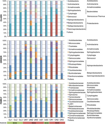
Clustering of bacterial sequences altogether resulted in 382 bacterial OTUs from 12 lichen samples. A taxonomic summary of lichen-associated bacterial communities (Fig. 1 and Table S2 found at http://dx.doi.org/10.1017/S0954102016000286) showed that Proteobacteria were typically dominant (54.7%). However, two samples, Cladonia borealis (CL1) and Ochrolechia parella (CR3), were dominated by Acidobacteria (22.9%). Other major phyla included Bacteroidetes (10.9%), Actinobacteria (6.1%), Armatimonadetes (3.4%) and Deinococcus-Thermus (1.7%). Minor phyla (0.3%) included Chloroflexi, Cyanobacteria, Firmicutes, Gemmatimonadetes, Planctomycetes, Verrucomicrobia and MATCR.
Relative abundances of major families of 11 lichen samples. Sequences that were not included in major families were pooled to ‘others’. Host lichen species are indicated by sample ID along the base of the figure. Substrates are indicated by the bar below the figure (green=moss, dark red=rock). Detailed taxonomic and abundance information for each OTU is presented in Table S2 found at http://dx.doi.org/10.1017/S0954102016000286.

The proportion of each phylum was highly variable depending on the sample. Cladonia and Usnea contained Alphaproteobacteria and Acidobacteria as major bacterial groups, and Armatimonadetes, Actinobacteria, Bacteroidetes, Gammaproteobacteria, Deltaproteobacteria and Betaproteobacteria as minor groups. In contrast, Umbilicaria contained Alphaproteobacteria, Acidobacteria, Bacteroidetes, Actinobacteria and Armatimonadetes as major bacterial groups with variation depending on the sample. Acidobacteria, Deinococcus-Thermus, Betaproteobacteria, Gammaproteobacteria and Deltaproteobacteria constituted the minor groups in Umbilicaria. The three crustose lichen samples showed highly variable microbial community structures. Buellia granulosa (CR1) and Amandinea coniops (CR2) contained Alphaproteobacteria, Gammaproteobacteria and Bacteroidetes as major groups. In contrast, most of the bacterial communities of O. parella (CR3) were occupied by Acidobacteria and Alphaproteobacteria. Betaproteobacteria, Deltaproteobacteria, Cyanobacteria, Chloroflexi, Firmicutes, Plantomycetes, Verrucomicrobia and SM2F11 were rarely recognized in the crustose samples.
The only bacterial class that was present as a major component in most of the samples was Alphaproteobacteria (13.9–67.6%). When Alphaproteobacteria was subdivided at order level, Rhodospirillales (76.4%), Rhizobiales (11.7%), Caulobacteriales (8.7%) and Sphingomonadales (2.5%) were the major orders observed. The dominant order, Rhodospirillales, was mostly occupied by Acetobacteriaceae (99.9% of Rhodospirillales sequences) and most of them (97.9%) could not be assigned to a known bacterial genus. The next major order in Alphaproteobacteria was Rhizobiales, but the frequency was highly variable depending on the sample. Rhizobiales was frequently found in the three Cladonia samples and the Usnea moss sample (US3). The most frequent OTUs in Rhizobiales belonged to Beijerinckiaceae, including the genera Beijerinckia and Methylorosula. The only three OTUs were included in LAR1 (7.8% of Rhizobiales sequences) which was defined by Hodkinson & Lutzoni (Reference Hodkinson and Lutzoni2009) and were found in one C. gracilis (CL3) and two Umbilicaria samples (UM1 and UM3).
Acidobacteria, the second major phylum, was abundant in the three Cladonia and Usnea samples, one Umbilicaria (UM3) sample and the A. coniops sample (CR2). Most of the Acidobacteria OTUs included Acidobacteriaceae (99.6% of acidobacterial sequences) and were classified as Acidobacterium, Granulicella and Terriglobus. Gammaproteobacteria was the major bacterial group in B. granulosa (CR1) and A. coniops (CR2) samples, and was mostly represented by major OTUs, Frateuria aurantia (ex Kondô & Ameyama) Swings et al. in CR1 and Rhodanobacter panaciterrae Wang et al. in CR2 (80.9% and 97.6% of Gammaproteobacteria, respectively). Bacteroidetes was the major phylum identified in the three Umbilicaria samples, and the CR1 and CR2 samples. Sphingobacteriales and Cytophagales were equally abundant in the three Umbilicaria and CR2 samples, but only Cytophagales was observed in CR1.
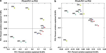
Based on a distance matrix calculated by weighted Fast UniFrac analysis for all bacterial OTUs, PCoA of 11 lichen samples, excluding US2 which contained a low frequency of bacterial sequences, resulted in close clustering of the same mycobiont genera (Fig. 2a). The three Cladonia samples and two Usnea samples with fruticose growth forms were closely related, and the three Umbilicaria samples were clustered together. In contrast, the three crustose samples of different mycobiont genera were not clustered together. When frequency data for alphaproteobacterial OTUs was used for PCoA analysis, the four moss inhabiting samples (CL1, CL2, CL3 and US3) were more closely related, and the three Umbilicaria samples, one Usnea sample (US1) and two crustose samples (CR1 and CR3) inhabiting rocks were closely related. One crustose sample (CR2) was not related to any of the other samples (Fig. 2b).
Principal co-ordinates analysis (PCoA) of all lichen-associated bacterial OTUs and b. alphaproteobacterial OTUs based on distance matrices generated by Fast UniFrac analyses.

Discussion
Lichens have long been recognized as symbiotic organisms between two major components, mycobiont and photobiont. However, it has also been recognized through culturing approaches that lichens harbour diverse bacterial species as components of the lichen thalli (González et al. Reference González, Ayuso-Sacido, Anderson and Genilloud2005, Cardinale et al. Reference Cardinale, Puglia and Grube2006, Liba et al. Reference Liba, Ferrara, Manfio, Fantinatti-Garboggini, Albuquerque, Pavan, Ramos, Moreira-Filho and Barbosa2006, Selbmann et al. Reference Selbmann, Zucconi, Ruisi, Grube, Cardinale and Onofri2010, Lee et al. Reference Lee, Kim, Lee and Hong2014). Recently FISH staining, DNA fingerprinting and high-throughput sequencing techniques have been applied to improve our understanding of the microbial community structures in lichen thalli (Cardinale et al. Reference Cardinale, de Castro, Müller, Berg and Grube2008, Grube et al. Reference Grube, Cardinale, de Castro, Muller and Berg2009, Bates et al. Reference Bates, Cropsey, Caporaso, Knight and Fierer2011, Hodkinson et al. Reference Hodkinson, Gottel, Schadt and Lutzoni2012). Studies on microbiomes in the human and animal gut suggest that microbiomes in animals affect health and body condition of host animals (Turnbaugh et al. Reference Turnbaugh, Ley, Mahowald, Magrini, Mardis and Gordon2006, Warnecke et al. Reference Warnecke, Luginbuhl and Ivanova2007). Similarly, microbiomes in lichens have ecological and physiological roles for all of the lichen thallus (Grube et al. Reference Grube, Cardinale, de Castro, Muller and Berg2009). Until now, most of the studies on microbial communities in lichen thalli have been conducted for lichens living in temperate climate zones (Grube et al. Reference Grube, Cardinale, de Castro, Muller and Berg2009, Hodkinson & Lutzoni Reference Hodkinson and Lutzoni2009, Bates et al. Reference Bates, Cropsey, Caporaso, Knight and Fierer2011) and rarely in the cold environment of the Arctic (Sigurbjörnsdóttir et al. Reference Sigurbjörnsdóttir, Heiðmarsson, Jónsdóttir and Vilhelmsson2014, Reference Sigurbjörnsdóttir, Andrésson and Vilhelmsson2015). Therefore, studies on lichens living in Antarctica will provide data complementary to the previous studies on bacterial communities in lichen thalli from temperate and Arctic areas. Fungal and algal community analyses have been performed on the same lichen samples (Park et al. Reference Park, Kim, Elvebakk, Kim, Jeong and Hong2015). Therefore, data on bacterial community structure will provide a balanced view on the microbiome composition in Antarctic lichen thalli.
Studies on bacterial communities repeatedly reveal Alphaproteobacteria as one of the major bacterial groups in lichen thalli comprising up to 60–70% of the total bacterial communities (Cardinale et al. Reference Cardinale, de Castro, Müller, Berg and Grube2008, Grube et al. Reference Grube, Cardinale, de Castro, Muller and Berg2009, Bates et al. Reference Bates, Cropsey, Caporaso, Knight and Fierer2011, Hodkinson et al. Reference Hodkinson, Gottel, Schadt and Lutzoni2012, Sigurbjörnsdóttir et al. Reference Sigurbjörnsdóttir, Andrésson and Vilhelmsson2015). This trend was continued in Antarctic lichens, although the first major bacterial group was Acidobacteria or Gammaproteobacteria in some cases (CL1, CR2 and CR3). Some crustose lichens that were in full contact with the substrate examined by Grube et al. (Reference Grube, Cardinale, de Castro, Muller and Berg2009) and Hodkinson et al. (Reference Hodkinson, Gottel, Schadt and Lutzoni2012) were shown to be different from the macrolichens. It was revealed that composition of all bacterial OTUs was dependent on the mycobiont genus from the PCoA (Fig. 2a). It was also suggested that growth form may affect the microbial community by showing that fruticose lichens, Cladonia and Usnea, were grouped together in PCoA based on bacterial OTU frequency (Fig. 2a). Substrate is proposed as another putative factor for alphaproteobacterial communities as the PCoA indicated that the composition of Alphaproteobacteria was closely related in the four lichen samples from mosses and in most of the lichens from rocks (Fig. 2b). The work of Printzen et al. (Reference Printzen, Fernández-Mendoza, Muggia, Berg and Grube2012) also suggested that the composition of lichen alphaproteobacterial communities was affected by environmental conditions. Therefore, host species, growth form and substrate might be putative factors that affect bacterial communities in lichen thalli. To understand the effects of growth form and habitat as a potential determinant of bacterial community composition more precisely, further studies with larger sample sizes are needed.
In previous studies of bacterial communities in lichen thalli, the LAR1 lineage of Rhizobiales has been recognized as an important symbiotic component that may provide fixed nitrogen to lichen ecosystems (Hodkinson & Lutzoni Reference Hodkinson and Lutzoni2009, Bates et al. Reference Bates, Cropsey, Caporaso, Knight and Fierer2011). However, LAR1 lineage was recovered from only three samples at low frequency (CL3, US3 and UM1; 7.8% of Rhizobiales). Instead, the dominant order from Alphaproteobacteria was Rhodospirillales and mostly found as Acetobacteriaceae. It is well known that many species of Acetobacteriaceae are involved in nitrogen-fixing processes (Pedraza Reference Pedraza2008, Saravanan et al. Reference Saravanan, Madhaiyan, Osborne, Thangaraju and Sa2008). Although direct evidence was not obtained in the current study, it is proposed that Acetobacteraceae might contribute to the lichen ecosystem as nitrogen fixers. Other well-known nitrogen fixers that were observed in the Antarctic lichen samples from this study included Beijerinckia, Bradyrhizobium, Burkholderia and Acinetobacter (Rösch et al. Reference Rösch, Mergel and Bothe2002, Grube et al. Reference Grube, Cardinale, de Castro, Muller and Berg2009, Bates et al. Reference Bates, Cropsey, Caporaso, Knight and Fierer2011).
To date, many researchers have speculated that lichens may be specific microecosystems (Cardinale et al. Reference Cardinale, de Castro, Müller, Berg and Grube2008, Grube et al. Reference Grube, Cardinale, de Castro, Muller and Berg2009, Bates et al. Reference Bates, Cropsey, Caporaso, Knight and Fierer2011, Park et al. Reference Park, Kim, Elvebakk, Kim, Jeong and Hong2015) and have studied the roles of lichen microbiomes. We examined bacterial diversity in Antarctic lichens to improve our understanding of the bacterial community structure. Although this study was conducted with a limited number of lichen samples, the analysis raised several interesting issues, including the complex composition of bacterial OTUs and their dependency on host species, growth form and substrate.
Acknowledgements
We are grateful for the valuable comments of the reviewers which improved this paper. This work was supported by Korea Polar Research Institute (Grant PE10050, PE11030 and PE15020).
Author contributions
C.H.P. performed the experiments, analysed the data and wrote the manuscript. S.G.H. conceptually designed the work and wrote the manuscript. K.M.K. and O.S.K. analysed the data. G.J. revised the article.
Supplementary Material
Two supplemental tables will be found at http://dx.doi.org/10.1017/S0954102016000286.