Introduction
Acute rheumatic fever is a systemic condition that impacts joints, skin, and heart following a throat infection caused by Group A beta-haemolytic streptococci. Chronic rheumatic heart disease represents the most severe long-term consequence, affecting 1.5 million patients in India. Acute rheumatic fever with severe carditis and sequelae is among the most prevalent acquired heart conditions in children. Recurrence can be prevented through secondary prophylaxis with benzathine penicillin. Educating patients and ensuring strict adherence to secondary prophylaxis can reduce morbidity and mortality. As Indians are a high-risk group, both governmental and non-governmental organisations should take proactive measures to prevent this debilitating disease.
Aim and objectives
To investigate the clinical and pathological characteristics and symptoms of children hospitalised with acute rheumatic fever and severe carditis at a tertiary care centre.
Materials and methods
This is a prospective study of 100 children admitted with acute severe rheumatic fever with severe carditis at Sri Jayadeva Institute of Cardiovascular Sciences and Research between 2015 and 2022. The study has been approved by the institutional ethics committee. Informed consent has been obtained from the patients enrolled in the study. Severe carditis was defined as the presence of signs and symptoms of heart failure and cardiomegaly on chest X-ray. Initial demographic details such as age, gender, and residence were documented. Socioeconomic status was assessed using the modified Kuppuswamy socioeconomic scale (2021). Information on previous episodes of acute rheumatic fever/chronic rheumatic heart disease, valvular involvement, and compliance with penicillin prophylaxis was gathered. Recurrence is defined as a new episode of rheumatic fever following another Group A beta-haemolytic streptococci infection, occurring >8 weeks after stopping treatment (1). Along with Jones’ criteria, symptoms like fatigue, exertional dyspnoea, recent deterioration in functional class, orthopnoea, abdominal swelling, pedal oedema and palpitations were inquired about. Physical examination findings like tachycardia, tachypnoea, low blood pressure, pedal oedema, and jugular venous pressure were noted. The presence of cardiomegaly, type, and cause of murmurs was investigated. Children with acute rheumatic fever without severe carditis were excluded. The children were divided into 3 groups based on severity of shock according to Society of Cardiovascular Angiography and Imaging classification (2).
All children underwent a haemogram, a C-reactive protein test, an erythrocyte sedimentation rate test, and an anti-streptolysin O titre test. Abnormal results were defined as C-reactive protein levels > 6 mg/dl, erythrocyte sedimentation rate > 30 mm/1st hour, and anti-streptolysin O titre > 200 Todd units. Chest X-ray, electrocardiogram, and transthoracic echocardiogram were performed. Additional factors such as presence of pulmonary hypertension, dimensions of left ventricle, ejection fraction, right ventricular performance, pericardial or pleural effusions were recorded.
The need for assisted ventilation, the duration of inotropes, diuretics, and vasodilators were recorded. Anti-inflammatory treatment included aspirin, oral prednisolone, and pulsed methyl prednisolone. The choice was based on the patient’s clinical and haemodynamic profile. Factors contributing to an extended stay in the ICU (> 96 hours) were examined. Outcomes such as mortality, persistent heart failure and necessity for immediate surgery were evaluated. The total duration of anti-inflammatory treatment was 12 weeks, with diuretics and vasodilators in selected patients.
The results are analysed using SPSS software. The categorical data is compared using the chi-square test or Fischer’s exact test (if frequencies are small), and continuous data is analysed using a one-way ANOVA test. A p-value of < 0.05 is considered significant.
Results
Incidence and demographics
The study included 100 consecutive children admitted with acute rheumatic fever and severe carditis in the paediatric cardiology unit. Acute rheumatic fever and chronic rheumatic heart disease accounted for the second highest cause (22%) of hospitalisation due to acquired heart disease in children. The majority of children were aged 10–15 years (52%), with 15–18 years comprising 35%. The median age was 13 years. Male children were 68%. A total of 63% of the children were from the upper-lower socioeconomic class, and 32% were from the lower middle class. As a tertiary care centre located in Bangalore, the institute also served children from other Indian states (Bihar, Jharkhand, Orissa, Uttar Pradesh, and Assam).
History and clinical findings
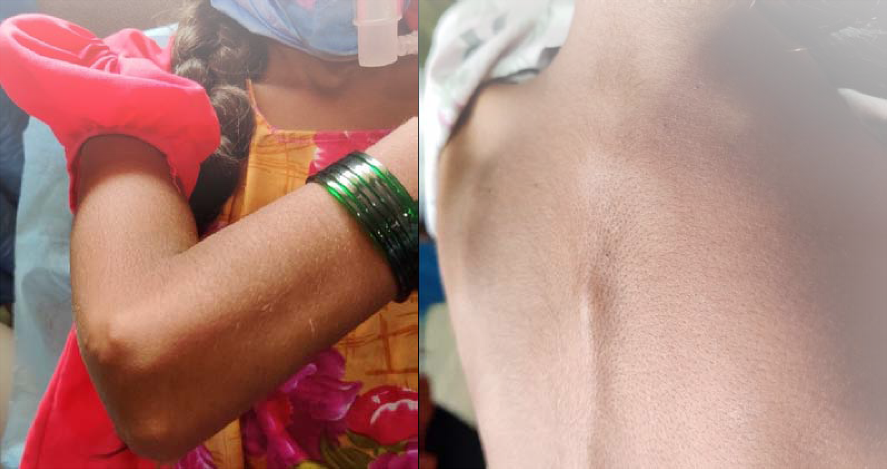
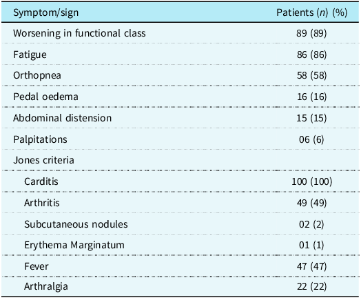
Ten children (10%) were hospitalised during the initial episode of acute rheumatic fever. Prior history of acute episode was noted in 30 (30%). Seventeen patients (56%) experienced reactivation or recurrence within the first year and 10 (33%) within 1–5 years of the initial episode. Five patients (5%) had previously undergone valve repair for chronic rheumatic heart disease and had recurrence. Rebound, defined as the return of symptoms during tapering or within 4–6 weeks after completing anti-inflammatory treatment, was observed in 4 patients (4%). A history of fever with joint pain not diagnosed as acute rheumatic fever was reported in 25%. Adherence to penicillin prophylaxis was seen in 50%. A total of 30% of the patients had no history of acute rheumatic fever or chronic rheumatic heart disease. The commonest symptom was exertional dyspnoea or a decline in functional class (89%), followed by fatigue (86%), orthopnoea (58%), pedal oedema (16%), abdominal distension (15%), and palpitations (6%). Among the Jones’ criteria, arthritis was the most prevalent, affecting 49 patients (49%). Polyarthritis was noted in 34 patients (69%), while monoarthritis in 15 patients (31%). The knee joint was most frequently affected (62%), followed by ankle (21%) and elbow (17%). Subcutaneous nodules were identified in 2 patients (Figure 1) and erythema marginatum in 1 patient. Among the minor criteria, fever was present in 47% of patients, with the commonest duration being < 10 days (43%). Ten patients who had a fever > 10 days were evaluated for infective endocarditis. Arthralgia was reported in 22%. The clinical symptoms and their frequency are illustrated in Table 1.
Subcutaneous nodules on elbow of right hand and thoracic spine in a 12 year old child.

Figure 1. Long description
The image consists of two separate photos. The left photo shows a child wearing a red shoe and a green bracelet on the wrist. The right photo shows a close-up view of the skin on the elbow of the right hand and the thoracic spine, revealing subcutaneous nodules. The nodules are visible as raised areas on the skin.
Clinical symptoms and frequency of their occurrence in all patients

The children were divided into three subgroups based on the severity of clinical signs. Eighteen patients with decompensated shock (Society of Cardiovascular Angiography and Imaging C [Classic: clinical evidence of hypoperfusion, hypotension requiring pharmacological support]) were in group A, Group B had 32 patients with compensated shock (Society of Cardiovascular Angiography and Imaging B [Beginning: clinical evidence of relative hypotension and tachycardia without hypoperfusion]), and Group C had 50 patients without shock (Society of Cardiovascular Angiography and Imaging A [At Risk: with risk factors without clinical symptoms]). All children exhibited severe carditis, tachycardia, and cardiomegaly.
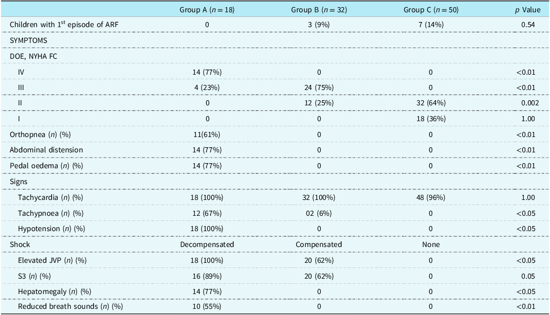
Table 2 presents the clinical characteristics of three patient subgroups, highlighting significant differences in both symptoms and signs among them. Group A exhibited a markedly more severe clinical presentation compared to Groups B and C. A large proportion of Group A patients had advanced dyspnoea on exertion (77% in NYHA Class IV), orthopnoea (61%), abdominal distension (77%), and pedal oedema (77%), all of which were absent in Groups B and C, differences that were statistically significant (p < 0.01 for each). Similarly, key clinical signs such as tachypnoea (67%), hypotension (100%), elevated jugular venous pressure (100%), hepatomegaly (77%), and reduced breath sounds (55%) were significantly more prevalent in Group A (p-values ranging from <0.05 to <0.01), suggesting acute haemodynamic compromise. The presence of an S3 gallop was also notably higher in Group A (89%) compared to B (62%) and completely absent in C (p = 0.05). In contrast, characteristics like tachycardia and the proportion of first-episode acute rheumatic fever cases did not differ significantly across groups (p = 1.00 and 0.54, respectively). Overall, these findings indicate that Group A patients had significantly worse cardiac symptoms and signs, reflecting a more severe disease state.
Clinical characteristics of the various subgroups

Investigations
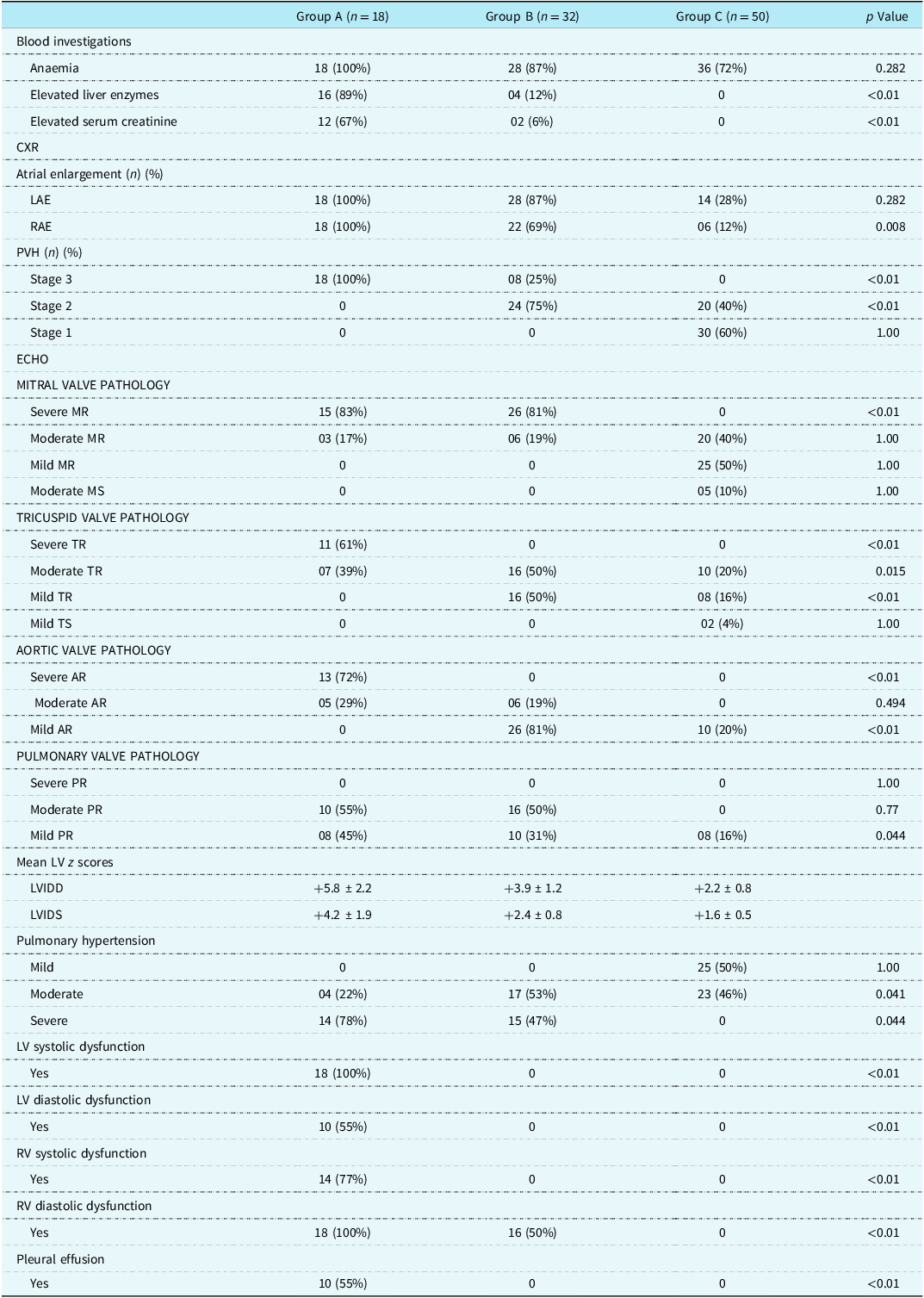
Table 3 compares the investigative and echocardiographic findings across three clinical subgroups and reveals several statistically significant differences, particularly highlighting the severity of disease in Group A. Elevated liver enzymes, and serum creatinine were significantly more frequent in Group A (89% and 67%, respectively) than in Groups B and C (p < 0.01 for both), indicating multisystem involvement. Radiologically, atrial enlargement, especially right atrial enlargement, was notably more common in Group A (100%) compared to B (69%) and C (12%), with a significant p-value of 0.008. Chest X-ray evidence of pulmonary venous hypertension was observed exclusively in Group A (Stage 3), whereas less severe stages or absence was noted in other groups (p < 0.01).
Investigations and echocardiographic characteristics of the subgroups

Echocardiography revealed significantly higher frequencies of severe valvular lesions in Group A. Severe mitral regurgitation was present in 83% of Group A patients versus none in Group C (p < 0.01). Similarly, severe tricuspid regurgitation and aortic regurgitation were significantly more prevalent in Group A (61% and 72%, respectively), with p-values < 0.01. Notably, Group A also exhibited larger left ventricular dimensions (mean LVIDD z-score + 5.8), and all patients demonstrated left ventricular systolic dysfunction (100%), a finding completely absent in Groups B and C (p < 0.01). Additionally, Group A had a significantly higher incidence of diastolic dysfunction (both left ventricular and RV), right ventricular systolic dysfunction, and pleural effusion (p < 0.01 for all). Pulmonary hypertension was also more severe in Group A (78% severe cases; p = 0.041).
Multivalvular involvement was predominantly observed in Group A patients. Among them, 11 children (61%) presented with severe regurgitation of the mitral, tricuspid, and aortic valves; these were the most critically ill. Two children (12%) exhibited severe aortic and mitral regurgitation along with moderate tricuspid regurgitation. Another two children (12%) had severe mitral regurgitation accompanied by moderate aortic and tricuspid involvement. Three children (15%) demonstrated moderate regurgitation across all the three valves.
Overall, these statistically significant findings underscore that Group A patients had far more advanced cardiac dysfunction and systemic involvement, as reflected by clinical, laboratory, radiographic, and echocardiographic abnormalities, compared to Groups B and C.
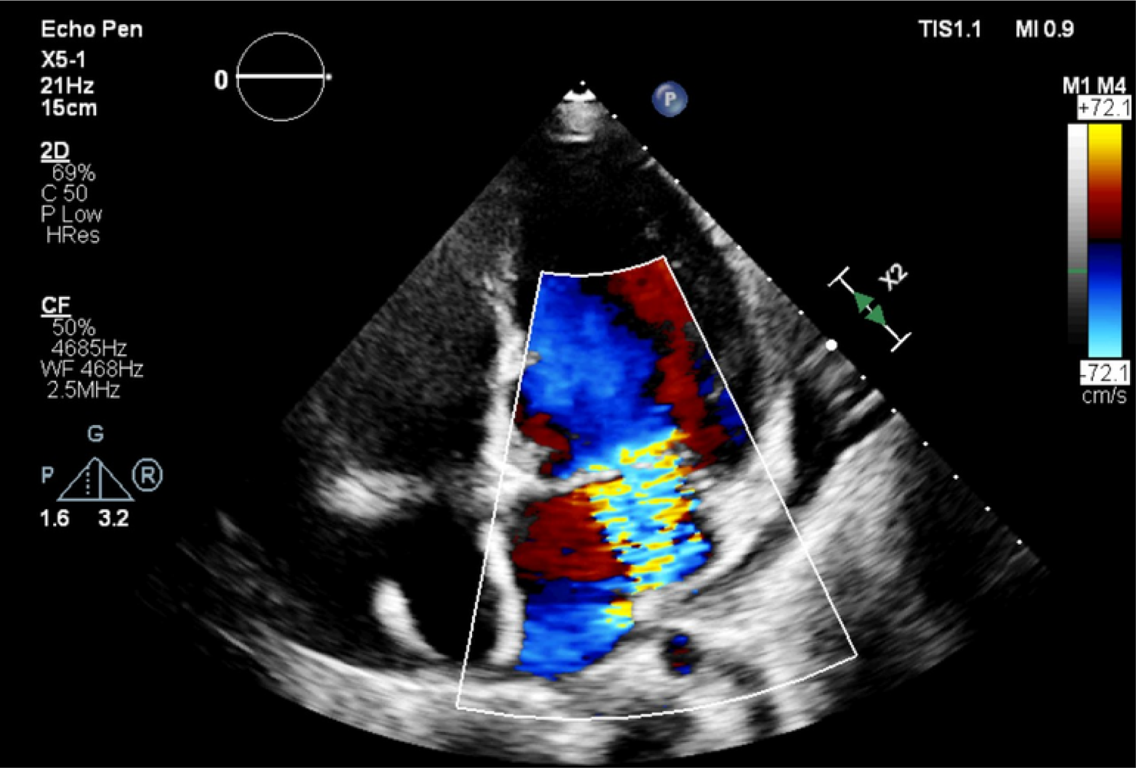
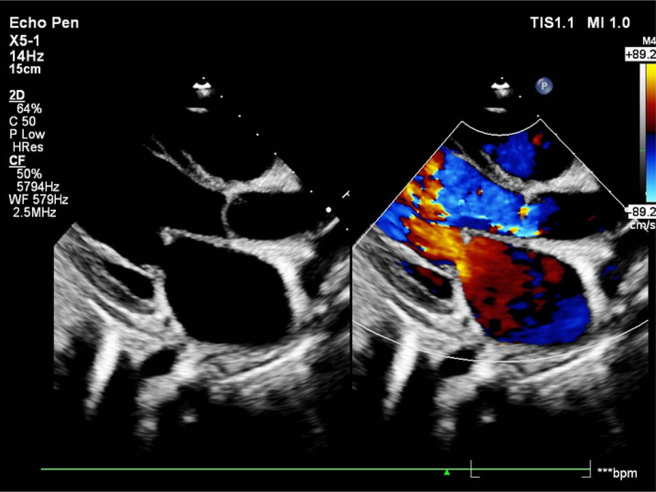
Figure 2 depicts a transthoracic echocardiogram image of a child with acute rheumatic fever, severe mitral regurgitation, and mild circumferential pericardial effusion. Figure 3 shows a transthoracic echocardiogram image of a child with chronic rheumatic heart disease and recurrent carditis with severe mitral regurgitation and severe aortic regurgitation.
Apical 4chamber view in TTE with colour interrogation of mitral valve showing severe MR. Circumferential pericardial effusion also can be noted.

Figure 2. Long description
An echocardiogram image displays a four-chamber view of the heart. Color Doppler is used to interrogate the mitral valve, revealing severe mitral regurgitation. The image also shows a circumferential pericardial effusion around the heart. The color Doppler highlights the direction and velocity of blood flow, with red and blue indicating different flow directions. The effusion appears as a dark border surrounding the heart.
PLAX view in TTE with simultaneous colour interrogation of mitral and aortic valves shows severe AR. The B mode image shows rheumatic mitral leaflets with AML thickening and leaflet tip prolapse. The restricted mobility of PML can be noted.

Figure 3. Long description
The image shows a transthoracic echocardiogram in the parasternal long-axis view with simultaneous color interrogation of the mitral and aortic valves. The B mode image reveals rheumatic mitral leaflets with anterior mitral leaflet thickening and leaflet tip prolapse. The posterior mitral leaflet shows restricted mobility. The color Doppler indicates severe aortic regurgitation.
Management
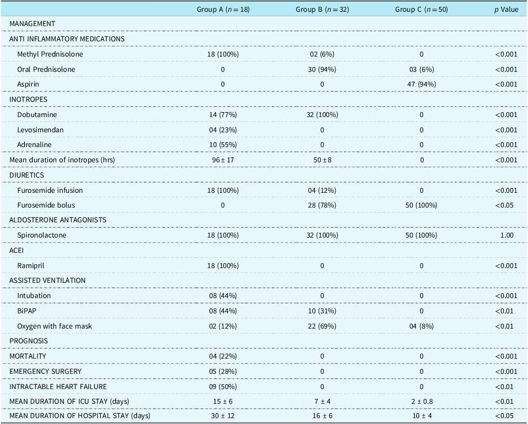
The Table 4 highlights notable differences in management and outcomes across Groups A, B, and C, with numerous statistically significant findings. The choice of initial anti-inflammatory therapy was guided by the clinical severity at presentation. Children in Group A received intravenous methylprednisolone, Group B received oral prednisolone, and Group C were initiated on anti-inflammatory doses of aspirin. Escalation of therapy was undertaken in patients who demonstrated clinical worsening, inadequate response, or intolerance to the initial treatment regimen. Multiple inotropic supports were used exclusively in Group A patients, with Dobutamine (77%) and Adrenaline (55%) usage (p < 0.001). The mean duration of inotrope use was exceptionally higher in Group A (96 ± 17 hours), showing high significance. Assisted ventilation via intubation and BiPAP was mostly confined to Group A, unlike the greater reliance on face mask oxygen in Group B (p < 0.001 to <0.01). Prognostically, Group A experienced higher rates of mortality (24%), emergency surgery (28%), and intractable heart failure (50%), all with significant p-values (< 0.001 to <0.01). Furthermore, ICU and hospital stays were prolonged in Groups B and A, respectively, with significant differences (p < 0.01 and <0.05).
Management and outcome characteristics of the subgroups

Upon admission, all children in Group A exhibited valvular morphologies consistent with chronic rheumatic heart disease, despite only a minority having a documented history of acute rheumatic fever. Among those with prior acute rheumatic fever, non-adherence to recommended secondary prophylaxis was observed. The failure to recognise initial episodes, coupled with poor compliance to secondary prophylaxis, was identified as a contributing factor to recurrent episodes within this cohort. The largest hearts were observed in children with severe regurgitation of three valves. The choice of anti-inflammatory agent in group A was methylprednisolone for 3 days, followed by an oral steroid. Nine children (50%) in group A had intractable heart failure, and five (28%) underwent emergency surgical repair without surgical mortality. A total of 3 children underwent mitral and aortic valve repair, and 2 children had replacement of both mitral and aortic valves. There were four deaths (24%) in the group. These children had severe regurgitation of both aortic and mitral valves, severe biventricular dysfunction, and intractable heart failure. The average ICU stay was 15 days, with a total hospital stay of 30 days. During the 1-year follow-up period, five children (55%) underwent surgery.
Group B received oral prednisolone at 2 mg/kg/day as the chosen anti-inflammatory treatment. Due to worsening shock symptoms, 2 (6%) children required an escalation to pulsed methylprednisolone. All children received aldosterone receptor antagonists, calcium, and magnesium supplements. All children recovered from acute decompensated heart failure, and none required emergency surgery. The average ICU stay was 7 ± 4 days, and the hospital stay averaged 16 ± 6 days. During the 1-year follow-up, 20 (62%) children underwent surgery.
Group C children were admitted to level I ICU for an average of 2 ± 0. 8 days and were administered anti-inflammatory doses of aspirin (75 mg/kg/day in 4 or 5 divided doses). All children received either a proton pump inhibitor or an antacid to prevent gastritis. Due to aspirin intolerance, 3 (6%) children were switched to oral prednisolone. None of the children in this group experienced intractable heart failure or mortality. The total hospital stay was 10 ± 4 days. During the one-year follow-up period, 18 children (36%) underwent surgery.
Discussion
Acute rheumatic fever, with its long-term sequelae, chronic rheumatic heart disease, remains a significant public health issue in developing countries. School surveys by the Indian Council of Medical Research indicate a prevalence of 2 per 1000. Reference Negi, Sondhi, Asotra, Mahajan and Mehta3 The societal impact, in terms of morbidity and economic burden, is considerable, particularly affecting adolescents and young adults. Global research funding for rheumatic heart disease is estimated at US $1.7 million, representing just 0.1% of total global health funding. Reference Shah and Narula4 Severity of carditis is a key determinant of long-term morbidity in acute rheumatic fever patients. Reference Watkins, Johnson and Colquhoun5 This study reflected that, with mortality confined to children presenting with severe lesions. While 10% of children required ICU admission during the initial episode, the remaining 90% had recurrent disease with decompensated heart failure. Recent research has focused on detecting subclinical carditis in school-aged children to better understand the community burden. Reference Sanyal, Berry, Duggal and Talwar6
There is a paucity of studies examining clinical presentation and outcomes in children with severe carditis in India. These children require close follow-up and continuous monitoring. Those who recover from severe carditis but retain more than mild residual lesions are particularly vulnerable. They are at increased risk for recurrent acute rheumatic fever, which may occur with atypical symptoms. Reference Saxena, Ramakrishnan and Roy7 Even minor haemodynamic changes can destabilise these patients. In contrast to adults with valvular heart disease, where anaemia, atrial fibrillation, thyroid disorders, and prosthetic valve dysfunction may cause decompensation, recurrent infection remains the predominant factor in paediatric rheumatic heart disease. Secondary prophylaxis with penicillin injections every 21 days can prevent recurrences. Reference Woldu and Bloomfield8 However, adherence is difficult due to the painful nature of injections and poor access to healthcare services in rural areas. Many children require a guardian to accompany them long distances for treatment.
International studies have mirrored these challenges. For instance, a cohort study in Uganda highlighted poor compliance with secondary prophylaxis as the strongest predictor of recurrence and progression to severe rheumatic heart disease, consistent with our findings. Reference Okello, Longenecker, Beaton, Kamya and Lwabi16 Similarly, data from Brazil and Pacific Island nations report high recurrence rates where community health programmes are weak or inaccessible. Reference Barik15,Reference Okello, Longenecker, Beaton, Kamya and Lwabi16 Compared to our cohort, studies in developed countries such as New Zealand and Australia have shown markedly better long-term outcomes, largely attributed to structured national registries, school-based injections, and follow-up by community nurses. Reference da Silva and Pereira17–Reference Webb and Wilson19 These programmes serve as models for countries like India, where decentralisation and outreach can make a substantial impact on compliance and morbidity.
This five-year study ensured at least one year of follow-up for all patients. Children were classified by clinical presentation and severity. Currently, no standard guidelines exist for selecting anti-inflammatory or supportive medications. The most severely affected children had moderate or greater involvement of two valves (with at least one being the aortic valve) or more than two valves. These cases required resuscitation and dual inotropic support. For such children, immediate anti-inflammatory intervention and circulatory support were essential. They received pulsed methylprednisolone for three days followed by oral steroids. Studies have demonstrated the efficacy of combining intravenous and oral steroids in managing severe carditis. Reference Manyemba and Mayosi9 A Cochrane review found no significant difference between intravenous methylprednisolone and oral prednisolone in reducing acute inflammation. Reference Torres, Lins and De Castro10 Additionally, the long-term morbidity outcomes were similar whether initial treatment involved aspirin or steroids. Reference Cilliers11
Children presenting with intractable heart failure and requiring Level II intensive care commonly had severe mitral regurgitation and aortic regurgitation. These patients also suffered hepatic and renal dysfunction due to reduced forward cardiac output. The mortality rate in this study was 4%, which is higher than previous reports. Reference Cilliers, Adler and Saloojee12 Key contributors to morbidity included NYHA functional class > II symptoms, severe AR, biventricular dysfunction (especially of the right ventricle), and shock necessitating dual inotropic support. Most patients were over 10 years of age and presented with recurrent heart failure.
From a public health perspective, our findings have critical implications. National health authorities should prioritise early detection through school screening, ensure consistent availability of benzathine penicillin, and develop community-based prophylaxis programmes. Training frontline healthcare workers in rural areas to recognise acute rheumatic fever symptoms early can prevent progression to severe carditis. Additionally, the establishment of regional rheumatic heart disease registries, modelled on programmes in New Zealand and Australia, would allow structured follow-up and surveillance. Reference Ralph, Fittock and Schultz20 Investment in mobile health technologies for appointment reminders and treatment adherence tracking may also enhance outcomes.
A strategic, multi-tiered approach is essential for children presenting with severe carditis and more than mild residual lesions. Efforts should focus on aggressive secondary prophylaxis, robust follow-up, and policies integrating acute rheumatic fever management into school and community health frameworks to reduce disease burden and improve survival.
Limitations
This study reflects a single-centre experience, with most patients hailing from south India. The precise burden and morbidity associated with paediatric acute rheumatic fever with severe carditis still require documentation.
Conclusions
In a stable child with chronic rheumatic heart disease, recurrent infection is the primary reason for decompensation. To reduce the morbidity and mortality associated with this serious disease, it is crucial to have multi-centre registries and strict adherence to secondary prophylaxis as part of national health initiatives.