Background
Tetralogy of Fallot is the most common cyanotic CHD, with an incidence of 4 of 10,000 live births.Reference Hoffman and Kaplan1 The definitive treatment for tetralogy of Fallot is complete surgical repair and relief of right ventricular outflow obstruction typically by transannular patch enlargement which inevitably can cause pulmonary insufficiency.Reference Hirsch, Mosca and Bove2 Severe pulmonary insufficiency may cause the right ventricle to dilate and stretch over time, which increases the risk of ventricular arrhythmias, heart failure, limited exercise capacity, and sudden cardiac death.Reference Carvalho, Shinebourne and Busst3–Reference Gatzoulis, Balaji and Webber6 Due to early surgical repair and revolutionary transcatheter interventions, there is a growing number of tetralogy of Fallot repair survivors.Reference Cuypers, Menting and Konings7 Despite improved outcomes, these patients are in frequent need of right ventricular outflow reinterventions.Reference Cuypers, Menting and Konings7 Transcatheter pulmonary valve implantation has revolutionised the non-surgical treatment of pulmonary insufficiency in selected patients with dysfunctional right ventricular-to-pulmonary artery conduits.Reference McElhinney, Hellenbrand and Zahn8
Case report
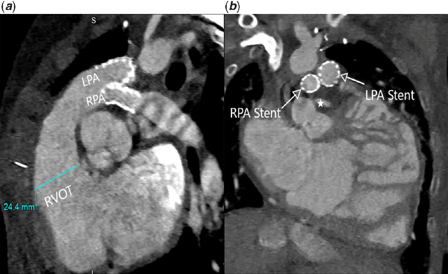
A 15-year-old female weighing 23 kg with a history of CODAS syndrome (cerebral, ocular, dental, auricular, and skeletal anomalies) and tetralogy of Fallot status-post transannular patch repair presented with class IV NYHA heart failure. She was also known to have partial anomalous right pulmonary venous return. She had bilateral branch pulmonary artery stenosis, which had previously been stented, as well as a severely dilated right ventricle and severe pulmonary insufficiency. Her other non-cardiac comorbidities include repaired intestinal malrotation, asthma, chronic respiratory failure on home oxygen as needed, and severe neuromuscular kyphoscoliosis status-post spinal fusion. Previous attempt at transcatheter pulmonary valve had evidence of coronary compression on balloon testing the main pulmonary artery. A cardiac CT scan confirmed findings of severely dilated right ventricle, bilateral branch pulmonary artery stenosis, and a left main coronary artery in close proximity to the existing right pulmonary artery stent (Fig 1). Due to lack of access and high surgical risks, she was referred for hybrid bilateral branch pulmonary artery Melody valve implantation.
CT oblique images (a, b) depicting dilated right ventricular outflow tract and branch pulmonary artery stenosis with stents in place; star depicts left main coronary artery in relation to right pulmonary artery stent. LPA = left pulmonary artery; RPA = right pulmonary artery; RVOT = right ventricular outflow tract.

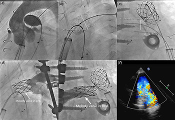
The procedure was performed under general anesthesia in the hybrid catheterisation laboratory room. Through a subxiphoid incision, direct access to the right ventricle was obtained, and an 8-Fr introducer was inserted after using pursestring suture. The sheath was used to obtain stable wire position prior to exchanging for a 22-Fr Gore DrySeal sheath (W. L. Gore & Associates, Flagstaff, AZ). Baseline right ventricular outflow angiogram confirmed dilation of the main pulmonary artery and branch pulmonary artery stenosis. Serial balloon angioplasty of the existing left and right pulmonary artery stents was performed using up to 18 mm and 14 mm Atlas Gold angioplasty catheter, respectively (Bard, Tempe, AZ). Compliance balloon testing of the right pulmonary artery was performed and revealed no evidence of coronary compression (Fig 2a). The first Melody valve was mounted on a 22 mm Ensemble delivery system and deployed across the left pulmonary artery stent. The distal diameter of the left pulmonary artery valve measured 17 mm and the proximal part measured 15 mm. The second Melody valve was shortened by folding the first zig outward due to the short landing zone; it was mounted on 14 mm balloon-in-balloon catheter (BIB, NuMED, Hopkinton, NY) and deployed across the right pulmonary artery stent (Fig 2a–e, Online video). The distal diameter of the right pulmonary valve measured 14 mm and the proximal part measured 12 mm. Post valves placement hemodynamics showed no gradient across either valve. The procedure was uncomplicated with significant reduction of pulmonary insufficiency (Fig 2f). At 2 months of follow-up, the patient was well and her echocardiogram showed no significant pulmonary insufficiency.
Serial angiography and echocardiography images. (a) No evidence of coronary compression on balloon testing the right pulmonary artery. (b, c) Post balloon angioplasty measurements of the left and right pulmonary artery. (d, e) Post Melody valve angiograms of the left and the right pulmonary artery. (f) Transthoracic echocardiography with Color Doppler demonstrating no pulmonary regurgitation post valve implantation.

Discussion
In the past 2 decades, transcatheter pulmonary valve implantation has become an increasingly well-established treatment for patients with stenotic or insufficient right ventricular outflow tracts.Reference McElhinney, Hellenbrand and Zahn8 However, many patients with dilated right ventricular outflow tracts can present a challenge for transcatheter pulmonary valve implantation due to the size limitation of the currently available valves (largest diameter Sapien XT valve 29 mm, Edwards Lifesciences, Irvine, CA). The largest non-randomised study by Qureshi et al. showed branch pulmonary artery valve implantation can be successful in patients with oversized right ventricular outflow tracts with potential benefit in the intermediate term.Reference Qureshi, Bansal and McElhinney9 Similar results by Bansal et al revealed successful implantation in seven patients with significant reduction of indexed right ventricular end diastolic volume on post-implantation cardiac MRI imaging.Reference Bansal, Kobayashi and Forbes10 Our patient demonstrated symptoms of combined right- and left-sided heart failure and was considered poor surgical candidate due to her multiple comorbidities. Additionally, she had bilateral femoral venous occlusion precluding standard femoral approach. Therefore, we elected to proceed with a hybrid technique. We were able to successfully place one valve into each pulmonary artery via hybrid technique without the need for cardiopulmonary bypass.
Conclusion
Bilateral branch pulmonary artery valve implantation via hybrid approach can be an alternative treatment for pulmonary insufficiency in certain high-risk patients with oversized right ventricular outflow tract and complex anatomy. A large multi-center study may be required to show broad applicability and long-term effects of this technique.
Supplementary material
To view supplementary material for this article, please visit https://doi.org/10.1017/S1047951120000827
Acknowledgements
None.
Financial Support
This research received no specific grant from any funding agency or from commercial or not-for-profit sectors.
Conflict of Interest
None.