Introduction
The bone morphogenetic protein (BMP) family is the largest within the TGF-β superfamily of growth factors. BMPs regulate growth, differentiation and apoptosis of a variety of tissues, including the ovary (Shimasaki et al., Reference Shimasaki, Moore, Otsuka and Erickson2004). In the ovary, the presence of BMP-6 mRNA (rat: Erickson & Shimasaki, Reference Erickson and Shimasaki2003) and protein (cow: Glister et al., Reference Glister, Kemp and Knight2004; sheep: Campbell et al., Reference Campbell, Souza, Skinner, Webb and Baird2006) has been shown across follicle development, but in caprine species, neither the levels of mRNA for BMP-6 at different stages of follicular development nor the presence of its protein have been described. In sheep, the expression of both BMP receptors, BMPR-II, BMPR-IA and BMPR-IB, has been observed in granulosa cells of follicles at all stages of folliculogenesis (Souza et al., Reference Souza, Campbell, McNeilly and Baird2002). Likewise, mRNA for BMPR-IA, BMPR-IB and BMPR-II is also detected in all types of goat follicles (Silva et al., Reference Silva, Van Den Hurk, Van Tol, Roelen and Figueiredo2004a), and also in bovine antral follicles (Glister et al., Reference Glister, Satchell and Knight2010). In vitro studies have demonstrated that BMP-6 regulates primordial follicle growth and viability (goat: Araújo et al., Reference Araújo, Magalhães, Silva, Chaves, Verde, Silva, Name, Báo, Campello, Silva, Tavares, Figueiredo and Ribeiro2010), as well as granulosa cell steroidogenesis and proliferation in antral follicles (cow: Glister et al., Reference Glister, Kemp and Knight2004; rat: Otsuka et al., Reference Otsuka, Kelly and Shimasaki2001). However, the in vitro effects of BMP-6 in the presence or absence of FSH on the development and differentiation of secondary follicles, particularly antrum formation and expression of BMP-6 and FSH-R, are unknown. Therefore the aims of the present investigation were to study: (i) the expression of BMP-6 mRNA and protein in different development stages of goat ovarian follicles; and (ii) the influence of BMP-6 on development and on BMP-6 and FSH-R expression in cultured secondary follicles.
Materials and methods
Messenger RNA quantification for BMP-6 in goat ovarian follicles
To evaluate mRNA expression, ovaries (n = 35) of adult (1–3 years old) and cycling cross-bred goats (Capra hircus) were collected and rinsed in saline (0.9% NaCl) that contained antibiotics (100 IU/ml penicillin and 100 μg/ml streptomycin). After this preparation, 10 ovaries were utilized for isolation of primordial, primary, and secondary follicles as described previously (Lucci et al., Reference Lucci, Amorim, Bao, Figueiredo, Rodrigues, Silva and Goncalves1999). After isolation, these follicles were washed and then placed by category into separate Eppendorf tubes in groups of 10. This procedure was completed within 2 h, and all samples were stored at –80°C until the RNA was extracted. From a second group of ovaries (n = 20), cumulus–oocyte complexes (COCs) were aspirated from small (1–3 mm) and large (3–6 mm) antral follicles. Compact COCs were selected from the follicle content as described by Van Tol et al. (Reference Van Tol, Van, Mummery, Van Den Hurk and Bevers1996). Thereafter, groups of 10 COCs were stored at –80°C until RNA extraction. Small (n = 10) and large antral follicles (n = 10) were isolated from the ovaries (n = 5) as described previously (Van Tol et al., Reference Van Tol, Van, Mummery, Van Den Hurk and Bevers1996). The follicles were then bisected and mural granulosa/theca tissue was collected and stored at –80°C.
Isolation of total RNA was performed using TRIzol® Plus RNA Purification Kit (Invitrogen); 1 ml of Trizol solution was added to each frozen sample and the lysate was aspirated through a 20-gauge needle before centrifugation at 10,000 g for 3 min at room temperature. All lysates were diluted 1:1 with 70% ethanol and subjected to a mini-column. After binding of the RNA to the column, DNA digestion was performed using RNase-free DNase (340 K units units/ml) for 15 min at room temperature. The column was washed three times, and the RNA was eluted with 30 μl RNase-free water.
The eluted RNA samples were incubated for 5 min at 70°C, and chilled on ice. Reverse transcription was then performed in a total volume of 20 μl, which comprised 10 μl of sample RNA, 4 μl 5× reverse transcriptase buffer, 8 units RNaseOUT™, 150 units Superscript III reverse transcriptase, 0.036 U random primers, 10 mM dithiothreitol (DTT), and 0.5 mM of each dNTP (Invitrogen). The mixture was incubated for 1 h at 42°C, for 5 min at 80°C, and then stored at –20°C. Negative controls were prepared under the same conditions but without the inclusion of the reverse transcriptase.
Quantification of the mRNA for BMP-6 was performed using SYBR Green. PCR reactions were composed of 1 μl cDNA as a template in 7.5 μl of SYBR Green Master Mix (PE Applied Biosystems), 5.5 μl of ultra-pure water, and 0.5 μM of each primer. The primers were designed to perform amplification of mRNA for BMP-6, housekeeping genes are shown in Table 1. The thermal cycling profile for the first round of PCR was: initial denaturation and activation of the polymerase for 15 min at 94°C, followed by 40 cycles of 15 s at 94°C, 30 s at 60°C, and 45 s at 72°C. The final extension was for 10 min at 72°C. All reactions were performed in a real-time PCR Mastercycler (Eppendorf). The ΔΔCT method was used to transform CT values into normalized relative expression levels.
Primer pairs used for real-time polymerase chain reaction (PCR)

Expression of BMP-6 protein in goat ovarian follicles
Ovaries (n = 10) of adult (1–3 years old) and cycling cross-bred goats (Capra hircus) were collected in a local slaughterhouse, washed with saline (0.9% NaCl) containing antibiotics (100 IU/ml penicillin and 100 μg/ml streptomycin) and transported to the laboratory at 32°C. Ovaries were fixed in paraformaldehyde (4%) for 18 h, dehydrated and embedded in paraffin. Then sections of 5 μm were obtained and mounted on glass slides. To demonstrate BMP-6, immunohistochemical reactions were performed according to a protocol described previously (Silva et al., Reference Silva, Van den Hurk, Matos, Santos, Pessoa, Moraes and Figueiredo2004b). Briefly, the epitopes were activated through incubation in citric acid to 98–100°C for 7 min, whereas non-specific binding was blocked through incubation in 5% normal goat serum diluted in phosphate-buffered saline (PBS). Then, the sections were incubated for 18 h at 4°C with monoclonal anti-BMP-6 (Wyeth Research) produced in mice (1:50). Subsequently, the sections were incubated for 45 min with secondary antibody biotinylated anti-mouse IgG (Vector Laboratories), diluted 200 times in PBS containing 5% normal goat serum. The sections were incubated for 45 min with avidin–biotin–HRP complex (1:600, Vectastain Elite ABC kits, Vector Laboratories). The location of the protein was demonstrated with diaminobenzidine (DAB) (0.05% DAB in Tris/HCl pH 7.6; 0.03% H2O2 – Sigma Chemicals). Finally, the sections were counterstained with haematoxylin, dehydrated and mounted in Canada balsam. The negative controls were carried out by replacing the primary antibody for IgG of the same species as the primary antibody was produced.
The ovarian follicles were classified as primordial, primary or secondary, as well as small (<3 mm) antral and large (>3 mm) antral follicles as described by Silva et al. (Reference Silva, Van Den Hurk, Van Tol, Roelen and Figueiredo2004a). The immunostaining was classified as absent, weak, moderate or strong in the different follicular compartments (oocyte and granulosa cells).
Effect of BMP-6 on growth of goat secondary follicles and expression of FSH-R and BMP-6 mRNA
Ovaries of adult and cycling goats (n = 10) were collected from a slaughterhouse and transported to the laboratory in minimum essential medium (MEM) that contained antibiotics (100 IU/ml penicillin and 100 μg/ml streptomycin) at 32°C within 1 h.
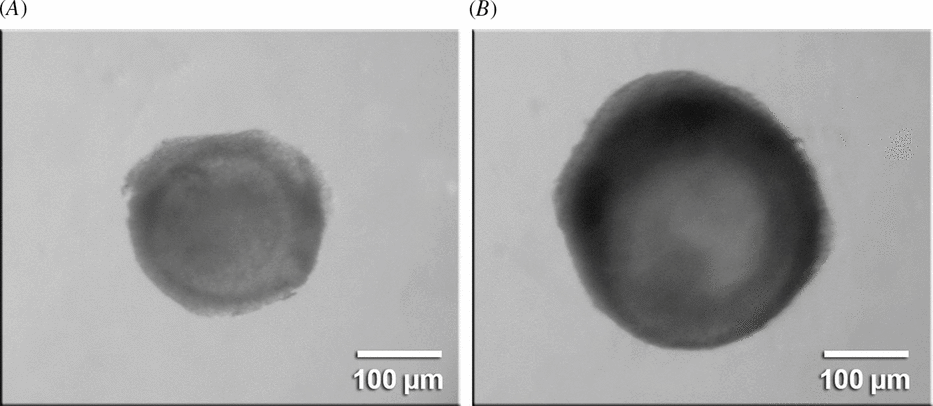
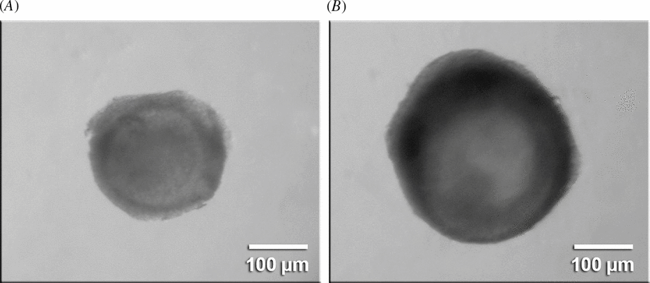
In the laboratory, surrounding fat tissue and ligaments were stripped from the ovaries. Ovarian cortical slices (1 to 2 mm in diameter) were cut from the ovarian surface using a surgical blade under sterile conditions. Subsequently the ovarian cortex was placed in fragmentation medium, consisting of MEM plus HEPES. Secondary follicles of approximately 200–230 μm in diameter were visualized under a stereomicroscope (SMZ 645 Nikon) and dissected manually from strips of ovarian cortex using 25-gauge (25G) needles. After isolation, secondary follicles were transferred to 100 μl droplets that contained fresh medium under mineral oil to further evaluate follicular quality. For secondary follicles with a visible oocyte, surrounded by granulosa cells, an intact basement membrane and no antral cavity were selected for in vitro culture (Fig. 1A).
Goat secondary follicle at day 0 (A) and antral follicle after 6 days of in vitro culture (B); oocyte surrounded by compact layer of cumulus cells.
After their selection, secondary follicles were cultured individually in 100 μl droplets of culture medium in Petri dishes (Corning). Control culture medium consisted of α-MEM (pH 7.2–7.4) supplemented with 1.25 mg/ml bovine serum albumin (BSA), ITS (insulin 10 μg/ml, transferrin 5.5 μg/ml and selenium 5 ng/ml), 2 mM glutamine, 2 mM hypoxanthine and 50 μg/ml of ascorbic acid under mineral oil (all from Sigma). For treatments, control culture medium was supplemented with 50 ng/ml of FSH (rFSH®, Nanocore,), 50 ng/ml of BMP-6 (Sigma) or both. Follicles were chosen randomly and cultured individually in 100 μl droplets of culture medium in Petri dishes (60 × 15 mm, Corning), under mineral oil, for 6 days with 5% CO2 in air at 39°C. Every other day, 60 μl of the culture medium were replaced. Morphology and follicular diameters of follicles were assessed at the beginning and end of culture with the aid of an inverted microscope. In addition, the per cent of secondary follicles that reached the antrum formation in vitro was determined.
To evaluate the effect of BMP-6 on follicular expression of mRNA for FSH-R and BMP-6 after a 6-day culture period, for each treatment, three groups of six secondary follicles, from three replicates, were collected at the end of the 6-day culture period and stored at –80°C until extraction of total RNA. Quantification of mRNA was performed as described previously and the primers for BMP-6 and FSH-R are shown in Table 1. Based on our findings that they are the most stable housekeeping genes in fresh and cultured caprine follicles (Frota et al., Reference Frota, Leitão, Costa, Brito, Van Den Hurk and Silva2010), glyceraldehyde 3-phosphate dehydrogenase (GAPDH), ubiquitin (UBQ) and β-actin were used as endogenous controls for normalization of gene expression (Table 1).
Statistical analysis
Data of mRNA expression in primordial, primary and secondary follicles were compared by non-parametric Kruskal–Wallis test, while t-test was used for comparisons of mRNA expression in small and large antral follicles (P < 0.05). Follicles diameters before and after culture were compared with the non-parametric Kruskal–Wallis test, while the chi-squared test was used to compare percentages of follicles that had reached antrum formation (P < 0.05).
Results
Messenger RNA and protein expression of BMP-6 in goat ovarian follicles
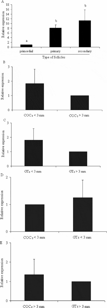
Quantification of mRNA demonstrated that primary and secondary follicles had significantly higher levels of BMP-6 than primordial follicles (P < 0.05). However, no difference in the levels of BMP-6 mRNA was observed between primary and secondary follicles (Fig. 2A). In antral follicles, the levels of mRNA for BMP-6 between COCs from small and large follicles did not differ significantly (Fig. 2B; P > 0.05). Similar results were obtained for mural granulosa/theca cells (Fig. 2C). When expression of BMP-6 mRNA was compared between COCs and granulosa/theca cell tissue of small or large antral follicles, no significant differences were observed (Fig. 2D, E; P > 0.05).
Expression of BMP-6 mRNA in goat ovarian follicles (mean ± SD). (A) Primordial, primary and secondary follicles. (B) Cumulus–oocyte complexes (COCs) from small (<3 mm) and large (>3mm) antral follicles. (C) Granulosa/theca cells (GTs) from small and large antral follicles. (D) COCs and GTs from small antral follicles. (E) COCs and GTs from large antral follicles.a,b,c Significant difference among follicles (P < 0.05).

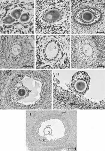
Immunohistochemical studies showed moderate immune reactivity for BMP-6 in oocytes of primordial, primary and secondary follicles (Fig. 3A–C). In addition, oocytes of small antral follicles had a strong immunoreaction (Fig. 3G), while in oocytes of large antral follicles the immunoreaction was weak (Fig. 3H). A weak immunoreaction for BMP-6 was observed in granulosa cells from primary and secondary follicles (Fig. 3B, C) and in mural granulosa cells of antral follicles (Fig. 3G, H). Cumulus and theca cells of small and large antral follicles did not immunostain for BMP-6 (Fig. 3G, H). In all types of follicles (Fig. 3D–F, I), immunoreaction was absent, when the BMP-6 antibody was replaced by IgG of the same species as the primary antibody was raised in.
BMP-6 immunoreactivity in oocytes (O) and granulosa cells (G) of primordial (A), primary (B) and secondary follicles (C), as well as in oocytes (O), mural granulosa (MGC), cumulus (CC) and theca cells (T) of small (G) and large antral follicles (H). Negative controls for each follicular category (D–F, I). Scale bars represent 30 μm (A–F) and 50 μm (G–I).

Growth of goat secondary follicles after culture
After culture of secondary follicles for 6 days, a significant increase (P < 0.05) in follicular diameter was observed in all treatments, when compared with day 0 (Table 2). Compared with culture in α-MEM alone, additional treatment with BMP-6, FSH or a mixture of BMP-6 and FSH significantly augment the follicular diameter (P < 0.05). The percentages of secondary follicles that reached antrum formation after culture in medium supplemented with BMP-6 (Fig. 1B) were significantly higher (P < 0.05) than those cultured in α-MEM alone, but no difference was observed with those cultured in presence of FSH or both FSH and BMP-6 (Table 2).
Follicular growth and antrum formation in goat secondary follicles cultured with BMP-6 in the presence or absence of follicle stimulating hormone (FSH)

a Significant difference between day 0 and day 6 (P < 0.05).
b, c Significant difference among treatments (P < 0.05).
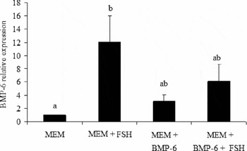
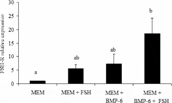
Evaluation of mRNA levels for FSH-R and BMP-6 in cultured follicles showed that α-MEM supplemented with FSH significantly enhanced the levels of BMP-6 mRNA compared with culture in α-MEM alone (P < 0.05; Fig. 4), while addition of BMP-6 alone or with FSH had no significant effect. In contrast, addition to α-MEM of both BMP-6 and FSH was necessary to increase FSH-R mRNA levels in cultured secondary follicles (P < 0.05; Fig. 5).
Steady state level of mRNA for BMP-6 in goat secondary follicles cultured for 6 days in MEM supplemented with FSH, BMP-6 or both compounds. MEM is considered calibrator and has level of mRNA equal to one in all repetitions. a,bSignificant difference among treatments (P < 0.05).

Steady state level of mRNA for FSH-R in goat secondary follicles cultured for 6 days in MEM supplemented with FSH, BMP-6 or both compounds. MEM is considered calibrator and has level of mRNA equal to one in all repetitions. a,bSignificant difference among treatments (P < 0.05).

Discussion
The present real-time PCR studies show BMP-6 mRNA expression in goat follicles from the primordial follicle stage onwards and an increase in the level of BMP-6 mRNA during the transition of caprine primordial into primary/secondary follicles. When using the in situ hybridization technique, in rat follicles, expression of BMP-6 mRNA was first detectable in secondary follicles (Erickson & Shimasaki, Reference Erickson and Shimasaki2003) but, in sheep, mRNA for BMP-6 was found in oocytes from all follicle classes (Juengel et al., Reference Juengel, Reader, Bibby, Lun, Ross, Haydon and McNatty2006). With use of RT-PCR, BMP-6 mRNA was demonstrated in ovine antral follicles (Xu et al., Reference Xu, Li, Han, Chen and Xie2010). In the goat antral follicles under study, COCs and mural granulosa cells/theca cells both express BMP-6 mRNA. In contrast, in mouse antral follicles, expression of BMP-6 mRNA was only restricted to oocytes (Elvin et al., Reference Elvin, Yan and Matzuk2000). In antral follicles, it has been shown that BMP-6 regulates granulosa cell steroidogenesis and proliferation (cow: Glister et al., Reference Glister, Kemp and Knight2004; rat: Otsuka et al., Reference Otsuka, Kelly and Shimasaki2001).
The present histochemical studies demonstrate BMP-6 protein in oocytes from all classes of caprine follicles, particularly in those from small antral follicles. In granulosa cells, BMP-6 staining was present in primary, secondary and antral follicles. Presence of BMP-6 immunoreactivity in granulosa cells indicate that mRNA was translated into functional protein that is involved in the control of steroidogenesis and proliferation (Glister et al., Reference Glister, Kemp and Knight2004). Similarly, immunohistochemical studies reported previously the presence of BMP-6 protein in bovine (Glister et al., Reference Glister, Kemp and Knight2004) and human (Shi et al., Reference Shi, Yoshino, Osuga, Koga, Hirota, Hirata, Yano, Nishii and Taketani2009) oocytes and granulosa cells, but only in those from collected antral follicles.
The increase of BMP-6 mRNA in caprine follicles beyond the primordial stage and the distribution of BMP-6 mRNA and protein in different follicle compartments may point to the importance of BMP-6 as an intracrine, autocrine or paracrine growth factor in developing follicles only. This opinion is strengthened by results of recent in vitro studies from our group that have shown the inability of BMP-6 to activate goat primordial follicles during their culture for 7 days (Araújo et al., Reference Araújo, Magalhães, Silva, Chaves, Verde, Silva, Name, Báo, Campello, Silva, Tavares, Figueiredo and Ribeiro2010). On the other hand, mRNAs for BMP receptors (BMPR-IA, BMPR-IB, and BMPR-II) are expressed in primordial follicles and all other classes of caprine ovarian follicles (Silva et al., Reference Silva, Van Den Hurk, Van Tol, Roelen and Figueiredo2004a) as well as in follicles of other mammalian species (murine: Elvin et al., Reference Elvin, Yan and Matzuk2000; ovine: Souza et al., Reference Souza, Campbell, McNeilly and Baird2002; Xu et al., Reference Xu, Li, Han, Chen and Xie2010; McNatty et al., Reference McNatty, Galloway, Wilson, Smith, Hudson and O'Connell2005; bovine: Glister et al., Reference Glister, Satchell and Knight2010; human: Abir et al., Reference Abir, Ben-Haroush, Melamed, Felz, Krissi and Fisch2008), but the possibility that mRNAs for BMP receptors are not translated into functional proteins in primordial follicles cannot be excluded.
This study furthermore shows growth of caprine secondary follicles that had been cultured with BMP-6 both in the presence and in absence of FSH. Besides, the percentage of cultured follicles starting antrum formation has been increased significantly only after treatment with BMP-6 alone. BMP-6 modulates proliferative and differentiative responses of granulosa cells to FSH stimulation (rat: Shimasaki et al., Reference Shimasaki, Moore, Otsuka and Erickson2004; sheep: Campbell et al., Reference Campbell, Souza, Skinner, Webb and Baird2006). Juengel et al. (Reference Juengel, Reader, Bibby, Lun, Ross, Haydon and McNatty2006) demonstrated that BMP-6 inhibited progesterone production and stimulated proliferation/survival of the rat granulosa cells. Thus, BMP-6 can have increased proliferation and permeability of goat granulosa cells, enhancing antrum formation in cultured secondary follicle. According to Rodgers & Irving-Rodgers (Reference Rodgers and Irving-Rodgers2010), production by granulosa cells of hyaluronan and the chondroitin sulfate proteoglycan versican generates an osmotic gradient during follicular fluid formation. Despite the relative permeability of the follicular wall, aquaporins are present in granulosa cells and could be actively involved in the transport of water into the follicle (McConnell et al., Reference McConnell, Yunus, Gross, Bost, Clemens and Hughes2002). Antrum formation in goat follicles cultured in medium supplemented with FSH or both FSH and BMP-6 were similar to those follicles cultured with BMP-6 alone. FSH is known to have divergent effects on follicular growth and differentiation in mammals, both in vivo and in vitro (Hirshfield, Reference Hirshfield1991). Previously, the combination of insulin and FSH induced earlier antrum formation than did follicles cultured with insulin alone (Gutierrez et al., Reference Gutierrez, Ralph, Telfer, Wilmut and Webb2000). Saraiva et al. (Reference Saraiva, Celestino, Araújo, Chaves, Almeida, Lima-Verde, Duarte, Silva, Martins, Bruno, Matos, Campello, Silva and Figueiredo2010) also demonstrated that FSH, in presence of insulin, induces antrum formation after long term culture of goat secondary follicles.
In cultured secondary follicles from this study, FSH enhances the level of BMP-6 mRNA mRNA, while FSH and BMP-6 synergize and augments the level of FSH-R mRNA. BMP-6 thus may interact with FSH to increase FSH-R expression in caprine secondary follicles. Recently, Saraiva et al. (Reference Saraiva, Celestino, Araújo, Chaves, Almeida, Lima-Verde, Duarte, Silva, Martins, Bruno, Matos, Campello, Silva and Figueiredo2010) demonstrated the expression of mRNA for rFSH in goat primary and secondary follicles. Thus, FSH could upregulates BMP-6 expression, which may explain the high level of BMP-6 found in goat primary and secondary follicles. Positive interactions between various BMPs and FSH were previously described in mammals and found to be important for the differentiation of antral follicles (Van den Hurk & Zhao, Reference Van den Hurk and Zhao2005). However, BMP-6 and FSH individually were able to increase the growth of cultured goat secondary follicles, but simultaneous use of them did not have any synergistic effect.
Based on the present findings, it is concluded that the levels of BMP-6 mRNA is enhanced during the transition from the primordial into primary and secondary follicles, and that BMP-6 protein induces antrum formation and expansion of secondary follicles after their culture in vitro. BMP-6 associated with FSH additionally increases the level of FSH-R mRNA in cultured secondary follicles, while FSH alone increases the level of BMP-6 mRNA.
Acknowledgements
This study was supported by CNPq (Grant No 477025/2009-9). The authors declare that there is no conflict of interest that would prejudice the impartiality of this scientific work.