Introduction
Endothelin is classified into three isotypes: endothelin 1, endothelin 2 and endothelin 3. The amino acid sequences of these three isotypes are highly homologous.Reference Rubanyi and Polokoff1 Endothelin is involved in many physiological processes, such as modulation of steroidogenesis, contraction of airway smooth muscle and enhancement of tumour cell proliferation. Endothelin signals are mediated by two major receptor subtypes, endothelin receptor A and endothelin receptor B, which belong to the G protein coupled family of receptors.Reference Arai, Hori, Aramori, Ohkubo and Nakanishi2, Reference Sakurai, Yanagisawa, Takuwa, Miyazaki, Kimura and Goto3 Endothelin receptor A and endothelin receptor B bind the three endothelin isotypes with varying degrees of affinity. Endothelins 1, 2 and 3 display similar affinity for endothelin receptor B, while endothelins 1 and 2 bind to endothelin receptor A with a higher affinity than does endothelin 3. The biological activities of endothelin can take effect only when it is combined with its receptor; thus, endothelin and its receptors should be regarded as a single system for research purposes.
The lateral wall of the cochlea is composed of the stria vascularis, spiral prominence epithelial cells, outer sulcus cells, spiral ligament fibrocytes and capillaries. The cochlear lateral wall is a very important inner ear structure, the functions of which include production and resorption of endolymph, ion transportation, and generation of the endocochlear potential. Endothelin 1 has been demonstrated to be extensively distributed in the marginal cells, spiral prominence epithelial cells, outer sulcus cells and spiral ligament fibrocytes of the guinea pig cochlea.Reference Xu, Tang, Liu and Liu4 Endothelin 3 is expressed in the stria vascularis, marginal cells, intermediate cells and basal cells of the rat cochlea, with no reactivity seen in the spiral ligament.Reference Franz, Hauser-Kronberger, Egerbacher, Böck, Stach and Quint5 Those data suggest that endothelin is involved in the biological functions of the cochlear lateral wall.
There are few reports concerning the distribution of endothelin receptors in the cochlear lateral wall. Endothelin receptor A has been demonstrated to be expressed in the marginal cells of the stria vascularis.Reference Fujimura, Furukawa, Doi, Makishima and Fujimoto6 Expression of endothelin receptor genes has been detected in the lateral wall of the mouse cochlea, using reverse transcription polymerase chain reaction.Reference Luo, Tang, Xia and Liu7 However, currently there are no available data on the general distribution of endothelin receptors in the cochlear lateral wall. Moreover, the importance of endothelin receptors to the function of the cochlear lateral wall requires further elucidation.
Therefore, we undertook the current study to investigate the expression of endothelin receptors in the lateral wall of the guinea pig cochlea, using immunohistochemical techniques.
Materials and methods
The reported procedures involving animals were approved by the animal care and use committee of the Huaxi Medical School of Sichuan University.
Healthy guinea pigs (weighing approximately 350–400 g) were used in this study. After injection with intraperitoneal pentobarbitone (30 mg/kg), the guinea pigs were sacrificed and their temporal bones removed and fixed overnight in formalin. The cochleae were dissected from the temporal bones and decalcified in 10 per cent ethylene diamine triacetic acid for two weeks.
The cochlear specimens were embedded in paraffin and serially sectioned into 3 µm slices. Immunohistochemical staining for endothelin receptor was performed with a Dako EnVisionTM kit (Dakocytomation, Glostrup, Denmark). Tissue sections were consequently deparaffinised, rehydrated, treated with 3 per cent H2O2 for 10 minutes, immersed in 0.01 mol/l citrate buffer (pH 6.0), heated at 100°C in a microwave oven for 10 minutes, washed three times with phosphate-buffered saline and blocked with 1 per cent bovine serum albumin for 30 minutes. The sections were then incubated at 37°C for 60 minutes with rabbit anti-endothelin receptor A polyclonal antibody (diluted 1:100; Chemicon International, Temecula, California, USA) and rabbit anti-endothelin receptor B polyclonal antibody (diluted 1:100; Chemicon International). A subsequent reaction was carried out using secondary antibodies (Dakocytomation) at 37°C for 30 minutes. The sections were then washed three times with phosphate-buffered saline, reacted in 3,3′-Diamino benzidine (DAB) (Dakocytomation) for 5 minutes, and then counterstained with haematoxylin.
In the negative control group, sections were processed using the same protocol except that the primary antibodies were replaced by phosphate-buffered saline.
In the positive control group, heart sections were incubated with the anti-endothelin receptor A antibody, and tracheal sections were incubated with the anti-endothelin receptor B antibodies; previous reports have demonstrated endothelin receptor A immunoreactivity in cardiac muscle cells and endothelin receptor B immunoreactivity in tracheal smooth muscle.Reference Fernandes, D'Aprile, Henry, Spalding, Pudney and Goldie8, Reference Isaka, Kudo, Imamura, Kawakami and Yasuda9
Results
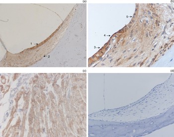
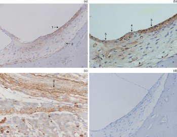
Endothelin receptor A like activity was found to be extensively distributed in the cells of the cochlear lateral wall. Strong staining for endothelin receptor A like activity was demonstrated in the marginal cells, spiral prominence epithelial cells, outer sulcus cells, stria vascularis capillaries and basal cells, while moderate endothelin receptor A immunoreactivity was present in spiral ligament fibrocytes (Figure 1). Endothelin receptor B like activity was also found to be distributed in the cochlear lateral wall, with moderate immunoreactivity found in the stria vascularis, spiral prominence cells and outer sulcus cells, and weak immunostaining detected in fibrocytes (Figure 1).
Photomicrographs showing endothelin receptor A activity analyses. (a) Endothelin receptor A like activity was found to be extensively distributed in all cells of the cochlear lateral wall, with strong immunoreactivity in the stria vascularis (arrow 1) and moderate immunoreactivity in the spiral ligament fibrocytes (arrow 2) (original magnification ×200). (b) Strong immunoreactivity for endothelin receptor A like activity was demonstrated in the marginal cells (arrow 3), spiral prominence epithelial cells (arrow 4) and outer sulcus cells (arrow 5) (original magnification ×400). (c) Positive control: endothelin receptor A immunoreactivity was observed in guinea pig cardiac muscle cells (original magnification ×400). (d) Negative control: no immunoreactivity was observed when phosphate-buffered saline was used instead of primary antibodies (original magnification ×400).

In the positive control sections, endothelin receptor A and endothelin receptor B immunoreactivity were observed in guinea pig cardiac muscle cells and tracheal smooth muscle cells, respectively (Figure 1c and Figure 2c). In the negative control sections, no staining was obtained when phosphate-buffered saline was used instead of primary antibodies (Figure 1d and Figure 2d).
Photomicrographs showing endothelin receptor B activity analyses. (a) Moderate endothelin receptor B immunoreactivity was found in the stria vascularis (arrow 1), with weak immunoreactivity in fibrocytes (arrow 2) (original magnification ×200). (b) Endothelin receptor B like activity was demonstrated in the marginal cells (arrow 3), spiral prominence epithelial cells (arrow 4) and outer sulcus cells (arrow 5) (original magnification ×400). (c) Positive control: endothelin receptor B immunoreactivity was observed in guinea pig tracheal smooth muscle cells (arrow 6) (original magnification ×400). (d) Negative control (original magnification ×400).

Discussion
In this study, we investigated the expression of endothelin receptors in the lateral wall of the cochlea. Immunohistochemical analysis showed that endothelin receptor A like activity was extensively distributed in all cells of the cochlear lateral wall. Endothelin receptor B like activity was also found in these areas. These results suggest that endothelin receptors may be involved in the physiology of the cochlear lateral wall.
The endolymph in the cochlear duct has an ionic composition characterised by large amounts of K+ ions and small amounts of Na+ ions. This unique ionic composition is essential for maintaining the high endocochlear potential, and hence normal hearing. The cells of the cochlear lateral wall play a very important role in maintaining this ionic composition. Marginal cells and outer sulcus cells are located on the surface of the cochlear lateral wall. The marginal cells transport K+ from the interstitial space and secrete it into the endolymph. The outer sulcus cells can absorb K+ and Na+ from the endolymph and transport K+ to the spiral ligament. Fibrocytes also play a role in ion transportation.
Ion transportation depends on the action of Na-K-adenosine triphosphatase (ATPase) and Na-K-2Cl presenting to the above cells. It has been demonstrated that endothelin 1 and endothelin 3 play roles in the regulation of the Na-K-ATPase/Na-K-2Cl transporter,Reference Gupta, Ruderman, Cragoe and Sussman10, Reference Prasanna, Dibas, Hulet and Yorio11 and are distributed in many cells within the cochlear lateral wall.Reference Xu, Tang, Liu and Liu4, Reference Franz, Hauser-Kronberger, Egerbacher, Böck, Stach and Quint5 These reports suggest that endothelin may regulate ion transportation through its effect on the activity of the Na-K-ATPase/Na-K-2Cl transporter in the cochlear lateral wall cells.
If endothelin is to be effective, endothelin receptors must be present on its target cells. The present study demonstrated the presence of endothelin receptor A and endothelin receptor B in marginal cells, spiral prominence epithelial cells, outer sulcus cells, basal cells and spiral ligament fibrocytes. These results provide evidence for the presence of an endothelin autocrine or paracrine loop in those cells, and support the suggestion that endothelin, via its receptors, may play a role in maintaining cochlear homeostasis.
• Endothelin has many biological activities, including regulation of the functions of the cochlear lateral wall
• This study aimed to analyse endothelin receptor expression in the cochlear lateral wall, and to investigate the significance of such receptors in maintaining the homeostatic environment of the cochlea
• Varying degrees of endothelin receptor A and endothelin receptor B like activity were found throughout the cochlear lateral wall
• These findings support the theory that endothelin, via its receptors, plays an important role in maintaining the homeostatic environment of the cochlea
Although the present study was not a quantitative analysis, differential distribution of endothelin receptor A and endothelin receptor B immunoreactivity was observed in the lateral wall of the cochlea. Stronger immunoreactivity for endothelin receptor A and endothelin receptor B was demonstrated in the spiral prominence epithelial cells, outer sulcus cells, stria vascularis capillaries and basal cells than in the spiral ligament fibrocytes. The observed variation in endothelin receptor expression in different cells, as assessed immunohistochemically, may mirror these cells' functional diversity. Luo et al. Reference Luo, Tang, Xia and Liu7 demonstrated higher levels of messenger RNA expression of the two endothelin receptor subtypes in the stria vascularis than in non-strial tissues; these results are similar to our findings.
In the sections analysed, we found stronger immunoreactivity for endothelin receptor A compared with endothelin receptor B. These two receptor subtypes share 64 per cent amino acid sequence identity, but show differences in their tissue expression and signal transduction.Reference Goldie, D'Aprile, Self, Rigby and Henry12 The fact that immunoreactivity was stronger for endothelin receptor A may indicate that this is the predominant endothelin receptor subtype in the cochlear lateral wall, and the main mediator of endothelin function in maintaining cochlear homeostasis.
Conclusion
Our study demonstrated the expression of endothelin receptors in the lateral wall of guinea pig cochleae. Endothelin receptor A may be the predominant endothelin receptor mediating the biological activities of endothelin at this site. The exact function of endothelin receptors in the cochlear lateral wall needs to be further investigated.
Acknowledgements
We thank Mr Hang-Bo Zhou for his excellent technical support. This work was supported by China's Post-doctoral Science Fund (grant number 20070421033).