Cardiac catheterisation plays a major role in the management of patients with functionally univentricular circulation early and late after surgical palliation.Reference Kreutzer and Rome1 Haemodynamic and angiographic data from diagnostic catheterisation can help identify reasons for failure and guide therapeutic interventions. Cardiac catheterisation with possible transcatheter intervention is indicated in patients with post-operative difficulties or unexpected changes in clinical status during long-term follow-up. There is wide variability in the interventions performed in these patients, ranging from treating stenosis along the pathway – balloon angioplasty and stent implantation in pulmonary arteries or systemic veins, closure of sources of right-to-left shunts – collaterals and fenestration, creation of right-to-left shunt – fenestration creation, fenestration dilation and stenting, closure of aortopulmonary collaterals, and more. We review the current role of catheter therapies in patients with hypoplastic left heart syndrome late after surgical palliation.
Fontan pathway obstruction
Maintenance of unobstructed flow is important for optimal long-term haemodynamics in the Fontan circulation. Late obstruction can occur in the superior and inferior caval veins, Fontan conduit or baffle, surgical anastomosis sites, or in the branch pulmonary arteries themselves. It has been reported to occur in 6.6–14.6% of all patients who underwent a Fontan operation.Reference Giannico, Hammad and Amodeo2–Reference Ovroutski, Ewert, Alexi-Meskishvili, Peters, Hetzer and Berger4 Clinical manifestations of obstruction can include ascites, pleural effusion, protein-losing enteropathy, desaturation, and low cardiac output.
Conduit or baffle stenosis
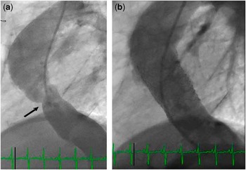
This can be seen after any of the variable surgical techniques used for Fontan completion. It is rare in patients with lateral intra-atrial tunnel type of procedures, given the growth potential of the lateral atrial wall. The late incidence of stenosis of extracardiac Fontan conduits is not well known. Obstruction in the Fontan conduit in the early post-operative period due to thrombus formation can be addressed with thrombolysis, followed by balloon angioplasty and stent placement.Reference Sreeram and Kammeraad5 The decrease in cross-sectional area of the conduit has been reported to be 14–17%, presumably because of neointimal growth within the conduit in the early post-operative period, although this did not result in significant haemodynamic changes.Reference Giannico, Hammad and Amodeo2, Reference Hannan, Zabinsky and Salvaggio3 Late obstruction can occur in 3% of patients, most commonly at the anastomosis of the inferior caval vein to the conduitReference Giannico, Hammad and Amodeo2 (Fig 1). This condition can be treated with balloon angioplasty and stent placement, which can obviate the need for surgical intervention and has been shown to relieve ascites, protein-losing enteropathy, and improve cardiac output.
Example of obstruction of the Fontan pathway in a 14-year-old male status-post extracardiac conduit. (a) Lateral projection demonstrating significant stenosis of the inferior portion of the Fontan conduit immediate to the anastamosis with the inferior caval vein (black arrow). There is approximately a 50% reduction in the luminal diameter at the area of stenosis. (b) Repeat angiogram performed after placement of a 26-millimetre-long Max LD stent (EV3) dilated to 20-millimetre diameter demonstrating no residual stenosis.

Pulmonary arterial stenosis
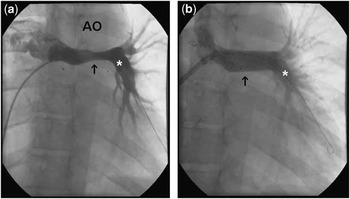
Obstruction in the pulmonary arteries can occur at the anastomosis with the caval veins, or in the pulmonary artery itself. Obstruction in the pulmonary arteries requiring intervention has been reported in 3–11% of Fontan patients.Reference Giannico, Hammad and Amodeo2, Reference Ovroutski, Ewert, Alexi-Meskishvili, Peters, Hetzer and Berger4 Narrowing of the left pulmonary artery is more common than the right in patients with hypoplastic left heart syndrome, which is most likely due to compression by an augmented neo-aorta (Fig 2). Although surgical staging often incorporates patch augmentation of the left pulmonary artery, this may not reach far enough given the location behind the aorta, such that stenosis continues to be relatively common at the left pulmonary artery. The absence of a pressure gradient across an area of narrowing does not rule out clinically significant obstruction, as this may result in increased flow to the contra-lateral lung, diversion of blood flow to other systemic venous channels, or increased pressure within the Fontan circulation. Therefore, stenosis in a pulmonary artery needs to be determined by imaging. A luminal narrowing of greater than 25% of the nominal vessel diameter is significant and usually requires stent placement. Care must be taken during stenting of the left pulmonary artery to avoid compression of the left main bronchus. Advance imaging with rotational angiography may identify areas of stenosis that are not identified in single-plane imaging, and delineate the relationship of the stenosis to other anatomic structures. Balloon dilation of these narrowings is commonly unsuccessful. Stent implantation is necessary.
(a) Stenosis (arrow) of the left pulmonary artery (asterisk) is demonstrated, secondary to compression from the augmented neo-aorta (AO). (b) Following stent implantation, there is resolution of the stenosis (arrow).

Systemic venous stenosis
Although sometimes signs and symptoms of venous obstruction can be obvious, the clinical presentation of this entity is often subtle. Patients with prior instrumentation from central venous catheters or peripherally inserted central catheter lines are at increased risk of venous stenosis, which occurs typically secondary to acute or subacute thrombosis. When the superior caval vein is affected, presentation may be that of superior vena cava syndrome, typically associated to effusions.Reference Kreutzer and Rome1, Reference Hanna, Truemper and Burton6 Sometimes, the presenting symptom is cyanosis related to decompressing venous collaterals (See below in the section on “Closure of decompressing veins and other venous anomalies”). When the inferior caval vein is affected, presentation can be that of ascites and lower extremity venous ectasia. Interventions aimed at restoring unobstructed flow, including angioplasty and stent implantation, are commonly successful.Reference Kreutzer and Rome1
Fenestration creation for Fontan failure syndromes
Baffle fenestration for Fontan failure syndromes
Fontan failure dichotomises into early – usually immediate post-operative – and late – years to decades later – syndromes. The patient who develops signs of low cardiac output or right heart failure immediately after Fontan completion should always undergo cardiac catheterisation for evaluation. In the absence of directly treatable causes, such as Fontan pathway obstruction or large systemic to pulmonary shunt lesions, fenestration creation or enlargement is often very effective in improving cardiac output, and relieving right heart failure. Immediately after creation or dilation of the fenestration, there is decreased saturation of arterial oxygen and pressure in the Fontan pathway, and an increase in the cardiac index.Reference Kreutzer, Lock, Jonas and Keane7 These haemodynamic changes often result in clinical improvement, that is, the ability to wean off mechanical ventilatory support, cessation of chest tube drainage, and hospital discharge. By contrast, the optimal treatment for Fontan failure occurring years to decades later is much less clear. Failure of the Fontan circulation will ultimately occur in most patients in whom Fontan palliation has been performed. This failure may manifest with the typical signs of right heart failure – oedema and effusions – or one of the syndromes more specific to Fontan physiology such as protein-losing enteropathy or plastic bronchitis. In these populations, a comprehensive diagnostic and therapeutic approach must be undertaken. Diagnostic evaluation includes assessment of pump and valvar function, heart rhythm disorders, and other organ system involvement including liver and kidney.Reference Rychik8 In addition, diagnostic catheterisation is required to identify remediable causes of Fontan failure as described above in the section entitled “Fontan pathway obstruction”. These include obstruction to flow anywhere in the cavopulmonary circuit, obstruction of pulmonary venous return at the pulmonary veins or the atrial septum, systemic outflow obstruction – most commonly from recurrent coarctation – or excessive systemic to pulmonary collateral flow. Therapy is then guided by diagnostic findings: clearly, when remediable factors are identified, specific treatments are undertaken. In many or even most cases, however, no specifically treatable cause can be identified. These patients require a comprehensive strategy of chronic disease management. Medical treatments may include phosphodiesterase inhibitors; for protein-losing enteropathy, minimally absorbed steroids (budesonide) has proven effective in many cases.Reference Reinhardt, Uzun and Bhole9, Reference Thacker, Patel, Dodds, Goldberg, Semeao and Rychik10 However, in a substantial percentage of patients, these treatments are either ineffective or result in unacceptable side effects. Palliation with fenestration creation has been demonstrated to be effective in alleviating symptoms at least temporarily and should be considered among the treatment options.Reference Vyas, Driscoll, Cabalka, Cetta and Hagler11 However, it is important to realise that, in contrast to children, adult Fontan patients may become quite symptomatic from the resultant decrease in saturation of arterial oxygen.
Regardless of whether fenestration creation is being considered for treatment of early or late Fontan failure syndromes, a careful evaluation of the baseline haemodynamic state must be undertaken. If significant cyanosis is present and not easily remediable, for example, by draining a large pleural effusion, fenestration creation is contraindicated. Furthermore, in patients with high venous pressure, minimal transpulmonary gradient, and elevated ventricular filling pressures, creation of a fenestration is unlikely to improve significantly the physiologic state; treatment should be aimed at improving pump function.
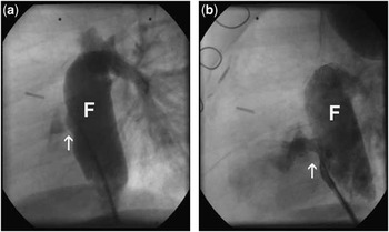
Several strategies have been described for creation of fenestrations in Fontan patients, an example of the maxim that the efficacy of a particular method is inversely proportional to the number of methods described. In current practice, the majority of Fontan connections requiring fenestration creation are either extracardiac or lateral tunnel types. Although in general the technique for fenestration creation is similar, the two differ in important ways. Transseptal puncture is the optimal way to create a connection from systemic to pulmonary venous circulation. However, in the extracardiac Fontan, the transseptal pathway must traverse the extravascular space then through the atrial free wall into the atrial chamber. This can only be safely accomplished if enough time has elapsed after surgical intervention to allow mature scar formation. Thus, transcatheter fenestration is not possible in the early post-operative extracardiac Fontan unless a small or occluded surgical fenestration can be crossed. Before the transseptal needle is used, an angiogram is performed in the Fontan conduit or lateral tunnel in biplane with levophase to identify the optimal puncture site. The best angle of approach may be from femoral vein, internal jugular, or less commonly a transshepatic route. Adding an additional bend to the transseptal needle will often aid in engaging the Gore-Tex for puncture. Although many advocate balloon dilation with either high pressure or cutting balloons for fenestration creation in lateral tunnel Fontans, it has been our experience that these methods do not achieve reproducible results: fenestration size tends to decrease rapidly and unpredictably over a short time. We usually prefer stent placement (Fig 3). In the patient with an extracardiac Fontan, a stent must be used for fenestration creation. Some prefer the “diablo” technique where a suture is used to constrict the central portion of the stent. In our experience, this is not necessary, and the absence of a constricting suture allows more freedom for expansion of the stent to increased diameters. Stent diameters of 6–8 millimetres are required to achieve effective fenestrations, and larger diameters are required for extracardiac connections where the pathway is longer.
(a) In the lateral projection Fontan conduit angiogram, (F) demonstrates a small Fontan fenestration (arrow) in a patient with protein-losing enteropathy. (b) Following balloon angioplasty and stent implantation, there is significant improvement in fenestration size (arrow).

A final important consideration in fenestration creation is thromboprophylaxis. Fenestration creation is in many ways a perfect capitulation of Virchow's triad. The failing Fontan patient is often in a hypercoagulable state, flow through the systemic venous pathway and fenestration is sluggish, the stent is thrombogenic, and in the case of the extracardiac fenestration, tissue factor is exposed in the pathway from Fontan conduit to atrium. Thrombosis may result not only in occlusion of the newly created fenestration, but also systemic embolisation of fresh thrombus with devastating consequences. The optimal strategy for thromboprophylaxis is not known. When practical, we evaluate the baseline state by measuring levels of proteins C, S, and antithrombin III, replacing these if the patient is deficient. In cases where the risk is deemed particularly high, we load patients with aspirin and/or clopidogrel before catheterisation. During the procedure, standard heparinisation protocols are used but with more frequent monitoring of activated clotting times – maintaining them over 250 seconds – as we have observed rapid loss of heparin effect. An attractive alternative is the use of a direct thrombin inhibitor such as bivalirudin avoiding dependence on antithrombin III. After the procedure, the patient is maintained on intravenous anticoagulation until converted to effective chronic anticoagulation.
Closure of fenestrations and baffle leaks
Fontan fenestration
Most surgeons continue to create a surgical fenestration at the time of Fontan procedure in patients with hypoplastic left heart syndrome.Reference Bridges, Lock and Castaneda12–Reference Atz, Travison and McCrindle14 At late follow-up, if the fenestration is still patent and cyanosis is present, transcatheter closure is considered.Reference Goff, Blume, Gauvreau, Mayer, Lock and Jenkins15, Reference Bridges, Lock, Mayer, Burnett and Castaneda16 The indications for elective fenestration device closure are controversial. Most would agree that it should be closed in patients with resting arterial oxygen saturation of less than 85%, if test occlusion in the catheterisation laboratory is well tolerated.Reference Bridges, Lock, Mayer, Burnett and Castaneda16 Often patients with patent fenestrations develop worsening cyanosis with exercise, and this can be considered an indication of referral for cardiac catheterisation with possible closure. Transcatheter fenestration closure is considered a highly successful procedure, performed with a variety of devices, including CardioSEAL, STARFlex, Amplatzer septal occluder,Reference Cowley, Badran, Gaffney, Rocchini and Lloyd17 Helex and Amplatzer Vascular Plugs II. Device deployment techniques have been well described.Reference Bridges, Lock and Castaneda12, Reference Bridges, Lock, Mayer, Burnett and Castaneda16
Although the device used depends partly on the underlying anatomy and surgical technique used, most fenestrations can be occluded with the 17-millimetre CardioSEAL device, which was traditionally preferred given its small profile and equal disc size on each side of the defect. However, this device is not currently further available. The Helex device, which shares the low profile features of CardioSEAL, is often challenging to deliver in a fenestration given the need for space on the left side of the septum to form the left-sided disc, which is most times lacking. Thus, many interventionalists implant Amplatzer septal occluders for this indication these days. If the fenestration has a significant length, such as those in extracardiac Fontan conduit with a Gore-Tex graft anastomosed from the extracardiac conduit to the atrium, a vascular plug and sometimes coils may be more appropriate.Reference Sommer, Recto, Golinko and Griepp18
Before closure, test occlusion of the fenestration is performed with the assessment of the haemodynamics. If an increase in venous pressure of greater than 16 millimetres of mercury is observed, or if the cardiac index decreases to less than 2 litres per minute per metre square, occlusion is not recommended.Reference Kreutzer and Rome1, Reference Cowley, Badran, Gaffney, Rocchini and Lloyd17
The immediate effects of fenestration closure are well known, including an increase in oxygen saturation, a slight decrease in cardiac index, and an increase in Fontan pathway pressures. At follow-up, transcatheter closure of Fontan fenestration increases the duration of exercise capacity and increases oxygen saturation at rest and after exercise.Reference Momenah, Eltayb, Oakley, Qethamy and Faraidi19 However, it has been demonstrated that Fontan fenestration closure does not significantly improve peak VO2 on stress test, but does lead to an improvement in arterial saturation and ventilatory abnormalities.Reference Meadows, Lang and Marx20 In a study of 181 patients undergoing fenestration closure, there was a persistent increase in oxygen saturation of an average of 9.4%.Reference Goff, Blume, Gauvreau, Mayer, Lock and Jenkins15 There was also a reduced need for anticongestive medication and an improvement in somatic growth at follow-up. Death (1.3%) or chronic decompensation (3.2%) was rare.
Closure of interatrial communications and baffle leaks
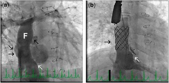
Following atriopulmonary anastomosis or lateral atrial tunnel Fontan procedure, decompressing connections may develop within the atrial wall between the higher pressure right atrium and the lower pressure left atrium. These defects can be of variable size and they are often multiple and tend to become larger over time. Transcatheter closure can be achieved successfully with a variety of devices or coils. Use of a covered stent may be indicated in patients with multiple such leaks and a relatively narrow Fontan baffle (Fig 4).
(a) Angiogram of the Fontan baffle (F) demonstrates multiple leaks of the baffle (black arrows), as well as a patent Fontan fenestration (white arrow) in a patient with marked cyanosis late after lateral tunnel Fontan procedure. (b) The right-to-left shunt is eliminated after implantation of a Cheatham Platinum-covered stent to cover the baffle leaks (black arrow) and fenestration closure with a 4-millimetre Amplatzer septal occluder (white arrow).

Closure of decompressing veins and other venous anomalies
Although the most significant potential decompressing veno–veno collaterals are often closed before surgical staged palliation, it is not uncommon to identify additional such vessels after Fontan procedure. They often drain directly to the atrium or the pulmonary veins. They cause a right-to-left shunt and progressive cyanosis, as they tend to get larger over time. Vessels that may have been diminutive at the time of Glenn procedure, and thus were missed on prior cardiac catheterisations, may become significant over time.
Anatomically, they may arise from variable sites.Reference Ro, Weinberg, Delrosario and Rome21, Reference McElhinney, Reddy, Hanley and Moore22 The most common among these are collaterals arising from the innominate vein, azygos vein, and venous connections between systemic and pulmonary veins. There can also be decompressing veins from below the diaphragm. The most commonly encountered collaterals are various azygos connections (58%), followed by persistent left superior caval vein to the coronary sinus (24%), and connections from innominate or superior caval veins to pulmonary veins (15%).Reference Ro, Weinberg, Delrosario and Rome21, Reference McElhinney, Reddy, Hanley and Moore22
The procedure consists of selective catheterisation of the vessel followed by venous embolisation with either vascular plugs or coils. Currently, only magnetic resonance compatible coils are used. It is important to position the closure device distal to the entry of the left hemiazygos or any other venous contributory. Otherwise, paravertebral veins entering the left hemiazygos will enlarge over time causing recurrent cyanosis. Once the left superior caval vein has been occluded, this pathway becomes much more difficult to access. In addition, before embolisation of a persistent left superior caval vein, coronary sinus atresia or stenosis should be ruled out.
Most veno–veno collaterals are amenable to closure by transcatheter techniques. There are rare instances when occlusive therapy is highly unsuccessful. Such is the case when there is chronic thrombosis of subclavian or innominate veins associated with the development of multiple venous collaterals decompressing to the paravertebral veins, or alternatively via a mesh of mediastinal vessels into the pulmonary veins.
Closure of arterial collaterals
Aortopulmonary collaterals are vessels that arise from the systemic circulation and supply the pulmonary circulation. These vessels are commonly found in patients with chronic cyanotic cardiac disease and functionally univentricular anatomy, and their presence have both potentially advantageous and deleterious effects. Strategies for the management of these vessels in patients with functionally univentricular physiology are varied.Reference Powell23, Reference Stern24
The development of arterial collaterals is thought to be secondary to chronic cyanosis, although the exact mechanism is not known. Collateral vessels augment pulmonary blood flow and thus may act to increase systemic oxygen saturation via an increase in fully saturated pulmonary venous flow returning to the single ventricle. In addition, as these vessels arise from the systemic circulation, they provide the perfused pulmonary segments with “hepatic factor”, and thus may inhibit the development of pulmonary arteriovenous malformations that are known to develop in some patients with cavopulmonary connections as the sole source of pulmonary blood flow. Thus, their presence may provide some physiologic advantages.
However, aortopulmonary collaterals exert numerous deleterious pathophysiologic effects as well. The increased pulmonary blood flow, and thus pulmonary venous return, represents a left-to-right shunt and thus imposes a chronic volume overload on the single ventricle, resulting in an increased workload on the ventricle. This volume overload potentially raises ventricular end-diastolic pressures and may exacerbate atrioventricular valve regurgitation, and in the long term may ultimately lead to ventricular systolic dysfunction. Furthermore, these vessels may cause pressure elevation in the pulmonary arteries and compete with flow with the cavopulmonary circulation. An increase in pulmonary arterial pressure may lead to prolonged pleural effusions and their associated morbidity. The competitive flow in the pulmonary arteries may result in loss of energy in the passive venous circulation and may thus decrease cardiac output in the Fontan circulation. Finally, collateral vessels in close proximity to the bronchial tree may dilate and erode over time, resulting in significant and potentially fatal haemoptysis (see section “Catheter management of haemoptysis”).
Although all these pathophysiologic consequences would seem to have deleterious effects on the Fontan circulation, data to correlate their presence to post-Fontan complications have been mixed. Early studies identified the degree of collateralisation as a risk factor for prolonged pleural effusion drainage post-Fontan procedure, although these studies were predominantly retrospective and subjectively quantified collateral flow via angiography alone.Reference Ichikawa, Yagihara and Kishimoto25–Reference Kanter, Vincent and Raviele27 More recent studies have attempted to objectively quantify collateral flow and have not replicated these findings. Bradley et alReference Bradley, McCall and Sistino28 used an intra-operative technique to quantify collateral flow by measuring the volume of blood returning from the pulmonary venous vent during the Fontan procedure. Although confounded by the non-physiologic state with non-pulsatile flow on cardiopulmonary bypass, no antegrade pulmonary blood flow in a hypothermic state, this study found no detectable effect of aortopulmonary collateral flow on the outcome post Fontan. Using a more physiologic state, Lim et alReference Lim, Graziano and Rocchini29 described a quantification technique using thermal indicator dilution at the time of the pre-Fontan catheterisation, measuring the degree of recirculation of iced saline injected into the ventricle. This study found no correlation between the degree of aortopulmonary collateral flow and post-operative Fontan pressure, duration of pleural effusions or mortality. Finally, a more recent multi-centre study compared 80 patients who underwent occlusion of aortopulmonary collaterals at the pre-Fontan catheterisation with 459 patients who did not and found no difference in hospital length of stay or post-operative complications after the Fontan procedure.Reference Banka, Sleeper and Atz30 Thus, the effect of arterial collaterals appears to be complex with regard to their impact on post-Fontan haemodynamics and outcome. As of now, therefore, the practice of routinely occluding all such vessels in patients before the Fontan does not appear to be supported. Recognising that a subset of patients may benefit from elimination of aortopulmonary collaterals, the practice of occlusion remains a reasonable approach for selected higher risk patients, for patients with very large collaterals, or for patients with prolonged effusions post Fontan to potentially reduce the ventricular volume overload and decrease pulmonary artery flow and pressure.
Adequate occlusion of collaterals requires an understanding of the pertinent anatomy. Most of the collateral vessels in patients with functionally univentricular anatomy originate from vessels off of the head and neck arteries, including the internal mammary arteries, thyrocervical trunk, and lateral thoracic trunk.Reference Triedman, Bridges, Mayer and Lock31 These vessels have extensive connections to the intercostal arteries, and thus occlusion of the collaterals solely at their origin is inadequate for complete occlusion. Thus, occlusion of the entire length of the vessel should be the goal of intervention. Distal cannulation of the artery, particularly large mammary or thoracic arteries, is often feasible with angled glide wires and a variety of end-hole catheters, and placement of embolisation coils is generally straightforward. Microcatheters and microcoils may facilitate accessing small or tortuous vessels, although a subset of patients may always have a network of small, tortuous vessels that may not be amenable to occlusion via a transcatheter approach. Embolisation particles offer a physiologic advantage over coils by occlusion of the distal microvasculature, and thus may be a better option to eliminate persistent flow from additional connections from the intercostals. However, their use for this purpose had been limited and not been systematically studied. Conversely, very large collateral vessels can be easily occluded with vascular plugs (Amplatzer, AGA Medical, Golden Valley, Minnesota United States of America) that can be accurately placed and are detachable and retrievable and thus provide an additional option for occlusion. Stainless steel coils produce a significant artefact in the magnetic resonance scanner, limiting the ability of thoracic magnetic resonance imaging diagnosis following extensive coil embolisation in these patients. Thus, all coils used these days are magnetic resonance imaging compatible.
In summary, aortopulmonary collaterals in patients with functionally univentricular physiology impart numerous physiologic disadvantages. Despite the lack of compelling data to support their routine occlusion in all patients, occlusion of these vessels remains a reasonable option for selected higher risk patients or those with inadequate Fontan haemodynamics. Further study, possibly with the use of magnetic resonance imaging to assist in the quantification of aortopulmonary collateral flow,Reference Grosse-Wortmann, Al-Otay and Yoo32 and trials randomising patients between occlusion and non-occlusion strategies, may help to identify patient populations that would benefit from routine occlusion before Fontan completion.
Catheter management of haemoptysis
Haemoptysis is an unpredictable complication in the Fontan population with a varied clinical presentation. Some patients will have occasional bouts coughing up small amounts of blood, the only consequence of which is anxiety. Others present with acute massive bleeding resulting in respiratory insufficiency, circulatory failure, and even death. There are several possible causes for haemoptysis in these patients. They are at risk for atypical mycobacterium, and this may result in pulmonary haemorrhage. Previously placed interventional devices – stents or closure devices – can erode into bronchi.Reference Pastuszko, Rome, Gleason, Bellah, Montengro and Gaynor33 By far, the most common cause of haemoptysis in this population is erosion of systemic – bronchial – arteries into the airway. Diagnostic evaluation is aimed at localising the bleeding site and identifying the cause. Interestingly, the patient often feels a sensation of gurgling and can tell the physician where the bleeding is coming from, a vignette worth enquiry. In addition to standard radiography, computerised tomography imaging may be useful in identifying the cavitary lesions of mycobacterial disease or help in identifying possible device erosion. Before catheter embolisation, we routinely perform bronchoscopy for localisation of bleeding site.
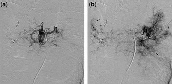
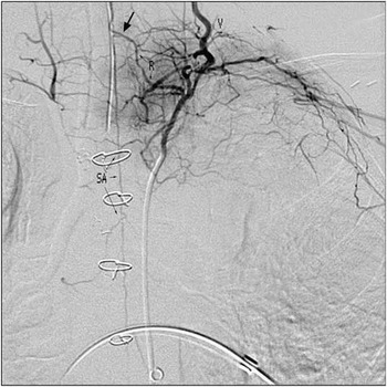
Catheter embolisation is the method of choice for treatment of pulmonary haemorrhage from systemic arterial collateralisation. Absolute indications for treatment include life-threatening bleeding or ongoing haemorrhage sufficient to result in a significant drop in haemoglobin concentration. In patients in whom there is recurrent or persistent bleeding even in the absence of evidence of significant blood loss, intervention may be indicated if the episodes are enough to result in a prolonged disruption of activities of daily living. Routine embolisation for minor haemoptysis is not recommended, as even effective embolisation will not prevent future episodes of bleeding. After the evaluations described above have lateralised the site of bleeding, angiography is performed to identify candidate vessels for embolisation. The approach here differs substantially from that for embolisation of systemic to pulmonary collaterals to decrease left-to-right shunt. Even small arteries can be the source of pulmonary haemorrhage. Selective angiograms of candidate vessels should be performed with digital subtraction looking for a characteristic blush of abnormally arborising small arteries (Figs 5 and 6). The most likely sources will be systemic arteries travelling along the walls of the bronchial tree. The true bronchial arteries should be visualised, as well as virtually all systemic arterial supply to the posterior thoracic circulation including deep cervical, thyrocervical, lateral thoracic, intercostal, and even phrenic arteries. Before embolisation of candidate vessels, they must be examined carefully to rule out supply to the anterior spinal artery (Fig 6). Embolisation for haemoptysis should be performed with particles rather than coils whenever possible. Coil embolisation often fails. Coils occlude only the most proximal portion of the feeder artery; however, the culprit vessels remain intact. Owing to the fact that there is tremendous redundancy of arterial supply, bleeding is often unabated. Even if transiently effective, abnormal vessels will parasitise other sources of arterial inflow resulting in recurrent or persistent bleeding. Having previously coil embolised the feeder, it is then difficult if not impossible to perform repeat embolisation. The angiographic catheter can be used as a guide catheter through which a microcatheter is advanced into stable position in the feeder artery. Particles greater than 500 microns in diameter, we prefer polyvinyl alcohol, mixed in contrast are slowly infused until arterial run-off is sluggish. Repeat subtraction angiography confirms successful vascular embolisation.
Digital subtraction angiogram performed in the bronchial artery in a Fontan patient with haemoptysis. (a) The early phase of the injection demonstrates a typical stenotic anterior origin from the aorta with subsequent dilatation and ectasia of the artery. (b) Late arterial phase of the injection highlights the characteristic abnormal areas of contrast enhancement (arrows), which appear like puffs of smoke.

Selective digital subtraction angiogram performed in a left superior intercostal artery. This injection demonstrates the anterior spinal artery (SA) filling via a thoracic radiculomedullary branch (R), which makes the characteristic hairpin turn as it feeds the spinal artery (arrow). The vertebral artery (V) is opacified on this injection because of extensive collateral formation between the intercostal and subclavian arteries.

Transcatheter therapies for thrombosis – Fontan pathway/pulmonary arteries
Fontan pathway thrombosis is a well-known complication early and late post-operatively.Reference Balling, Vogt, Kaemmerer, Eicken, Meisner and Hess34, Reference Jacobs35 Accurate diagnosis can be difficult via non-invasive tests, as visualisation of the pathway is often incomplete on echocardiography, and streaming of contrast during computerised tomography angiography may falsely diagnose the presence of thrombus. Although extensive Fontan thrombosis may demand surgical thrombectomy and Fontan re-do, catheter-directed therapies can be considered,Reference Kudumula, Mathur and Bu'Lock36–Reference Gossett, Rocchini and Armstrong38 particularly when there is no significant risk of systemic embolism – thrombus localised in systemic veins or pulmonary arteries and not associated with the site of a fenestration. Catheter-directed therapies aim to relieve obstruction and remove as much clot as possible, minimising the chances of distal pulmonary artery emboli. Treatment typically involves a combination of mechanical techniquesReference Sreeram, Emmel and Trieschmann37 and pharmacological thrombolytics.Reference Robinson, Fellows, Bridges and Rome39 When thrombosis is acute, the clot can be disrupted mechanically with either angioplasty balloons, wires, and basket catheters, or using specific thrombectomy and/or thrombolytic catheter systems such as the X-SIZERReference Gossett, Rocchini and Armstrong38 or the Angiojet. The latter uses powerful saline jets that create a low-pressure zone around the catheter tip causing a vacuum effect. Thrombus is drawn into the catheter, where it is fragmented by the jets, and removed from the body. Depending on the diameter of the vessel affected, there are different catheters that can be used. Thrombolytic agents can be administered following thrombectomy. It is not uncommon that thrombosis occurs at a site of stenosis. Thus, transcatheter therapy of the underlying obstruction is essential to prevent recurrence.
If the thrombus is old and organised, catheter management is aimed at purely treating the obstruction. When the occlusion is complete, radiofrequency perforation allows access through the clot, followed by sequential balloon angioplasty and stent implantation to regain vessel patency.
Pulmonary arteriovenous malformations
Pulmonary arteriovenous malformations developing late after Fontan palliation can be a cause of significant morbidity. In most cases, they occur in the lung with inadequate return of hepatic venous effluent as a result of an uneven distribution of hepatic venous return. Pulmonary arteriovenous malformations after Fontan are most common in patients with heterotaxy syndromes. Several approaches have been reported for addressing this problem by redistributing hepatic venous return evenly to both lungs.Reference Pike, Vricella, Feinstein, Black and Reitz40 In most instances, surgical Fontan revision is required to achieve this goal.Reference AboulHosn, Danon, Levi, Castellon, Child and Moore41 However, catheter-directed therapy may occasionally be effective. When severe pulmonary artery stenosis or discontinuity results in maldistribution of hepatic venous return, stenting to treat the anatomic abnormality may result in more equitable distribution of hepatic venous return and lead to regression.Reference McElhinney, Kreutzer, Lang, Mayer, del Nido and Lock42 We have encountered other patients in whom, with the use of combinations of covered and uncovered stents, it was possible to redirect hepatic venous return such that this flow mixed more effectively distributing to each lung. Each of these cases requires a unique solution. We have found that complex flow modelling using magnetic resonance imaging-derived velocity mapping and three-dimensional anatomic data are useful in planning this type of intervention.
There are patients in whom incorporation of hepatic venous flow may still not be effective in resolution of the pulmonary arteriovenous malformations. This can be due to the advanced severity of the disease, but can also be due to the presence of persistent congenital portocaval shunts, which can be found in patients with heterotaxy.Reference Newman, Feinstein and Cohen43
Late aortic arch obstruction
Coarctation of the aorta is a well-recognised complication following aortic arch reconstruction for hypoplastic left heart syndrome, with an incidence of between 9% and 37%.Reference Ballweg, Dominguez and Tabbutt44–Reference Larrazabal, Tierney and Brown46 Arch obstruction imposes an increased afterload on the functionally univentricular heart. Early in the surgical palliation, in the presence of a Blalock–Taussig shunt, the obstruction distal to the shunt may lead to an increase in shunt flow, imposing a volume load on the ventricle as well. These alterations may therefore impact ventricular function, worsen tricuspid regurgitation, and limit systemic blood flow. Recurrent arch obstruction is associated with worse right ventricular function and may be a cause of interstage mortality.Reference Larrazabal, Tierney and Brown46 Thus, timely treatment of the obstruction may result in improvement in right ventricular function.Reference Larrazabal, Tierney and Brown46
The aetiology of aortic arch obstruction following Norwood palliation is likely multifactorial. Residual ductal tissue has the potential to cause late obstruction, and thus resection of this tissue is necessary to reduce the risk of recurrent obstruction. In the light of this, some have advocated performing an extensive coarctectomy involving resection of the aorta at the level of the coarctation, wide resection of all the ductal tissue and posterior shelf, extended counterincision distally in the anterolateral aspect of the descending aorta, mobilisation of the supra-aortic trunks, and direct end-to-end anastomosis between the greater curvature of the native aortic arch and the descending aorta.Reference Bautista-Hernandez, Marx and Gauvreau47 Using this approach, a reduction in the risk of late obstruction can be observed.Reference Bautista-Hernandez, Marx and Gauvreau47 Others have advocated an interdigitating arch reconstruction to produce a similar improvement in the incidence of recoarctation.Reference Burkhart, Ashburn and Konstantinov48 Furthermore, the type of tissue used in the aortic arch augmentation may be an important factor as well, with data supporting the use of autologous tissue rather than aortic/pulmonary homograft patches to help reduce the incidence of late obstruction.Reference Bautista-Hernandez, Marx and Gauvreau47 Nonetheless, surgical technique and material will likely remain contributing factors in the occurrence of late arch obstruction.
Identification of residual obstruction in patients following Norwood palliation may be difficult. The presence of adequate lower extremity pulses on physical examination can be misleading with increased pulsatility in the aorta from the diastolic run-off secondary to the Blalock–Taussig shunt. Furthermore, upper to lower limb blood pressure gradients may underestimate the degree of anatomic obstruction as well, particularly in the light of any alteration in ventricular function, as well as due to the frequent stenosis of the right subclavian artery associated with the take-off of the shunt. Using echocardiography, Lemler et alReference Lemler, Zellers and Harris49 identified a coarctation index – ratio of the most narrow aortic diameter to the descending aortic diameter – of less than 0.7 and a maximum instantaneous gradient of greater than 30 millimetres of mercury across the arch to be predictive of recurrent coarctation. Thus, the anatomic appearance of the arch by angiography or non-invasive imaging, including computational tomography and magnetic resonance, can be an important determinant of severity in addition to the blood pressure gradient alone, and may be helpful in determining the indication for intervention.
Although surgical revision of the reconstructed aorta remains an option for treatment, balloon angioplasty is generally considered the first line of treatment for recurrent obstruction, particularly within the first year following Norwood. It is important to recognise that the immediate reduction in right ventricular and ascending aortic pressure following balloon angioplasty can result in decreased coronary perfusion pressure and decreased flow across the Blalock–Taussig shunt, and thus may result in haemodynamic instability due to acute alterations in the physiologic state. Numerous studies have shown that this procedure is not without risk and that the need for cardiopulmonary resuscitation measures due to significant haemodynamic instability may be as high as 25%.Reference Chessa, Dindar and Vettukattil50–Reference Zellers52 Thus, the procedure should be performed with general anaesthesia, and the use of inotropic support both during and after intervention is wise. Owing to the potential risk for femoral arterial occlusion with larger sheaths needed for the appropriate sized balloons, the procedure is generally performed in an antegrade manner via the femoral or internal jugular vein. This approach does carry a risk of further haemodynamic instability with relatively stiff wires traversing and stenting the tricuspid valve. Furthermore, injury to the aortic valve, likely form the balloon itself, is possible and has been reported. The size of the balloon chosen for dilation is generally equal to the diameter of the isthmus or descending aorta at the diaphragm and should not exceed three times the narrowest portion of the coarctation.
Despite the risks described above, the mortality of this procedure remains very low with substantial acute improvement in the degree of arch obstruction. Multiple studies have demonstrated excellent results with this technique. Chessa et alReference Chessa, Dindar and Vettukattil50 found 22 of 95 Norwood patients (22%) to have an aortic arch gradient greater than 10 millimetres of mercury with evidence of discrete narrowing by angiography following stage I palliation. Of the 22 patients, 17 underwent balloon angioplasty with a significant relief of obstruction in 16 of 17 (94%). In a larger, more recent series, Zeltser et alReference Zeltser, Menteer and Gaynor45 described an incidence of late obstruction of 9.2%. There were 35 patients who underwent balloon angioplasty at a median age of 6.6 months, and a post-dilation gradient of less than 10 millimetres of mercury was achieved in 92% of these patients. Furthermore, recurrent obstruction was rare in this series, occurring in 17% of patients, and nearly all of these were successfully treated with repeat balloon angioplasty. Most recently, Ballweg et alReference Ballweg, Dominguez and Tabbutt44 reviewed 176 patients who underwent stage I palliation between 2002 and 2005, with an incidence of late arch obstruction of 23%. In the 43 patients who underwent balloon angioplasty for this late obstruction, there was a mean reduction in the aortic arch gradient from 28.5 millimetres of mercury to 1 millimetre of mercury, with the need for repeat intervention in 16% of patients. Patients requiring intervention were more likely to have diminished ventricular function and had higher filling pressures at the time of the catheterisation as compared with those without arch obstruction. Interestingly, the presence of arch obstruction before stage II did not affect interstage mortality between stages I and II and was not associated with morbidity or mortality at the Fontan procedure. Nonetheless, these data all emphasise the importance and efficacy of early transcatheter intervention in patients with functionally univentricular hearts with recoarctation of the aorta.
Most literature on arch obstruction after Norwood refers to interventions performed at early stages of surgical palliation. The incidence of arch obstruction after Fontan is thought to be low, but has been poorly described. Following early angioplasty, remodelling of the vessel with restoration of growth is most commonly observed. It is known, however, that these patients share long-term morbidity as those who had isolated coarctation of aorta repair, and have decreased distensibility and increased stiffness,Reference Cardis, Fyfe and Mahle53 which may play a role in the late incidence of systemic hypertension and need for long-term afterload-reducing agents. Late arch obstruction in the older patient following Fontan procedure is managed with stent implantation.Reference Holzer, Qureshi, Ghasemi and Vincent54
In conclusion, aortic arch obstruction in patients with hypoplastic left heart syndrome is not uncommon and has detrimental effects on ventricular function and tricuspid regurgitation. Timely identification of this obstruction is critical and can be difficult. Balloon angioplasty is very effective at relieving early arch obstruction with a low likelihood of recurrence and is considered the first line of treatment. With timely relief of the obstruction, recoarctation does not appear to affect outcome following the Fontan procedure, although these data reflect early and limited results.
Miscellaneous interventions
Pulmonary vein stenosis
Even small gradients in the pulmonary veins can be associated with severe symptomatology in patients with a Fontan circulation. If stenosis is intrinsic and not due to external compression, prognosis is often poor, except for rare instances when discrete stenosis is present associated with a large intrapulmonary vein. In such a case, angioplasty and stenting achieving a large diameter vein can be effective. However, this is a rare situation. For most, therapeutic options available are limited, of high risk and with poor efficacy. Transcatheter therapy – angioplasty and stent implantation – tends to be only palliative and could be of use as a bridge to transplantation. Although quite uncommon after lateral tunnel or extracardiac Fontan, a special consideration should be made for patients with extrinsic compression of normal sized right pulmonary veins by the right atrium, or Fontan conduit. This problem demands surgical revision of the Fontan pathway and has a good prognosis after the obstruction is released.Reference Kreutzer, Keane and Lock55
Restrictive atrial septal defect
Late intracardiac restriction across the atrial septal defect can be a cause of Fontan failure. To achieve complete resolution of the gradient, surgical resection of all septal tissue is the best approach. Transcatheter therapies can be used, but the efficacy of such interventions in these patients situation is low, given the limited access to the left atrium – via a restrictive fenestration or retrograde via the atrioventricular valve, both suboptimal courses for aggressive dilation or stenting of the atrial septum.