A persistently patent arterial duct, one of the most common congenital heart defects, occurs in about 10% of children with congenital heart disease.Reference Mullins and Pagotto1 The female-to-male ratio is reported to be 2:1. Although the exact aetiology is unclear and is likely to be multi-factorial, at least one causative gene has been identified. This gene, TFAP2B, encodes a transcription factor that is expressed in the cardiac neural crest during cardiac development.Reference Satoda, Zhao and Diaz2
The natural history of an arterial duct is variable. In the worst case, a large unrestrictive arterial duct causes pulmonary overcirculation and systemic pulmonary artery pressures, usually with severe symptoms. Left untreated, these patients will likely develop pulmonary vascular obstructive disease resulting in premature death. Patients with a smaller arterial duct, although less symptomatic, carry a lifelong risk for bacterial endocarditis. Consequently, closure of all but the smallest, so-called silent arterial duct, is recommended for most patients.
The original treatment for an arterial duct consisted of surgical division or ligation through a thoracotomy.Reference Celermajer, Sholler, Hughes and Baird3 Although surgical ligation can be performed successfully and safely, less invasive approaches including catheterisationReference Moore, George and Kirkpatrick4 and thorascopic techniquesReference Burke, Wernovsky, van der Velde and Hansen5 have been developed. These techniques minimise morbidity, specifically eliminating the need for surgical thoracotomy, thereby decreasing pain, potential complications of surgery, and duration of hospitalisation. In western countries, the most common approach for closure of the arterial duct in term infants and children is by delivery of an occlusive device through a catheter, and most patients are diagnosed and treated in the first year of life. The procedure can be performed as an outpatient with excellent short-term and long-term results. For smaller infants, surgical ligation is still the standard of care.
In contrast to western countries, third world countries usually lack the necessary human and economic resources to diagnose and treat patients with congenital heart disease. For most of those patients, access to paediatric tertiary care, specifically with a cardiopulmonary bypass programme and/or a cardiac catheterisation laboratory, is limited. Unfortunately, many children with haemodynamically significant and potentially curable congenital cardiac defects receive only palliative care and frequently die prematurely.
In contrast to most haemodynamically significant congenital cardiac defects, patients with arterial ducts can potentially undergo surgical closure without the need for cardiopulmonary bypass and/or a catheterisation laboratory. Despite this possibility, most third world countries lack experienced healthcare professionals that have the expertise to diagnose and perform surgical closure.
We had previously performed closure of a clinically significant arterial duct on 37 patients at Angkor Hospital for Children by either surgical ligation or clipping, from which we have refined our surgical and medical approaches. In this report, we describe our most recent experience on 18 additional patients with clinically significant arterial ducts. All 55 patients had excellent outcomes. Taken together, we show that with an experienced team of healthcare professionals, safe and successful surgical closure of an arterial duct, with minimal complications, can be performed on children in an underdeveloped and underserved third world country.
Methods and results
Facilities
The Angkor Hospital for Children is a 50-bed charity-based paediatric hospital located in Siem Reap, Cambodia. The hospital is staffed by 21 paediatricians, three general surgeons, two anaesthesiologists, and 86 nurses. It serves Siem Reap and surrounding provinces, with a population base of over 100,000. It is one of the only two paediatric cardiac centres serving all of Cambodia, the population of which is 13 million. The hospital has a single general surgery theatre where all surgical procedures are performed. Adjacent to the theatre is a post-anaesthesia care unit where patients receive initial post-operative assessments and care, followed by transfer to a 12-bed post-surgical care unit. The hospital also has a four-bed paediatric intensive care unit and a 20-bed general ward.
Medical team
Our medical team consisted of two cardiothoracic surgeons with extensive experience in performing paediatric cardiac surgery, one paediatric cardiologist, two paediatric anaesthesiologists, one paediatric echocardiography technician, two paediatric intensive care nurses, two operating room nurses, and one respiratory therapist.
Patients
A total of 21 patients underwent a history and physical examination, chest X-ray, and a transthoracic echocardiogram using a General Electric Logic Analyzer 500 ultrasound machine (General Electric, Fairfield, Connecticut, USA). Complete echocardiograms were performed using cross-sectional, colour, and spectral Doppler from the parasternal long and short axes, apical four-chamber, subcostal, and suprasternal notch views. Left ventricular systolic function was estimated by M-mode analysis of the parasternal short-axis view. In one patient with a history of an arterial duct, it was found to have closed. Another patient’s arterial duct was tiny and deemed not necessary for closure. A third patient, with a history of human immunodeficiency virus infection and pulmonary hypertension, had bidirectional flow across the arterial duct and suprasystemic pulmonary artery pressures. This patient was started on medical therapy (sildenafil) and will be re-assessed in the future. Of note, four patients with a history of an arterial duct who were selected for pre-operative assessment died during the 2 months preceeding our arrival. At least three died of respiratory infections.
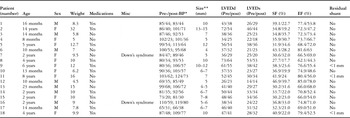
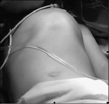
The remaining 18 patients had a clinically significant arterial duct with exclusively left-to-right flow, no evidence of increased pulmonary vascular resistance (Table 1), a left-sided aortic arch, and were selected for surgery. There were eight male and ten female patients, with an age range of 10 months to 14 years. The weight ranged from 4.5 to 32 kilograms. Several patients had additional cardiac defects: Five patients had mitral regurgitation, including one with moe than four regurgitations and severe left atrial enlargement (Fig 1), and one with two to three or more regurgitations. All patients had enlargement of the left atrium and ventricle, diagnosed by chest X-ray and echocardiography. Post-ductal oxygen saturations were normal in all patients, pre- and postoperatively. Ten patients were treated with anti-congestive medications before surgery. When asked, parents of all patients that were ambulatory stated that they had significant exercise intolerance, in most cases only able to walk no more than about 10 metres before having to rest because of dyspnoea. In all, six patients had recent upper respiratory symptoms and/or pneumonia, but were afebrile with a normal white blood cell count before surgery. Two patients had dysmorphic features consistent with Down’s syndrome. All patients undergoing surgery had pre-operative laboratory tests, including a serum sodium, potassium, blood urea nitrogen, creatinine, ionized calcium, complete blood count with differential, platelet count, prothrombin/International Normalised Ratio, and were typed and crossed.
Clinical and echocardiographic data.

LVEDd, left ventricular end diastolic dimension; LVEDs, left ventricular end systolic dimension; BP, blood pressure; SF, shortening fraction; EF, ejection fraction
*Blood pressures were obtained by manual cuff in the operating room, just before and immediately following placement of the clips
**Arterial duct size was determined by direct inspection intraoperatively, and by echocardiography postoperatively
The following patients had additional associated cardiac lesions:
4: Functional bicuspid aortic valve with mild stenosis (Peak gradient 36 mmHg)
8: Severe mitral regurgitation, with severe left atrial enlargement, moderate tricuspid regurgitation with mild dilation of the right atrium and ventricle
9: Mild mitral and aortic regurgitation
14: Moderate mitral regurgitation
15: Mild mitral regurgitation
17: Mild mitral regurgitation
Patients 2, 8, 9, and 18 had surgical ties placed around the duct in addition to placement of the clips
Chest asymmetry in a patient (no. 8) with severe left atrial enlargement and severe mitral valve regurgitation with a 10 millimetre arterial duct.

Anaesthesia protocol
All patients were evaluated and assigned the American Society of Anesthesiologists’ classification before surgery. Intravenous access was obtained in the patients before entry into the operating theatre. After placement of routine monitors, anaesthesia was induced using propofol, 2–4 milligrams per kilogram, and rocuronium, 0.6 milligram per kilogram. After tracheal intubation and transfer to the right lateral position, anaesthesia was maintained with a combination of sevoflurane, air, and oxygen, with fentanyl, 1–2 micrograms per kilogram as needed. At the conclusion of surgery, muscle relaxant was reversed with neostigmine, 0.05–0.07 milligram per kilogram, and glycopyrrolate, 0.02 milligram per kilogram, and spontaneous ventilation was allowed to resume. In all, seven patients were given a single dose of hydralazine, 0.05–0.1 milligram per kilogram, in the operating theatre, with subsequent doses of hydralazine and/or labetalol, 0.25 milligram per kilogram, over the ensuing 2 hours, as needed, until the patients remained normotensive. No patient required hypertension medications more than 2 hours after the completion of surgery.
Surgical technique
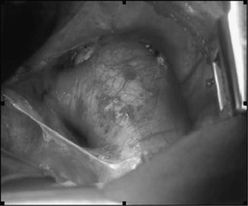
With the left chest prepared and draped, access to the left thoracic cavity was obtained through a left limited posterolateral thoracotomy incision located over the fourth intercostal space, just below the tip of the scapula (Supplementary Fig 1). The incision was extended through the skin, subcutaneous tissue, and the latisimus dorsi muscle, with sparing of the serratus anterior muscle. The intercostal muscles were then divided in the fourth intercostals space and a chest retractor was placed. The left lung was gently retracted medially and the posterior mediastinal pleura was incised over the aorta with electrocautery (Supplementary Fig 2). The courses of the vagus and recurrent laryngeal nerves were identified, and extreme care was taken to avoid injury to either of these nerves (Supplementary Fig 3). Once the mediastinal pleura was incised over the descending aorta, it was gently retracted medially to expose the duct. The dissection of the tissues surrounding the duct and the aorta were then initiated. The duct was dissected both superiorly and inferiorly, as was the aorta to ensure adequate control in case of bleeding (Supplementary Fig 4). In most patients, the duct was relatively short and large in diameter (Fig 2), and therefore required maximal exposure and control before ligation. Using an appropriate size clip applier, the duct was then doubly clipped (Supplementary Fig 5). In most patients, the arterial duct was able to be closed using two large size hemoclips. In four patients, heavy silk ties were placed in addition to the clips due to the extraordinarily large size of the duct. In some patients, it was necessary to temporarily clamp the descending aorta just past the take off of the subclavian to relieve the pressure within the duct during placement of the clips.
The ductus and descending aorta shown just before placement of the clip.

After haemostasis was assured, the chest was irrigated with a solution of saline containing Ancef. Before closure, 0.25% marcaine solution was injected in the intercostal spaces above and below the incision line to achieve intercostal nerve block and within the muscles layers and the incision line to achieve adequate local anaesthesia. The left lung was then allowed to expand fully, and the ribs were approximated using multiple 2.0 Tycron sutures. A left pleural thoracostomy tube was placed in two patients; one because of the presence of adhesions, in anticipation of potential increased bleeding, and one due to an inadvertent slight breech into visceral pleura. In both cases, the tube was removed the following day. The closure of the muscle layers and subcutaneous tissues were performed in routine manner and the skin was closed with a subcuticular suture. After placement of an appropriate dressing, the patients were returned to supine position, extubated and transferred to the surgical unit after a brief period of monitoring for haemodynamic stability in the post-anaesthesia care room. For the 14-year-old patient with adhesions, the total operating time was 37 minutes. For all patients, the total operating time was less than 30 minutes. A total of five to seven cases were performed each day over 3 consecutive days.
Post-operative care
After awakening, pain was managed with paracetamol, 20 milligrams per kilogram q6 hours for 24 hours, and with morphine sulphate, 0.1 milligram per kilogram, q4 hours for breakthrough pain. All patients received prophylactic antibiotics, including a single dose of cefazolin, 25 milligrams per kilogram, at the completion of surgery, followed by oxacillin, 50 milligrams per kilogram per day, for the first 24 hours after surgery.
Post-operative echocardiograms
All patients underwent a limited follow-up transthoracic echocardiogram on the first or second post-operative day to assess for left ventricular function, residual flow through the arterial duct, and pericardial effusion. Three patients, who had the largest arterial duct, had a haemodynamically insignificant residual shunt through the duct, <1 millimetre. In all, 13 patients had decreased left-ventricular systolic function (estimated by M-mode), with at least a seven percentage point decrease in ejection fraction and/or a five percentage point decrease in shortening fraction. There was no correlation between decreased left ventricular systolic function and size of the duct.
Complications
No patients had significant bleeding, and no patient required transfusion of blood products. No wound infections occurred. Two patients had sinus bradycardia in the immediate post-operative period, but without any haemodynamic compromise. One patient was treated with glyopyrrolate for bradycardia, heart rate in the 40s, with a subsequent immediate improvement. There were no other post-operative arrhythmias. No patients showed any clinical evidence of recurrent laryngeal nerve injury. All 18 patients were able to be discharged by 2 days postoperatively. One patient (no. 12) remained hospitalised for non-cardiac reasons, in order to optimise nutritional intake and weight gain.
Discussion
In this report, we describe our experience in performing surgical closure of clinically significant arterial ducts in 18 children at the Angkor Children’s Hospital in Siem Reap, Cambodia in October, 2008. All 18 ducts were successfully closed with no significant complications. Those with the largest arterial duct had a tiny, less than 1 millimete, residual left to right shunt.
Our team consisted of 11 medical professionals with significant experience in caring for children with congenital cardiac disease, including two cardiothoracic surgeons, one paediatric cardiologist, two paediatric anaesthesiologists, one paediatric echocardiography technician, one paediatric respiratory therapist, two operating theatre nurses, and two paediatric critical care nurses. We believe that there were several factors that contributed to our successful results, including the following:
• Careful patient selection, which included a history, physical examination, transthoracic echocardiogram, as well as comprehensive pre-surgical labs.
• The use of surgical clips, with additional surgical ties used only on four patients with an exceptionally large arterial duct.
• Minimal use of chest tubes.
• Aggressive use of anti-hypertensive medication in the immediate post-operative period.
• Extubation immediately postoperatively, with the presence of a respiratory therapist to optimise airway management before and after extubation.
• Aggressive pain management, with encouragement of ambulation (when age-appropriate) by the first post-operative day, and avoidance of medications that inhibit platelet function.
• On-site presence of the team until all patients were able to be discharged home.
No patients had persistent hypertension. In our two previous missions, nearly half of the patients had hypertension, which persisted up to 2 days postoperatively. The administration of hydralazine to patients with even mild hypertension in the immediate post-operative period, followed by aggressive medical management during the first 2 hours postoperatively, appears to have prevented the occurrence of persistent systemic hypertension in the post-operative period. All 18 patients were normotensive within 2 hours after surgery and did not require additional blood pressure medication more than 2 hours postoperatively. The aetiology of post-operative hypertension is unknown, but is possibly related to the renal response to hypoperfusion in the presence of significant left to right shunting. It is also likely due to the redistribution of previously shunted blood into the systemic intravascular space after closure of the arterial duct.
In all, 13 out of 18 patients had a decrease in their left ventricular systolic function in the immediate post-operative period, that is, shortening fraction decreased by at least five percentage points and or ejection fraction decreased by at least seven percentage points. Of the five patients who did not have decreased left ventricular systolic function postoperatively, there was no correlation with age, sex, or size of the ductus; four of these five patients had not received cardiac medications before surgery.
The reasons for decreased left ventricular function postoperatively may be multiple. One likely possibility is the decrease in the volume load of the left ventricle (Table 1), causing a left shift on the Frank–Starling curve. In addition, most patients had an increase in blood pressure after arterial duct closure (Table 1), thereby causing an increase in afterload to the volume-reduced left ventricle. The increased afterload could have also contributed to the decreased left ventricular function.
In summary, we performed successful surgical closure of a clinically significant arterial duct without complications in 18 patients at the Angkor Hospital for Children in Siem Reap, Cambodia. Although resources are limited for the majority of children with congenital cardiac disease in the third world countries, targeting those patients with arterial ducts for surgical closure is both safe and feasible. We hope that as a result, more teams from western countries will be inspired to perform similar missions on patients in third world countries.
Acknowledgements
This medical mission was entirely funded by Variety Children’s Lifeline of Solana Beach, California. The authors thank all of the staff from Angkor Hospital for Children, the additional surgical support team from Rady Children’s Hospital of San Diego, and Mr Jon Morgan for their support and kindness. The authors also thank all of the patients and their families for their unwavering support and appreciation.
Supplementary material
The online figures can be viewed at http://journals.cambridge.org/cty