Introduction
The bone-anchored hearing aid (BAHA) implantation surgical technique has evolved in the last few years to avoid subcutaneous tissue trimming. This step in the procedure (i.e. tissue trimming) was related to post-operative morbidity, such as local pain, loss of sensation and peri-abutment skin reactions. The majority of skin lesions associated with BAHA implantation (Holgers' skin reaction gradesReference Holgers, Tjellström, Bjursten and Erlandsson1 1 and 2) resolve with conservative therapy. However, some patients need resection of the granulation tissue (Holgers' grade 3), and, in some cases, replacement of the abutment is needed (Holgers' grade 4). This paper describes the use of the Z-plasty technique for resection of peri-abutment granulation tissue.
Case report
We present the case of an eight-year-old boy with left microtia who underwent BAHA implantation in April 2012. Subsequently, the patient presented, over a period of 12 months, with sporadic skin reactions (Holgers' grade 2) that resolved with topical corticosteroid therapy. In the latter six months, the episodes became more frequent and the skin reaction became persistent (Holgers' grade 3), showing no response to topical corticosteroid therapy.
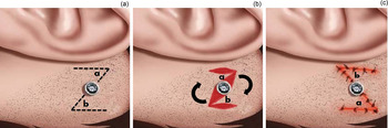
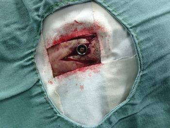
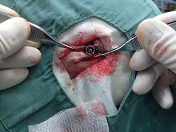
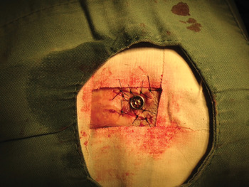
We opted to surgically resect the peri-abutment granulation tissue. To avoid recurrence of granulation tissue, we applied the principles of Z-plasty (Figure 1). By inverting the position of the skin flaps, the affected edge can be distally dislocated and the disease-free edges can then be proximally dislocated and fixed in close contact with the abutment (Figures 2–4).
Z-plasty technique, indicating the Z-shape skin incision (a), rotation of flaps (labelled ‘a’ and ‘b’) (b) and suturing (c).

Z-shape skin incision.

Skin flap rotation.

Suturing and fixation of skin flaps.

Discussion
Bone-anchored hearing aid implants were introduced in 1977 by Tjellström and colleagues (as cited in Holgers et al.Reference Holgers, Tjellström, Bjursten and Erlandsson1). Since then, BAHAs have become a well-established tool for satisfactory management of several conditions associated with hearing loss. The BAHA system comprises a speech receptor and processor, which processes the received noise and transmits it as vibratory energy to the abutment. This abutment is attached to the titanium implant, which in turn is embedded in the mastoid bone.Reference Hobson, Roper, Andrew, Rothera, Hill and Green2
The surgical procedure for BAHA implantation is safe, and the majority of adverse reactions are local reactions and are related to the implant itself. Most of the complications associated with BAHA implants concern the subcutaneous tissue reduction and the consequent skin reaction.Reference Husseman, Szudek, Monksfield, Power, O'Leary and Briggs3 The most common complications reported in the literature are skin overgrowth and infection of the metallic implant site.Reference Hobson, Roper, Andrew, Rothera, Hill and Green2 Unsuccessful skin grafting, cellulitis, granulation, eczematous dermatitis, hypertrophic scarring and keloid formation, and even loss of the implant, can also occur.Reference Shirazi, Marzo and Leonetti4–Reference Gillett, Fairley, Chandrashaker, Bean and Gonzalez8 The incidence of skin reaction varies between 5 and 50 per cent in different studies, depending on the surgical technique used and the method of inflammation classification.Reference Granström and Tjellström9–Reference Tamarit Conejeros, Dalmau Galofre, Murcia Puchades, Pons Rocher, Fernández Martínez and Estrems Navas12
The Holgers' classification is used to describe peri-abutment skin reactions.Reference Holgers, Tjellström, Bjursten and Erlandsson1 The skin reactions are graded as follows: 0 = no tissue irritation, 1 = small erythema, 2 = erythema and humidity, 3 = granulation, and 4 = infection leading to removal of the abutment. The majority of cases referred for complications can be conservatively treated, although 1 to 2 per cent of patients need surgical intervention, and, in some cases, removal of the implant is required. Recent studies have shown that the application of a corticosteroid gel can be effective at reducing the occurrence of skin reaction.Reference Falcone, Kaylie, Labadie and Haynes13 This achieves better results in cases with Holgers' grade 1 and 2 skin reactions. In Holgers' grade 3 cases, resection of the granulation tissue is often indicated. In Holgers' grade 4 cases, in addition to resection of the granulation tissue, replacement of the abutment with a longer one is recommended.Reference Holgers, Tjellström, Bjursten and Erlandsson1
In our patient, the episodes of skin reaction became very frequent and he was unable to use his BAHA for 2 to 3 days in a week. The response to the topical treatment was poor. Furthermore, the child had moved to a distant location with limited access to clinical care. We opted for surgical treatment using a Z-plasty technique for management of the skin complications.
The classic Z-plasty technique involves a central incision and two peripheral incisions made in a ‘Z’ shape, creating two skin flaps of identical size with a triangular shape. This technique allows the surgeon to change the direction of the scar, interrupt the linear component of the scar and lengthen its cicatricial contracture.Reference Frodel14 In the present case, the Z-plasty technique enabled removal of the granulation tissue at the edges of the incision from the surroundings of the implant, therefore ensuring that only disease-free skin would be in contact with the implant after the procedure.
• Bone-anchored hearing aid implantation is a well-known approach for successfully managing conditions associated with hearing loss
• The procedure is frequently associated with skin reactions, which cause discomfort and may necessitate abutment replacement
• The use of Z-plasty is a novel approach to overcome this complication
• It involves removal of granulation tissue from abutment edges and replacement with disease-free skin to ensure proper wound closure
• The Z-plasty technique is recommended for management of peri-abutment dermal reactions
Several BAHA implantation surgical techniques have been described. These range from a skin flap creation with a surgical dermatome (a technique currently proscribed given the high levels of adverse reactions),Reference Van Rompaey, Claes, Verstraeten, van Dinther, Zarowski and Offeciers15 to the technique of simple incision with subcutaneous tissue reductionReference de Wolf, Hol, Huygen, Mylanus and Cremers10, Reference de Wolf, Hol, Huygen, Mylanus and Cremers16 and recently without subcutaneous tissue reduction.Reference Hultcrantz17 A recent clinical trial by Hultcrantz showed that a BAHA implantation technique using simple incision methods, without subcutaneous tissue trimming, was associated with reduced surgical time, faster wound healing and no complications in the studied population, when compared to the conventional dermatome technique and subcutaneous tissue reduction.Reference Hultcrantz17
New techniques aiming to reduce skin reactions focus on the use of longer abutments (8.5 mm)Reference Pelosi and Chandrasekhar18 and hydroxyapatite-coated abutments.Reference Larsson, Wigren, Andersson, Ekeroth, Flynn and Nannmark19 In our department, after a discussion at the Osseo workshop in Newcastle, UK, in June 2013, we abandoned the technique of subcutaneous tissue resection. Instead, we now perform linear incision with local removal lateral to the incision using a 0.6 mm punch. While the hydroxyapatite-coated abutments await the Brazilian National Health Surveillance Agency (‘ANVISA’) licence, we have been using the 8.5 mm abutments in adults and older children, and the 4.5 mm abutments in younger children. After adopting this technique, we observed the results described in the literature in our patients. These gains included reduced surgery time, the increased use of local anaesthetic and sedation (rather than general anaesthetic), the absence of pain or loss of sensation, and the absence of skin complications.Reference Larsson, Wigren, Andersson, Ekeroth, Flynn and Nannmark19
Conclusion
Patients who undergo BAHA implantation with subcutaneous tissue resection have an increased risk of experiencing skin reactions. In Brazil, this technique is applied in a large number of patients, some of whom will eventually develop Holgers' grade 3 and 4 skin reactions, and thus require surgical treatment. We recommend the use of Z-plasty, as the granulation tissue is removed from the edges of the abutment and instead disease-free skin makes contact with the implant. We also advise that the surgeon performs the simple linear incision technique for BAHA implantation when possible in order to prevent skin reactions.