Introduction
Graphidaceae at present contains more than 50 genera and comprises a vast micro-lichen diversity in the tropics (Rivas Plata et al. Reference Rivas Plata, Lücking and Lumbsch2008, Reference Rivas Plata, Lücking, Sipman, Mangold, Kalb and Lumbsch2010, Reference Rivas Plata, Lücking and Lumbsch2012; Lücking et al. Reference Lücking, Archer and Aptroot2009, Reference Lücking, Seavey, Common, Beeching, Breuss, Buck, Crane, Hodges, Hodkinson and Lay2011). During the course of recent work on the taxonomy of crustose lichens in Vietnam (Lücking & Vězda Reference Lücking and Vězda1998; Aptroot & Sparrius Reference Aptroot and Sparrius2006; Sparrius et al. Reference Sparrius, Saipunkaew, Wolseley and Aptroot2006), we found some interesting and noteworthy species of Graphidaceae. Although most of the graphidaceous lichens collected during expeditions in the tropical rainforests of Vietnam were subsequently identified, the huge collection also conceals unidentified and undescribed thelotremoid taxa. Here we consider the material of lirellate taxa, collected from northern and central highland national parks in Vietnam. Compared with other South Asian countries, the number of lirellate taxa in Vietnam is relatively low, probably because the country has not been exhaustively surveyed; the number has been estimated to be c. 20 accepted species (Aptroot & Sparrius Reference Aptroot and Sparrius2006). In this contribution, two new species belonging to the genera Acanthothecis and Diorygma are described.
Acanthothecis Clem. is a pantropical genus and c. 40 species are known worldwide (cf. CABI Bioscience et al. 2010; Sharma et al. Reference Sharma, Makhija and Khadilkar2010; Tripp et al. Reference Tripp, Lendemer and Harris2010). The new taxon Acanthothecis salazinica, which differs from other species of Acanthothecis in containing salazinic acid, is the only species of this genus known so far from Vietnam.
Diorygma includes c. 50 species distributed in tropical to subtropical regions of the world (Kalb et al. Reference Kalb, Staiger and Elix2004; Archer Reference Archer2006, Reference Archer2009; Sharma & Makhija Reference Sharma and Makhija2009a , Reference Sharma and Makhija b ; Makhija et al. Reference Makhija, Gayatri and Bharati2009; Sharma & Khadilkar Reference Sharma and Khadilkar2012). The genus is poorly studied in Vietnam. Indeed, with the exception of Diorygma tinctorium Eschw., no other species has been recorded from the country (Aptroot & Sparrius Reference Aptroot and Sparrius2006). The material of Diorygma examined is distinguished by thallus and ascomatal morphology, ascospore size and chemistry, and described here as Diorygma archeri, new to science.
Materials and Methods
The specimens observed were collected in the Chu Yang Sin, Tam Dao and Yok Don National Parks, and at the Dray Nur Waterfall area of Vietnam, in April 2012. The national parks and waterfall are situated in the northern and central highlands of the country and provide moist, humid conditions suitable for graphidaceous taxa. Fresh samples, preserved in the lichen herbarium of the Korean Lichen Research Institute (KoLRI), were observed under a SMZ 168 dissecting microscope and Olympus BX50 compound microscope for morpho-taxonomic and anatomical characteristics. Methods for colour spot reaction tests and thin-layer chromatography (TLC) were based on Orange et al. (Reference Orange, James and White2001). Thin-layer chromatography was performed in solvent systems A [toluene (180): 1, 4-dioxane (45): acetic acid (5)] and C [toluene (170): acetic acid (30)]. Lugol's solution (I) was used to check the amyloidity of the asci, interascal filaments and ascospores. Anatomical measurements were made from 15–20 apothecial sections, and the lower and higher values show the minimum and maximum of the range. Various publications (Staiger Reference Staiger2002; Nakanishi et al. Reference Nakanishi, Kashiwadani and Moon2003; Kalb et al. Reference Kalb, Staiger and Elix2004; Archer Reference Archer2006, Reference Archer2009; Qing-Feng & Jiang-Chun Reference Qing-Feng and Jiang-Chun2008; Makhija et al. Reference Makhija, Gayatri and Bharati2009; Sharma & Makhija Reference Sharma and Makhija2009a , Reference Sharma and Makhija b ; Sharma & Khadilkar Reference Sharma and Khadilkar2011, Reference Sharma and Khadilkar2012) were consulted for species identifications. Images were taken using ZEISS Scope A1 (AX10) and Axio cam ERc 5s, and illustrations were prepared using Coral Draw (version 12).
The New Species
Acanthothecis salazinica S. Joshi & Hur sp. nov.
MycoBank No.: MB 803518
Differing from Acanthothecis clavulifera in the leprose thallus, the slit-like ascomatal disc, and the presence of salazinic acid as a secondary compound.
Type: Vietnam, Dak Lak Province, Yok Don National Park, 12°517′20·0″N, 107°45′58·1″E, alt. c. 760 m, on tree, 22 April 2012, Hur & Oh 120211 (KoLRI—holotype).
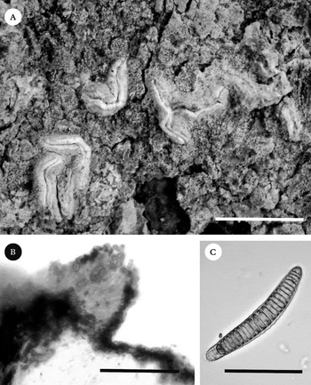
Acanthothecis salazinica (holotype). A, habit and habitat; B, cross-section of ascoma showing periphysoids; C, ascospore. Scales: A=3 mm; B=100 µm; C=50 µm.

Thallus corticolous, epiperidermal, greenish grey to olive-grey or whitish grey, ecorticate, thin, up to 150 µm, ±granular, leprose, rough, continuous, integrated with crystals; prothallus indistinct to whitish.
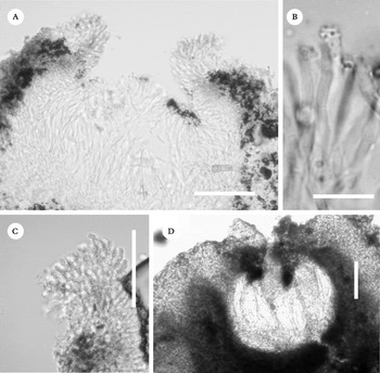
Ascomata numerous, lirellate, emergent to sessile, bent, scattered on thallus; lirellae simple to branched, sinus with acute ends, 1–3×0·7–1·0 mm, cream to whitish, entire, epruinose, 0·34–0·45 µm thick; disc closed or rarely slightly open, slit-like, ±creamy pruinose, later on ±reddish in colour; thalline margin entire, apically thin, 30–65 µm thick; proper exciple convergent, entire, non-carbonized, hyaline to reddish brownish outside, 45–65(–124) µm thick; epithecium indistinct to hyaline, appearing dark, crystalline to granular, 15–20 µm high; hymenium hyaline, clear, 140–210 µm high, I−; hypothecium indistinctly hyaline to reddish brown in lower side, up to 45 µm high; paraphyses simple, apically warty, 2–3 µm thick; periphysoids distinct, long to short, warty at apices, apices covered with crystalline layer, 25–90 µm long; asci clavate, 6–8-spored, 150–170×30– 40 µm, gelatinous matrix becoming brownish upon maturity, I−; ascospores fusiform, elongate, with ±rounded ends, hyaline, transversely septate, appearing submuriform due to disintegration of luminal content, transversely 28–30-locular, with gelatinized wall, 70–120×7–14 µm, I−.
Chemistry
Thallus K+ yellow-red, PD+ yellow-orange, C−, KC−; salazinic acid and traces of norstictic acid detected by TLC.
Etymology
The specific epithet refers to the secondary compound produced by the new taxon.
Ecology and distribution
The corticolous Acanthothecis salazinica is known only from its type locality in a tropical, evergreen forest of Yok Dou National Park, Vietnam, at an altitude of c. 760 m. It was collected from a tree trunk above breast height, where it was growing luxuriantly with Diorygma, Graphis and Letrouitia species.
Remarks
The new taxon is characterized by an ecorticate, thin, leprose, greenish grey to olive-grey or whitish grey thallus, lirellate, emergent to sessile apothecia, creamish to whitish, entire, epruinose labia, a slit-like, closed disc with entire thalline margin, hyaline to brown, entire proper exciple, warty periphysoids, clear hymenium, 8-spored asci and elongate, 28–30-loculate, hyaline, I− ascospores of 70–120×7–14 µm.
Acanthothecis salazinica is the first species described in the genus to contain salazinic acid as the main secondary metabolite. No other species of the genus, except rare forms of Acanthothecis peplophora (M. Wirth & Hale) E. Tripp & Lendemer, contain salazinic acid (Tripp et al. Reference Tripp, Lendemer and Harris2010). The latter species shows an entirely different morphology, having a thick, greenish, corticate thallus and large, elongate lirellae with a crumbling margin and striate exciple, and muriform ascospores.
Acanthothecis clavulifera (Vain.) Staiger & Kalb closely resembles the new taxon in having ascocarps (1·0–3·5×0·5–0·7 mm) and ascospores of similar size range (85–115×8–13 µm), and an entire excipulum, but it produces protocetraric acid as the major compound (Staiger & Kalb Reference Staiger and Kalb1999). Moreover, the bumpy, cracked thallus, bulging margins of the ascocarp and the usually exposed disc of A. clavulifera separate it from A. salazinica. Similarly, A. subclavulifera Staiger & Kalb shows proximity to the new species in having 1–3×1 mm large lirellae, 8-spored asci and ascospores 75–110×11–15 µm, but it has a striate excipulum and a corticate thallus containing protocetraric acid. Acanthothecis africana Staiger & Kalb also has comparable ascospores, but differs in producing norstictic acid as secondary compound.
An Indian species, Acanthothecis archeri B. O. Sharma et al., is close to the new taxon in having emergent ascomata, creamy, pruinose, apothecial discs, entire excipula, and ascospores 75–120 µm long, but differs in lacking thallus compounds and having I+ weakly blue, muriform ascospores (Sharma et al. Reference Sharma, Makhija and Khadilkar2010). Acanthothecis aquilonia A. W. Archer & Elix has 80–100 µm long, transversely sepate ascospores similar to our new taxon, but lacks secondary metabolites, and has a different thallus morphology (Archer & Elix Reference Archer and Elix2008).
Diorygma archeri S. Joshi & Hur sp. nov.
MycoBank No.: MB 803519
Similar to Diorygma pruinosum, but differing in the thick, distinctly corticate, greyish green thallus having a well-developed medulla, scarcely open apothecial disc, non-carbonized proper exciple and large ascospores.
Type: Vietnam, Dak Lak Province, Yok Don National Park, 12°51′20·0″N, 107°45′58·1″E, alt. c. 760 m, on tree, 22 April 2012, Hur & Oh 120390 (KoLRI—holotype).
(Figs 2A & B, 3D)
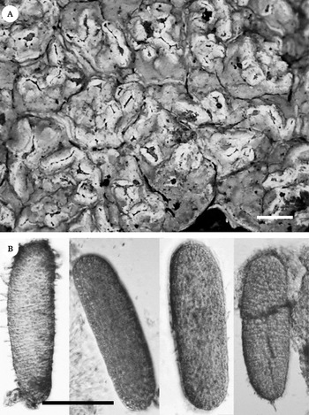
Diorygma archeri (holotype). A, habit and habitat; B, ascospores. Scales: A=2 mm; B=100 µm.

A–C, Acanthothecis salazinica (holotype ); A & C warty periphysoids at the tip of exciple; B, warty paraphyses tip. D, Diorygma archeri (holotype), cross-section of ascoma. Scales: A, C & D=100 µm; B=5 µm.

Thallus corticolous, epiperidermal, whitish to greenish grey or light grey to greyish green, rimose, continuous, smooth, dull, up to 800 µm thick; pseudocortex distinct, reaching up to 30 µm; algal layer well developed, continuous, 50–60 µm thick; medulla white, thick, inspersed with crystals, epi- to endoperidermal; prothallus indistinct, white.
Ascomata numerous, irregular, shortly lirellate, immersed in periderm, ±fused, aggregate; lirellae short to sinuous, curved, branched, flexuous, 2·0–3·5×0·55–0·93 mm; disc initially closed, later exposed, brown to dark brown or reddish brown, covered by a dense, granular to coarse, crystalline, white pruina; thalline margin raised, straight to incurved, prominent, irregular, 0·11–0·46 mm thick; proper exciple poorly developed, convergent to scarcely divergent in mature ascomata, entire, non-carbonized, ±hyaline to pale brown, surrounded by periderm in mature ascomata, 20–30 µm thick; epithecium brown, studded with crystals, granular, 10–15 µm high; hymenium hyaline, clear, 157–215 µm high, I+ blue or weakly blue, mostly in lateral parts; hypothecium distinct, hyaline, up to c. 40 µm high; paraphyses reticulately interwoven at tips, c. 1·0–1·5 µm thick, gelatinous; asci broadly clavate, 1-spored, c. 160–255×56–87 µm, I−; ascospores oval to elliptic, hyaline, muriform, multicelled, peripheral and central cells equal in size, gelatinous sheath not seen, 150–255×53–85 µm, I+ violet-blue.
Chemistry
Thallus K− or K+ slightly yellow, PD+ yellow-orange, C−, KC−; only protocetraric acid detected by TLC.
Etymology
The specific epithet is named in honour of the eminent Australian lichenologist Dr Alan W. Archer.
Distribution and ecology
The new species is widely distributed in the tropical rainforests of Yok Don National Park, Vietnam, where it was collected from tree trunks at elevations of 700–800 m. The other associated lichens are members of Graphidaceae and Letrouitiaceae.
Remarks
The new taxon is distinguished by a whitish grey to greenish grey, rimose, dull, smooth thallus, a poorly developed, hyaline to pale or brownish proper exciple, 1-spored asci, colourless, multicelled, muriform, oval to elliptic, I+ blue ascospores and the presence of protocetraric acid in the thallus. Other protocetraric acid-containing species of Diorygma are: D. africanum Kalb et al., D. hololeucum (Mont.) Kalb et al., D. pruinosum (Eschw.) Kalb et al., D. rufopruinosum (A. W. Archer) Kalb et al., D. reniforme (Fee) Kalb et al. and D. agumbense B. O. Sharma & Khadilkar (Kalb et al. Reference Kalb, Staiger and Elix2004; Sharma & Khadilkar Reference Sharma and Khadilkar2012). The new taxon closely resembles D. pruinosum, which has a greenish white, smooth, dull thallus, conspicuous, immersed, white, irregular apothecia, a densely pruinose disc, 150–200 µm high hymenium, 1-spored asci and multicelled, muriform ascospores, but it can be distinguished by an open and ±convex apothecial disc, laterally and basally carbonized proper exciple, and smaller ascospores, 100–150 (–175)×20–30 µm (Kalb et al. Reference Kalb, Staiger and Elix2004). Diorygma africanum sometimes also has a carbonized exciple, but the peripheral ascospore locules are distinctly smaller than the central ones. Diorygma hololeucum is distinguished by smaller ascospores (130–160×30–36 µm) and comparatively large, sessile lirellae (up to 8 mm) exposing the disc. Diorygma rufopruinosum has a reddish brown pruina on the apothecial disc, a non-amyloid hymenium, lacks a proper exciple, has slightly smaller ascospores (105–170×30–42 µm), and produces norstictic, salazinic, stictic, subvirensic, virensic acids and methyl virensate as thallus compounds. Diorygma reniforme differs from D. archeri in having a ±carbonized exciple and its thallus containing norstictic and salazinic acids in addition to protocetraric acid. Diorygma agumbense is completely different from the new taxon in having a convergent proper exciple, 8-spored asci, 87–125×20–37 µm large ascospores, and producing cryptostictic and stictic acids.
Additional specimens examined. Vietnam: Dak Lak Prov.: Yok Don National Park, alt. c. 760 m, on tree, 2012, Hur & Oh 120211-1, 120214 (KoLRI).
New Records
Acanthothecis corcovadensis (Redinger) Staiger & Kalb
Mycotaxon 73: 98 (1999).
(Fig. 4A)
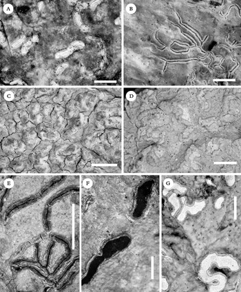
New records of Graphidaceae in Vietnam. A, Acanthothecis corcovadensis; B, Diorygma erythrellum; C, Diorygma junghuhnii; D, Diorygma macgregorii; E, Platygramme platyloma; F, Pliariona montagnei; G, Thecaria quassiicola. Scales: A, C, D & G=2 mm; B & F=1 mm; E=5 mm.

This corticolous species is characterized by a green to olive-green or brownish green, smooth and ±dull thallus; emergent, solitary, scarcely branched, lirellate apothecia, 6×1 mm; open to ±closed, densely white pruinose, flesh-coloured disc; brown, marginally crenate proper exciple mostly lined with crystals, I+ blue inside; thin, conglutinate paraphyses lacking warty tips; periphysoids mostly indistinct (due to eroded ascomata), apically lined with dense crystals; clear and hyaline, I− hymenium of 200–230 µm; 1-spored, 170–180×40–45 µm asci; hyaline, muriform, 120–150×29–37 µm, I− ascospores, and the presence of stictic, constictic and hypostictic acids as thallus compounds. The material examined closely resembles Hemithecium stictilabiatum (Patw. & C. R. Kulk.) Chitale & Makhija, which has similar morpho-anatomical and chemical characteristics, but this species differs in its apically smooth paraphyses and amyloid ascospores (I+ blue). Given the taxonomic importance of amyloidity in the Graphidaceae (Lücking et. al. Reference Lücking, Seavey, Common, Beeching, Breuss, Buck, Crane, Hodges, Hodkinson and Lay2011), the reported material agrees well with Acanthothecis in having warty paraphyses. It has been previously reported from South America (Staiger & Kalb Reference Staiger and Kalb1999).
Specimen examined. Vietnam: Dak Lak Prov.: Chu Yang Sin National Park, alt. c. 763 m, on tree, 2012, Hur & Oh 120103 (KoLRI).
Diorygma erythrellum (Mont.) Kalb et al.
Symb. Bot. Ups. 34(1): 150 (2004).
(Fig. 4B)
This species is characterized by a cream to pale grey, or whitish grey, smooth and ±glossy thallus; immersed to ±emergent, numerous, lirellate, ±flexuous, branched, 2–3×0·1–0·3 mm ascomata; open, reddish or reddish brown disc covered by thin, white pruina; raised, incurved, entire thalline margin; poorly developed, divergent, uncarbonized, yellowish to brownish proper exciple; hyaline and clear, I+ blue hymenium 120–140 µm; 8-spored asci; oval to elliptic, hyaline, muriform, I+ violet-blue ascospores, 40–50×15–20 µm, and a thallus containing norstictic and connorstictic acids. This species is similar to Platythecium colliculosum (Mont.) Staiger and P. dimophodes (Nyl.) Staiger, but differs in its much larger ascospores and nature of the paraphyses, typically of Diorgyma (Kalb et al. Reference Kalb, Staiger and Elix2004).
Specimen examined. Vietnam: Dak Lak Prov.: Chu Yang Sin National Park, alt. c. 780 m, on tree, 2012, Hur & Oh 120193 (KoLRI).
Diorygma junghuhnii (Mont. & Bosch) Kalb et al.
Symb. Bot. Ups. 34(1): 157 (2004).
(Fig. 4C)
This species is characterized by a whitish grey to grey-white, rimose, ±verrucose thallus; numerous, shortly lirellate, irregularly branched and aggregated lirellate ascomata, 2·0–3·5×0·5–1·0 mm; brown to dark brown, exposed disc covered with dense, granular or crystalline, white pruina; raised, straight to incurved, crenate thalline margin; poorly developed, non-carbonized, divergent, hyaline proper exciple surrounded by periderm; hyaline and clear, I+ blue hymenium of 70–130 µm, 1-spored asci of 97–107×20–25 µm; oval to elliptic, hyaline, muriform, I+ violet-blue ascospores of 73–114×22–33 µm, and thallus containing norstictic acid and traces of constictic acid. It differs from the similar Diorygma soozanum (Zahlbr.) M. Nakan. & Kashiw. by having a distinct amyloid hymenium. Diorygma junghuhnii has a pantropical distribution and has been reported from Australia, Fiji, China, India, Japan, the Philippines, Indonesia, Togo, Tanzania, the Solomon Islands, New Caledonia, Costa Rica, Guatemala, Brazil and Paraguay (Nakanishi et al. Reference Nakanishi, Kashiwadani and Moon2003; Kalb et al. Reference Kalb, Staiger and Elix2004; Archer Reference Archer2006, Reference Archer2009; Qing-Feng & Jiang-Chun Reference Qing-Feng and Jiang-Chun2008).
Specimen examined. Vietnam: Vinh Phuc Prov.: Tam Dao National Park, Tay Thien Mt, alt. c. 77 m, on tree, 2012, Hur & Oh 120379 (KoLRI).
Diorygma macgregorii (Vain.) Kalb et al.
Symb. Bot. Ups. 34(1): 159 (2004).
(Fig. 4D)
This species is characterized by a pale green to green, crystalline, ±verrucose thallus; numerous, ±emergent, scattered, short lirellate, oval to oblong, ±flexuous ascomata 1·5–3·5×1·4–1·6 mm; white to slightly pale pruinose, open disc; straight to incurved, raised, prominent, crenate, irregular thalline margin; poorly developed, divergent, non-carbonized, hyaline to ±brownish proper exciple; hyaline and clear, I+ blue hymenium, 195–250 µm; 1-spored, 180–226× 44–57 µm asci; hyaline, muriform, oval to ellipsoidal, I+ violet-blue ascospores, 175–202×40–55 µm, and a thallus containing stictic, cryptostictic, connorstictic, norstictic and traces of hypostictic acids. This species might be confused with Diorygma hieroglyphicum (Pers.) Staiger & Kalb, but differs in the prominent ascomata and larger ascospores. Diorygma macgregorii is most common in eastern palaeotropical countries and endemic to South-East Asia (Kalb et. al. Reference Kalb, Staiger and Elix2004).
Specimens examined. Vietnam: Vinh Phuc Prov.: Tam Dao National Park, alt. c. 990 m, on tree, 2012, Hur & Oh 120390 (LWG). Dak Lak Province: Chu Yang Sin National Park, alt. c. 780 m, on tree, 2012, Hur & Oh 120187 (KoLRI).
Platygramme platyloma (Müll. Arg.) M. Nakan. & Kashiw.
Bull. Natl. Sci. Mus., Tokyo, Ser. B. 29(2): 89 (2003).
(Fig. 4E)
This species is characterized by a pale green, greyish green to brownish green, thin, glossy thallus; numerous, long, curved or sinuous, scarcely branched, sessile, lirellate, faintly sulcate, black ascomata, 6–20×0·5–1·0 mm; convergent, apically carbonized proper exciple; hyaline and inspersed hymenium 170–200 µm; 1-spored asci; hyaline, muriform, I+ faintly amyloid to reddish brown ascospores, 125–130×25–30 µm, and lack of secondary metabolites. This species might be confused with Platygramme pudica (Mont. & Bosch) M. Nakan. & Kashiw., which differs by the entire lirellae and presence of echinocarpic acid. Platygramme platyloma has previously been reported from Australia, Japan and Indonesia (Archer Reference Archer2009).
Specimens examined. Vietnam: Dak Lak Prov.: Chu Yang Sin National Park, alt. c. 780 m, on tree, 2012, Hur & Oh 120188, 120190, 120191 (KoLRI).
Pliariona montagnei (Bosch) A. Massal.
Atti Inst. Veneto Sci. lett., ed Arti, Ser. 3. 5: 318 (1860).
(Fig. 4F)
This species is characterized by a green to olive-green or dull green, smooth to warty (due to the texture of the bark), glossy thallus; numerous, sessile, lirellate, mostly scattered, simple or infrequently branched ascomata up to 6·0×0·5 mm; open, red, epruinose disc; well-developed, entire, completely carbonized, 130–250 µm proper exciple, covered by thick, complete thalline margin; red pigmented, densely inspersed, 150–200 µm hymenium; 1-spored asci; muriform, ellipsoidal to oval, straight, I−, hyaline ascospores, 125–155×25–40 µm, that become ±brown or mottled when mature, and the presence of isohypercrelline. A recent phylogenetic study proclaimed distant lineages of several morphologically similar species within Graphidaceae that were traditionally treated under the same genus (Rivas Plata et al. Reference Rivas Plata, Hernández, Lücking, Staiger, Kalb and Cáceres2011). Due to parallel evolution, Thecaria montagnei and T. quassicola showed a similar superficial resemblance but are indeed not congeneric, and cannot be treated under the same genus. The strong molecular evidence, supported by some major contrasting taxonomic characters such as an epruinose apothecial disc and a dark red hymenium, strongly advocates the independent existence of Thecaria montagnei and it is placed here under the reinstated genus Pliariona. The species has a wide distribution in Australia, West Africa, Japan, the Philippines, India, Indonesia, the Solomon Islands, Vanuatu and Norfolk Island (Archer Reference Archer2009; Singh & Sinha Reference Singh and Sinha2010).
Specimen examined. Vietnam: Dak Lak Prov.: Chu Yang Sin National Park, alt. c. 780 m, on tree, 2012, Hur & Oh 1201871–1 (KoLRI).
Thecaria quassiicola Fée
Essai Crypt. Écorc. 97 (1825).
(Fig. 4G)
This species is characterized by a green to olive-green or grey-green, smooth and glossy thallus; numerous, sessile, lirellate, mostly scattered, simple or infrequently branched ascomata, c. 10·0×1·5 mm; an open, black disc covered with dense white pruina; completely carbonized, 100–200 µm proper exciple covered by a thick, complete thalline margin; hyaline and densely inspersed hymenium, 200–260 µm; 8-spored asci; muriform, ellipsoidal to oval, straight to curved or reniform, I−, hyaline ascospores, 85–100×19–22 µm, that become ±reddish brown when mature, and a lack of secondary metabolites. Thecaria quassiicola is widespread in Australia, Brazil, India, Indonesia, Japan, Sri Lanka, the Solomon Islands, New Caledonia, Fiji and Norfolk Island (Archer Reference Archer2009; Singh & Sinha Reference Singh and Sinha2010).
Specimen examined. Vietnam: Vinh Phuc Province: Tam Dao National Park, alt. c. 990 m, on tree, 2012, Hur & Oh 120307 (LWG).
This work was supported by a grant from the National Research Foundation of Korea (#2012-0031494), the Korea National Research Resource Center Program, and the Korean Forest Service Program. Santosh Joshi thanks Dr A. Aptroot for providing a lichen checklist of Vietnam, and J.-S. Park for considerable help with lichen collections and preservation.