Abnormalities of the coronary arteries are present in about 1–2% of the population undergoing cardiac catheterisation.Reference Yamanaka and Hobbs 1 This increases to about 5% with patients who undergo a diagnosis of tetralogy of Fallot.Reference Carvalho, Silva, Rigby and Shinebourne 2 Recent use of dual-source multislice CT provides high accuracy-defining coronary artery supply in CHD.Reference Vastel-Amzallag, Le Bret and Paul 3
Case 1
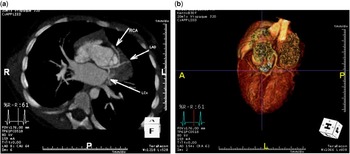
A baby with antenatal diagnosis of tetralogy of Fallot was reviewed and confirmation of the diagnosis was made; however, imaging of the coronary arteries was difficult. Subsequent reviews failed to confirm the origin of the right coronary artery, and thus a CT scan was requested to confirm the coronary course. The baby (9.9 kg) was fed and lightly wrapped before the scan, 18 ml of contrast was injected at 1 ml/second in the right arm via an intravenous line, and images were acquired. The scan showed a single coronary artery with the left main stem arising normally from the posterior sinus of the valsalva, but the right coronary artery being a branch of the left anterior descending coronary artery passes anterior to the right ventricular outflow tract to reach the anterior atrioventricular groove (Fig 1a and b). In view of the coronary course, surgical repair was planned using a homograft rather than a transannular patch.
( a ) Scan showing single coronary artery – left anterior descending artery (LAD) – originating from the posterior sinus of the valsalva with subsequent branching to the left circumflex (LCx) and the right coronary arteries (RCA). ( b ) Three-dimensional iterative reconstruction showing single origin of the coronary artery in infant with subsequent branching. The RCA passing anterior to the right ventricular outflow tract.

Case 2
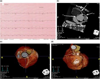
A 38-year-old gentleman with learning difficulties and a relatively high body mass index was admitted for assessment before pulmonary valve replacement. He had worsening exercise tolerance and broadening QRS complex (Fig 2a). His initial operation was the placement of a Blalock–Taussig shunt at the age of 12 years and subsequent full repair with a transannular patch at the age of 14 years. On review, his echocardiogram showed suboptimal imaging. A cardiac magnetic resonance scan showed right ventricular dilatation with poor function, and although a single coronary was suspected, the details were unclear. A CT scan was performed using a time-density curve, and 50 ml of contrast confirmed a single coronary artery arising from the anterior sinus of the valsalva, trifurcating into the right coronary artery, left anterior descending artery, and left circumflex artery (Fig 2b and c). There were also significant flow-limiting plaques estimated to be >50% in the mid left anterior descending artery – the largest diagonal branch and the origin of the circumflex coronary artery (Fig 2d). This detail was not apparent on cardiac magnetic resonance scan. The patient had his medical management optimised and, in view of the widening QRS complex, a defibrillator was implanted. Pulmonary valve replacement was planned after these interventions.
( a ) Echocardiogram showing broad QRS complexes in the adult. ( b ) Scan of adult showing single origin of the coronary artery from the anterior sinus of the valsalva and trifurcating to the right coronary artery (RCA), left anterior descending artery (LAD), and left circumflex artery (LCx). ( c ) Three-dimensional (3D) iterative reconstruction showing single origin of the coronary artery from the anterior sinus of the valsalva with subsequent trifurcation. ( d ) 3D iterative reconstruction of adult showing plaque in the mid LAD.

Discussion
CT angiography has the benefit of showing both the coronary artery and intracardiac anatomy in great detail, and the advent of advanced multislice scanners has improved both the spatial and temporal resolution significantly. Both our patients benefited from the short acquisition time as even technological advances still require cooperation. In both cases, the study was conducted without the need for sedation. Although CT angiography is frequently used to delineate the coronary artery course in adults, it is less frequently used in small children/babies. Coronary artery arrangement has important implications in therapeutic options,Reference Fellows, Freed, Keane, Van Praagh, Bernhard and Castaneda 4 as it determines the approach at surgery and ultimate method of correction. As a result, it is crucial to get preoperative anatomical detail of coronary artery course correct.
The latest technology in CT has minimal radiation exposure for patients, which is particularly important in children. In the case of the infant, the entire scan exposed the child to only 0.2 mSv (millisievert), whereas in the case of the adult the exposure amounted to 3 mSv. To put it into context, background radiation for those living in the United Kingdom is 2.5 mSv in a given year, and similarly the background radiation for living in the United States is 6.2 mSv. 5 A chest X-ray is ~0.02 mSv, 5 and the typical exposure for a diagnostic cardiac catheter is 7 mSv. Furthermore, the possibility of post-processing such as three-dimensional iterative reconstructions adds further weight in the use of CT, as this not only gives accurate anatomy but also reduces radiation exposure further.Reference Vastel-Amzallag, Le Bret and Paul 3 , Reference Leber, Johnson and Becker 6
We suggest that in cases where coronary artery anatomy may have significant surgical implications and is not adequately defined, state-of-the-art CT can be used to define the anatomy in patients, from baby to adult.
Acknowledgements
The authors would like to thank Dr Lorna Swan in reviewing the article before submission.
Financial Support
This research received no specific grant from any funding agency, commercial, or not-for-profit sectors.
Conflicts of Interest
None.