




No CrossRef data available.
Article contents
Stage-specific alternative splicing of the heat-shock transcription factor during the life-cycle of Schistosoma mansoni
Published online by Cambridge University Press: 05 October 2004
Abstract
Stage-specific alternative splicing of the heat-shock transcription factor of Schistosoma mansoni (SmHSF) generates isoforms with structural diversity that may modulate the activity of SmHSF at different life-stages, and thus may regulate the expression of different genes at different developmental stages. RT-PCR, cloning and DNA-sequence analyses showed stage-specific alternative splicing inside the DNA-binding domain (DBD) involving introns I1 and I2, and beyond the DBD involving introns I4a and I7. Retention of introns I2 and I4a would inactivate SmHSF since they contain termination codons. Retention of intron I1 would add 11 amino acids inside the DBD and may change the DNA-binding specificity of SmHSF; intron I7 would add 13 amino acids to the effector region of HSF. Retention of introns was more pronounced in cercariae (larval stage living in water) than in adult worms (parasitic form in mammals). The isoforms were expressed in bacteria, but functional evaluation was not feasible, because only the isoform lacking introns was soluble while isoforms with introns were insoluble. However, stage-specific alternative splicing that changed HSF function in vivo was evidenced in intact cercariae. The cercarial SmHSF mRNA was enriched with introns I2 and I4a that contain termination codons. Therefore, translation of the SmHSF mRNA was impaired, and the SmHSF protein was undetectable. Consequently, the HSP70 gene could not be transcribed, and the HSP70 mRNA was missing. Alternative splicing was observed for short DNA segments (33–45 bp) bound by splice signals, located in the coding region. These are not bona fida exons since they are not flanked by introns. Yet, they are not regular introns since they are often found in mature mRNA. Alternative splicing of these DNA segments caused structural diversity that could modulate the function of the gene product.
Keywords
Information
- Type
- Research Article
- Information
- Copyright
- © 2004 Cambridge University Press
INTRODUCTION
Schistosomes are parasitic helminths that cause bilharzia, which is a major health problem in developing countries. The parasites have a complex life-cycle in 2 hosts (mammal and snail) and short periods of larvae living freely in water (Cherfas, 1991). During transformation from one stage to another schistosomes are exposed to different environments and temperatures (20 °C or 37 °C), and undergo profound changes in size (0·1 to 15 mm), shape, physiological and biochemical properties (e.g. aerobic or anaerobic metabolism). It is conceivable that distinct features of the parasite at different developmental stages are determined to a large extent by the activation/inactivation of a selected group of genes, termed stage-specific genes. Many stage-specific genes were identified in schistosomes (see Grossman et al. 1990; Neumann, Lantner & Schechter, 1993), yet, nothing is known on the regulation of stage-specific gene expression.
Alternative splicing of pre-mRNA can generate a diversity of protein isoforms that play an important role in tissue differentiation and developmental processes. Of special interest for control of the life-cycle of the parasite are transcription factors subjected to alternative splicing that change DNA-binding specificity, activate or suppress transcription activity (Foulkes & Sassone-Corsi, 1992; Rio, 1993). For example, alternative splicing of the CF2 transcription factor of Drosophila (changing the number of zinc fingers, Hsu et al. 1992) and of the Wilms tumour associated wt1 gene product (addition/deletion of 3 amino acids between zinc-fingers, Bickmore et al. 1992) generates proteins with distinct DNA-binding specificities, capable of regulating different sets of genes in different tissues and developmental periods.
In eukaryotes, the expression of heat-shock proteins is controlled mainly at the level of transcription by heat-shock transcription factors (HSFs). The HSF molecule (about 600 amino acids long) contains a DNA-binding domain (DBD, about 100 amino acids long) that reacts with the heat-shock responsive element (HSE) composed of 5 bp modules (nGAAn) arranged as inverted repeats in the promoter of HSP genes. Current models propose that HSF binds HSE as a homotrimer in which the DBD of each monomer contacts a single nGAAn of HSE. Homotrimer formation is mediated by the oligomerization domain composed of three leucine zippers (LZ123), and a fourth leucine zipper (LZ4) involved in the maintenance of HSF in a non-DNA binding state (Perisic, Xiao & Lis, 1989; Rabindran et al. 1993). Earlier studies in our laboratory demonstrated alternative splicing of the heat-shock transcription factor of S. mansoni (SmHSF), generating isoforms with structural diversity at the C-terminus of LZ4 that may modulate the DNA-binding activity of SmHSF, and all isoforms originated from a single SmHSF gene (Lantner et al. 1998).
Here we report on the cloning of SmHSF isoforms generated by stage-specific alternative splicing of small introns (33 bp or 39 bp) located inside and outside the DBD of HSF. We demonstrate that the inactive cercarial SmHSF mRNA is generated by alternative splicing leading to the incorporation of small introns and of a larger DNA segment (279 bp from the 2 kb intron I4) that contain termination codons. Alternative splicing was observed in DNA segments located in the coding region (not flanked by introns) and inside introns, and these events affected the gene products.
MATERIALS AND METHODS
Parasites
The Puerto Rican strain of S. mansoni was maintained by passage through Biomphalaria glabrata snails and ICR mice. Adult worms were obtained by perfusion of the hepatic portal system of infected mice. Infected snails were induced by bright illumination to shed cercariae that were collected from water (Levi-Schaffer et al. 1984).
RNA and DNA methods
Total RNAs of adult worms and cercariae of S. mansoni were prepared as described (Kirby, 1968). cDNA libraries of cercariae and adult worms were kindly donated, respectively, by Dr Johnston from the WHO Schistosome Genome Network and by Dr Simpson (Sau Paulo). Other procedures, including the screening of the cDNA libraries by a 32P-labelled probe of 2·3 kb SmHSF cDNA (clone c-257, Lantner et al. 1998), RT-PCR, Western blots and Northern blots in formaldehyde gels, were done according to published procedures (Sambrook, Fritsch & Maniatis, 1989). DNA nucleotide sequences were determined from both strands by automated sequencer (ABI DNA sequencer, model 377) using primers of the plasmid vector flanking the cloned DNA, and primers complementary to internal segments of the cloned DNA. The cDNA of S. mansoni HSP70 (1071 bp, clone 98) used for the development of Northern blots, was described (Neumann et al. 1993). The oligomer that was used as a hybridization probe to detect intron I1 was composed of 5 nucleotides from the 3′ end of exon 1, 33 nucleotides of intron I1 and 5 nucleotides from the 5′ end of exon 2, altogether 43 nucleotides: 5′-CGTGCTGAACGAAGTTACTAACCAAACTTAAACTTACCGGATC-3′. The single-strand probe was end-labelled by γ-32P dATP (Sambrook et al. 1989).
RT-PCR and cDNA cloning
Primers used for RT-PCR to amplify RNA segments flanking introns I1 and I2 were: 5′ primer with Nco1 site 5′-CGCCATGGATGGTTTCACATCTGGACC-3′, 3′ primer with BamH1 site 5′-CCGGATCCGAATTAGCCTGAAC-3′. The PCR products were digested with Rsa1 at internal sites of the cDNA, cloned into KS Bluescript vector digested with Sma1, and sequenced using KS primers. Primers used to amplify the region flanking intron I7 were: 5′ primer 5′-CCCCGTTGTATCGCACC-3′, 3′ primer 5′-GACCATTTGAAGAACTGCG-3′.
To clone the DNA-binding domain into the pJC40 expression vector (Clos & Brandau, 1994) we used the two primers listed above to clone segments flanking introns I1 and I2. The cloned RT-PCR products encode the DBD of SmHSF, from amino acids Met 1 to Ser 143.
In order to tag the DBD with a tail of 10 histidines, the 5′ primer was 5′-CCGAATTCACATCTGGACCTCC-3′ with EcoR1 site. The 3′ primer was the same primer used above for cloning the DBD. The cloned RT-PCR products were ligated into pJC40 to yield the DBD protein segment from amino acid Phe 4 to Ser 143, tagged with a His-tail at the N-terminus.
Antibodies to SmHSF
The 73 kDa full length molecule of SmHSF was synthesized in bacteria as insoluble inclusion bodies (Lantner et al. 1998) that were washed repeatedly in phosphate-buffered saline (PBS). The insoluble pellet was dissolved in reducing buffer, resolved on SDS–PAGE, a band containing the 73 kDa SmHSF (approx. 95% pure) was cut from the gel, it was suspended in Freund's adjuvant and injected into rabbits at 10-day intervals (Markovics et al. 1994). Antisera were collected 12 days after the third injection. Purified antibodies were prepared by adsorption of the antisera onto a lysate of the PlysS bacterial host immobilized on a nitrocellulose filter, followed by affinity purification, i.e. second adsorption on purified 73 kDa SmHSF protein resolved on SDS–PAGE, electro-transferred and immobilized on nitrocellulose filter. Purified antibodies were obtained from the filter by elution with 0·1 M acetic acid, neutralization with Tris base to pH 7·2, and dialysis against PBS. Western blot of parasite lysates developed with the purified antibodies (2 μg/ml) revealed a distinct ~67 kDa protein in the adult worm. The size of the protein (~67 kDa) conformed with the expected size from the cDNA sequence (73 kDa), though it was somewhat smaller, possibly due to altered electrophoretic mobility (Maizel, 1972). The purified antibodies did not detect any protein in the control of mouse kidney lysate (Fig. 5).
RESULTS
Stage-specific alternative splicing of small introns located inside and outside the DNA-binding domain of SmHSF
It was previously shown that adult worms express 3 isoforms of SmHSF mRNA generated by alternative splicing inside the coding region at the C-terminus of LZ4 located in exon 5 (Fig. 1, see AS in exon 5). The coding region contained in-frame splice signals, and introns were not involved in the process since the deleted segments (36 or 45 bp) were not flanked by introns in the gene (Lantner et al. 1998). The SmHSF gene contains 9 introns, 4 of which are rather small (33–39 bp, Fig. 1). Therefore, we tested whether these introns may be included in the mRNA, similar to retention of the 9 or 45 bases downstream of LZ4 (Lantner et al. 1998).

Fig. 1. Structures of the Schistosoma mansoni HSF gene and of products generated by alternative splicing. Scheme of gene on top. Exons (open boxes), region encoding the DNA-binding domain (hatched), the site of alternative splicing at the end of LZ4 within exon 5 (AS, dark box, see Lantner et al. 1998), introns (I1–I9), the initiator Met (M1) codon, the C-terminal Glu658 codon, the polyA-addition site (pAad), and positions of primers for RT-PCR of RNA (arrows), are indicated. Exons are drawn to scale. Introns are not drawn to scale, but the sizes (in bp) are given. Isoforms generated by alternative splicing are shown below. Diagrams of four RT-PCR products (I-0, I-1, I-2, I-1+2) spanning introns I1 and I2 (left). Relative sizes of the RT-PCR products are shown (437 bp for I-0, 470 bp for I-1 and I-2, 503 bp for I-1+2). Numbers below open boxes represent exon numbers. Introns retained in the mRNA are shown as dark boxes. The asterisk marks the position of a translation termination codon. Translated and untranslated regions are indicated by wide and narrow boxes, respectively. Diagrams of 2 RT-PCR products with and without intron I7 are shown on the right. Central scheme shows expanded intron I4 (1964 bp, GenBank Accession no. AY434014) from which a segment of 279 bases (I4a, nucleotides 370–648 of intron I4) is incorporated into the mRNA by alternative splicing.
In genomic DNA the DBD of SmHSF is split by 2 small introns I1 and I2. Cercariae and adult worm RNAs were subjected to RT-PCR with primers from exons 1 and 3 that span the DBD (Fig. 1). Product analyses on agarose gels revealed different patterns of isoforms in cercariae and adult worms, suggesting stage-specific alternative splicing (Fig. 2A). RT-PCR products were further analysed by cloning into Bluescript KS. Recombinant plasmids were characterized by restriction enzymes. Digestion by EcoR1 and XbaI released the intact insert (Fig. 3A) while EcoR1 and BamH1 released fragmented insert cleaved at internal BamH1 sites (Fig. 3B). The digested clones were hybridized with 2 probes: a cDNA probe encoding exons and a probe of intron I1 (Fig. 3C,D). The results (Fig. 3) enabled us to define unambiguously 4 types of PCR products, that were further confirmed by DNA sequencing. (1) I-0, product identical to cDNA without any intron (GenBank Accession no. AF043418, nucleotides 29–457), (2) I-1, product contains all 33 bp of intron I1 and none of intron I2 (GenBank Accession no. AY346308), (3) I-2, product contains all 33 bp of intron I2 and none of intron I1 (GenBank Accession no. AY346309), (4) I-1+2, product identical to gene containing both introns I1 and I2 (GenBank Accession no. AY346307). Cloned cDNAs (Fig. 3A) were shorter than the PCR products (Fig. 2A) by 24 bp due to cleavage at 2 internal Rsa1 sites adjacent to the 5′ and 3′ ends of the RT-PCR products (−54 bp), and addition of a few bases (+30 bp) from vector cleaved by Xba1 and EcoR1.

Fig. 2. Stage-specific alternative splicing of SmHSF. Ethidium bromide-stained agarose gels of RT-PCR products of RNA from cercariae and adult worms. (A) Primers from exons 1 and 3 yield four RT-PCR products corresponding to: (1) I-0, product identical to cDNA, (2) I-1, product contains all 33 bp of intron I1 and none of intron I2, (3) I-2, product contains all 33 bp of intron I2 and none of intron I1 (products 2 and 3 are of identical size), (4) I-1+2, product identical to gene, contains introns I1 and I2. Two different RNA preparations of cercariae and adult worms are shown (same results were obtained with additional RNA preparations, 2 of cercariae and 2 of adult worms). (B) Primers from exons 7 and 8 yielded 2 RT-PCR products: (1) I-0, product identical to cDNA, (2) I-7, product contains all 39 bp of intron I7. Four different RNA preparations of cercariae and adult worms are shown. RNA preparations were treated with RNase-free DNase to remove possible contamination of genomic DNA. Two reactions with and without reverse transcriptase were run in parallel. As expected, PCR products were formed only with (+), but not without (−) reverse transcriptase. Similar results were obtained for introns I1 and I2 (A) and I7 (data not shown). The RT-PCR products were of correct size, and their authenticity was ascertained by cloning and sequencing. Positions of DNA size markers (bp) are given on the right.

Fig. 3. Characterization of mRNA isoforms of the DNA-binding domain of SmHSF. RT-PCR products, prepared from adult worms RNA serving as template and primers flanking the DBD, were cloned into the KS Bluescript and characterized by restriction enzyme digestions and hybridizations. Ethidium bromide-stained agarose gels (A, B) of clones digested by EcoR1/XbaI (A) to release the intact DBD insert, and by EcoR1/BamH1 (B) to release fragmented insert cleaved at internal BamH1 sites. (C) Autoradiogram of (B) reacted with 32P-labelled probe of SmHSF cDNA (2·3 kb) hybridizing with all fragments of all DBD isoforms. (D) After removal of radioactivity from (C) the blot was rehybridized with a 32P-labelled intron I1 probe hybridizing with a single fragment only in DBD isofroms containing intron I1. Representative clones corresponding to the 4 DBD isoforms are shown. (1) I-0, product identical to cDNA composed of exons only and no introns, (2) I-1, product contains intron I1, (3) I-2, product contains intron I2, (4) I-1+2, product contains introns I1 and I2. Positions of DNA size markers (bp) are given on the left. (E) Scheme of cloned cDNA encoding DBD I-0. RT-PCR product digested by Rsa1 was cloned into Sma1 cleaved pJC40 expression vector (R, cloning site). Numbering of nucleotides of DBD is according to the SmHSF cDNA sequence deposited in GenBank (Acc. no. AF043418). BamH1 (B) sites in DBD, locations of introns I1 and I2 in DBD, positions of EcoR1 (E) and BamH1 sites of the vector, short DNA segments of the vector flanking DBD (black bar), and sizes of DNA fragments (bp) between restriction sites, are given.
The I-1 isoform may influence DNA binding because intron I1 has no termination codon and its reading frame is continuous with that of the flanking exons. Therefore translation of this isoform would add a segment of 11 amino acids to the DNA-binding domain. The I-2 and I-1+2 isoforms that retain intron I2 would pre-terminate translation, because intron I2 contains an in-frame translation termination codon (see Figs 1 and 4).
Primers from exons 7 and 8 that span intron I7 (39 bp) yielded PCR products with and without intron I7 as indicated from the size of the products (Fig. 2B), and confirmed by cloning and sequencing. Intron I7 has no termination codon and its reading frame is continuous with that of the flanking exons. Therefore retention of intron I7 would add a segment of 13 amino acids to the effector region of the HSF molecule. Here again, the pattern of PCR products differed in cercariae and adult worms (Fig. 2B).
Visual inspection of RT-PCR products spanning introns I1, I2 and I7 revealed that isoforms without introns were more abundant in adult worms than in cercariae, while isoforms with introns were more pronounced in cercariae than in adult worms. Densitometer scan of isoform bands (Fig. 2) showed that isoforms without introns I1 and/or I2 comprised 36·4% (±0·7%) in adult worms, and 18·5% (±0·6%) in cercariae (average values calculated from scan of total RT-PCR products). Isoforms without intron I7 comprised approximately 95% in adult worms, and 29·5% (±7%) in cercariae. These findings demonstrate different patterns of alternative splicing at different developmental stages (Figs 2 and 3).
To ascertain that the PCR products originated from mature mRNA and not from incomplete processing of pre-mRNA or genomic DNA, it was necessary to demonstrate the retention of distinct intron(s) in the full (or nearly full) length SmHSF mRNA. Accordingly, cDNA libraries of cercariae and adult worms were screened with the 2·3 kb SmHSF cDNA probe. We isolated SmHSF clones that were analysed by restriction enzyme mapping, and PCR with appropriate primers, to detect clones that retained introns I1, I2 and I7. Several clones positive for the presence of introns were identified, and sequenced (Table 1). All cDNA clones retained 1 intron, while other introns (2–7) were correctly spliced, except in clones CE32 and CE52 in which a portion of intron I4 was retained (see below). The cDNAs ranged in size from 800 bp to almost full size cDNA (2·5 kb, Lantner et al. 1998), and 2 cDNA clones contained polyA-tail characteristic of mature mRNA. These findings established that cDNA clones with introns represent true SmHSF mRNA isoforms, and not unrelated species like gene fragments.
Table 1. Clones of SmHSF mRNA isoforms containing introns (Clones were isolated from cDNA libraries of adult worms (WO) and cercariae (CE). All clones were sequenced.)

Expression of isoforms of the DNA-binding domain
To evaluate the significance of alternative splicing of introns I1 and I2 located in the DBD of SmHSF, we cloned all 4 isoforms (Figs 2A and 3) in the pJC40 expression vector. It was previously reported that the recombinant full length (~73 kDa) and half length (~40 kDa) SmHSF formed insoluble inclusion bodies (Lantner et al. 1998). Therefore, the DBD fragment (~18 kDa) and not the entire SmHSF molecule, were cloned. Also, it was shown that the DBD of SmHSF binds 32P-HSE probes, and binding specificity could be analysed by inhibition studies (Lardans et al. 2001). The pJC4O plasmid has 2 cloning sites; (1) EcoR1/BamH1 for the expression of proteins with a tail of 10 His residues, (2) Nco1/BamH1 for the expression of proteins without added histidines.
Schistosome RNA treated with DNase, and primers with built-in restriction sites were used for RT-PCR. The RT-PCR products were ligated into pJC4O. Many clones were isolated and characterized by restriction enzymes, hybridization with exon and intron I1 specific probes, and DNA sequencing. All 4 isoforms, with and without His-tail, were identified. cDNAs of the DBD isoforms were of similar size, as expected, since they differ only by the presence or absence of the short introns I1 and/or I2. However, the DBD protein isoforms programmed by the cDNAs should differ markedly in size because the retention of intron I1 would add in-frame 11 amino acids, while retention of intron I2 would yield a short DBD protein fragment because it contains a translation termination codon approximately in the middle of the DBD (see Fig. 1).
BL21 or PlysS bacteria transfected with pJC4O constructs encoding the DBD isoforms synthesized proteins of expected size, as seen by SDS–PAGE of total bacterial lysates (Fig. 4A). DBD I-1 containing intron I1 (~21 kDa, lane 3) was a little bigger than DBD I-0 that lacks introns (~20 kDa, lane 2); DBDs with intron I2 were smaller than above DBDs due to translation termination in intron I2 (lanes 4, 5); DBD I-1+2 containing intron I1 (~13 kDa, lane 4) was a little bigger than DBD I-2 (~12 kDa, lane 5). The lysate of bacteria transfected with pJC40 vector control (without DBD cDNA insert) did not show any protein induced by IPTG (Fig. 4A, lane 1). The same sizes were observed for recombinant DBD isoforms without (data not shown) and with His-tail (Fig. 4A).

Fig. 4. Expression of protein isoforms of the DNA-binding domain of SmHSF. Coomassie blue staining of SDS–PAGE of lysates from PlysS bacteria transfected with the pJC40 vector (1), and with pJC40 constructs expressing DBD isoforms with His-tails: DBD I-0 (2), DBD I-1 (3), DBD I-1+2 (4) and DBD I-2 (5). (A) Total bacterial lysate. (B) Total bacterial lysate (T), supernatant (S) and insoluble pellet (P) of the lysate. Positions of the protein size markers (kDa) are indicated.
Total bacterial lysates were spun to separate supernatant and insoluble pellet fractions. SDS–PAGE analyses showed that only DBD I-0 was soluble (found in supernatant and not in pellet, Fig. 4B, panel 2), while the other 3 DBD isoforms were insoluble (found in pellet but not in supernatant, Fig. 4B, panels 3, 4, and 5). This was true for DBD isoforms without (data not shown) and with His-tails (Fig. 4B).
The recombinant full length SmHSF molecule (73 kDa) is insoluble. However, when the bacteria were grown at 20 °C (instead of 37 °C), small amounts of the SmHSF protein conceivably remained in solution, enough to test DNA-binding activity by the gel shift assay (Lantner et al. 1998). Accordingly, bacteria transformed with DBDs (with and without His-tail) were grown at 20 °C, but in this case supernatants of the lysates of insoluble DBDs did not show any binding activity of the 32P-HSE probe.
The soluble DBD I-0 isoform (with and without His-tail) showed specific binding of the 32P-HSE probe (data not shown), in agreement with earlier studies (Lardans et al. 2001). Further efforts to obtain soluble molecule centered on the insoluble DBD I-1 isoform since it is composed of the entire DBD I-0 plus 11 amino acids inserted between Pro-37 and His-38 of DBD I-0, potentially it can bind the 32P-HSE probe, and DNA-binding specificity could be analysed using HSE-related probes as inhibitors. The two isoforms with intron I2 are fairly short, corresponding to about half of the DBD (see Figs 1 and 4), and conceivably can not bind the 32P-HSE probe. Additional efforts (denaturation–renaturation experiments using guanidinium-HCl or 8 M urea, various detergents, different growth conditions, yeast-one-hybrid system) to obtain soluble DBD I-1 protein with DNA-binding activity have failed. Therefore, it was not possible to study DNA-binding specificity of DBD I-1, and to evaluate if it differs from that of DBD I-0.
Stage-specific inactivation of cercarial HSF by alternative splicing inside the 2 kb intron I4
Soluble DBD isoforms containing introns are not yet available to evaluate the significance of alternative splicing on SmHSF function, such as, effect on DNA-binding specificity. However, earlier studies indicated a change in HSF function due to alternative splicing under physiological conditions in vivo, i.e. in intact cercariae. It was previously shown that adult worms contained both HSF and HSP70 mRNAs. In contrast, cercariae expressed fairly high levels of larger HSF mRNAs (up to 3 kb) that were missing in adult worms, and the HSP70 mRNA was undetectable (Fig. 5, see Lantner et al. 1998). This inverse relation suggested that the HSF mRNA species of cercariae were inactive, probably due to alternative splicing (Lantner et al. 1998). This notion is further supported here by addressing two issues regarding the cercarial HSF mRNA species: increased size of the mRNA, and translation of mRNA into protein.

Fig. 5. Expression of HSF mRNA and protein, and of HSP70 mRNA, in cercariae and adult worms. Northern blots (10 μg RNA/lane) hybridized with 32P-labelled 2·3 kb SmHSF cDNA (top) or 1 kb HSP70 cDNA of Schistosoma mansoni (bottom). Western blot (50 μg protein/lane) developed with rabbit purified antibodies to SmHSF protein (2 μg/ml) followed by 125I-labelled goat purified antibodies to rabbit Ig. Total RNA and proteins were prepared from cercariae (CE), adult worms (AW), and mouse kidney (MK) serving as control. Positions of the 18S rRNA and protein size markers (kDa) are indicated.
The SmHSF gene contains four short introns (I1, I2, I3 and I7) that add up to 139 bp (Fig. 1). The retention of all short introns would cause a relatively small change in the size of the 2·5 kb SmHSF mRNA, that would be barely (if at all) detected on Northern blots. Yet, Northern blots of cercariae showed multiple bands of SmHSF mRNA in the range of 2·5–3 kb. That is, cercariae express SmHSF mRNA species larger by up to about 0·5 kb than the 2·5 kb mRNA observed in adult worms (Lantner et al. 1998). Therefore, we searched the cercariae cDNA library for SmHSF cDNA clones containing introns larger than the short introns. Two relevant independent clones encoding about 2·2 kb of SmHSF mRNA were identified (Table 1, clones CE32 and CE52; Fig. 1). Sequence analyses revealed that they contain a portion of intron I4 and none of the 6 introns that flank intron I4. These clones had short polyA-tails, thus establishing retention of a portion of intron I4 in mature SmHSF mRNA. Intron I4 is 1964 bp long (complete sequence of intron I4, GenBank Accession no. AY434014). In the mRNA we found only 279 nucleotides (termed I4a) of intron I4 (Fig. 1 and Table 1). The I4a segment was flanked by consensus sequences of acceptor and donor splice signals, the reading frame was continuous with exon 4 till nucleotide 87 of I4a, where a translation termination codon was found. Therefore, SmHSF mRNA isoforms containing I4a would encode a truncated SmHSF protein lacking exons downstream of exon 4, and a new unrelated peptide segment of 29 amino acids encoded by I4a till the termination codon (see Fig. 1).
The cercarial SmHSF mRNA is enriched with species retaining intron I2 (Fig. 2A) and I4a (Fig. 1) which contain termination codons that may impair mRNA translation into protein. Therefore, antibodies to the SmHSF protein were prepared and used to detect the protein in parasite lysates. Northern and Western blots showed that adult worms express the SmHSF mRNA and the SmHSF protein, and the protein was functional in HSP70 gene transcription to yield the HSP70 mRNA (Fig. 5, AW right lanes). In contrast, cercariae had abundant SmHSF mRNA larger than the adult worms mRNA species due to the retention of introns. Since the introns contained termination codons the SmHSF protein was undetectable, consequently the HSP70 gene was not transcribed, and the HSP70 mRNA was missing (Fig. 5, CE central lanes).
DISCUSSION
The present study supports the notion that alternative splicing controls the expression of stage-specific genes during the life-cycle of the schistosome. Transcription factors would be preferred targets since their modification may affect the transcription (activation/suppression) of different genes at different developmental stages. RT-PCR, cloning and DNA-sequencing demonstrated stage-specific alternative splicing of short introns (33, 39 bp) located in the DBD (introns I1, I2), and in the effector region of the HSF molecule (intron I7). Northern blots and cDNA sequencing revealed preferential incorporation of intron I4a (279 bp from the 2 kb intron I4) in cercariae.
Two cercarial clones (CE32 and CE52) spanned intron I4 and both retained the I4a segment (2 other cercarial clones, CE22 and CE74 did not span intron I4, and as such did not provide any information on intron I4a). On the other hand, 2 adult worm clones spanning intron I4 (WO18 and WO12) did not contain intron I4a. In addition, the sequences of 5 long cDNA clones of SmHSF of adult worms were reported previously (Lantner et al. 1998). These clones spanned the region of intron I4, but none retained intron I4a. Altogether, 2 relevant cercarial clones were analysed and I4a was found in both. However, intron I4a was not found in any of 7 relevant clones isolated from adult worms.
The retention of introns in nearly full length SmHSF mRNA, and the finding of polyA-tail in some of them, established that these molecules represent mature SmHSF mRNA isoforms. SmHSF mRNA species retaining introns were more abundant in cercariae than in adult worms. These findings established stage-specific alternative splicing of SmHSF.
It was previously shown that adult worms express 3 isoforms of SmHSF mRNA generated by alternative splicing inside the coding region of exon 5, leading to structural diversity at the C-terminus of LZ4. Exon 5 contained splice signals in the coding region, and the reading frame of the involved segments (designated AS) was continuous with that of exon 5. Introns were not involved in the process since the deleted segments (36 or 45 bp) were not flanked by any intron (Lantner et al. 1998). The SmHSF gene contains 9 introns, 4 of which are rather small (33 to 39 bp). Therefore, we tested whether these introns may be included in the mRNA similar to retention of the AS segment downstream of LZ4. Three introns were studied (I1, I2 and I7) and all of them were incorporated into mature mRNA. Thus, the status of the AS segment in exon 5 is identical to that of introns I1, I2 and I7. Bases retained/deleted by alternative splicing in exon 5 were not initially regarded as intron, since structural diversity involving this region was found before the gene was cloned, and before we found the retention/deletion of introns I1, I2, and I7. The short introns and AS are deleted by alternative splicing using splice signals. Yet, they are not regular introns as they are often found in mature mRNA and, as such, they function as exons. On the other hand, they are not bona fida exons since they are not flanked by any intron. Alternative splicing of the ‘short introns’ may affect gene function, yet better designation/definition of these introns/units remains for the future.
The I4a segment can be viewed as an exon, because it is flanked by introns that have legitimate splice signals and it is subjected to alternative splicing (it was found in some but not all SmHSF mRNAs). However, it is not a regular exon because it contains a stop codon and retention of I4a would generate a truncated protein, about 40% the size of the regular SmHSF protein. Therefore I4a can be regarded as a regulatory exon/unit.
Short introns were found in many schistosome genes (~40 bp, see Markovics et al. 1994; Craig et al. 1989) and most introns of C. elegans are short (~50 bp, Blumenthal & Thomas, 1998). The significance of the short introns was not known. The studies on SmHSF showed that alternative splicing of such introns caused structural diversity that could modulate the function of the gene products. It will be of interest to evaluate if short introns of C. elegans are subjected to alternative splicing, and whether this occurs at distinct developmental stages of the worm.
Alternative splicing may change the function of the isoforms in different ways. The retention of introns that contain termination codons (I2, I4a) would generate truncated, probably inactive HSF molecules. Alternative splicing at the C-terminus of LZ4 added hydrophobic amino acid residues that were in register with the hydrophobic heptad repeats of LZ4. These modifications may affect the interaction of LZ4 with LZ123, influence the oligomerization state of HSF and thus modulate the activity of HSF (Rabindran et al. 1993). Retention of intron I1 (11 residues added inside the DBD) may change the DNA-binding specificity, and of intron I7 (13 residues added beyond DBD) may influence effector function of HSF.
The heat-shock response is induced by a large variety of stresses, and heat-shock proteins are also evident in non-stress conditions, such as constitutive expression in certain tissues and during development (Bienz, 1985; Lindquist, 1986). It was shown that the HSP70 gene of S. mansoni is regulated by two mechanisms. Stress induction, specific to HSP70, refers to transient and high levels of HSP70 mRNA observed during cercariae – schistosomulum transformation, and in heat shocked (42 °C) adult worms. The developmental programme, common to HSP70 and other genes (e.g. paramyosin, Grossman et al. 1990), refers to constitutive expression of HSP70 mRNA in miracidia, sporocyst and adult worms, but not in cercariae (Neumann et al. 1993).
We propose that stage-specific alternative splicing of SmHSF plays a role in the regulation of HSPs during the life-cycle of the parasite. Structural diversity of the HSF isoforms would enable them to differentially recognize ideal- and variant-HSE sequences present in the promoters of HSP genes, and thus activate different sets of HSPs.
HSP promoters contain multiple HSEs that often deviate from the ideal HSE consensus sequence (Bienz, 1985; Lis, Xiao & Perisic, 1990). For example, the Xenopus HSP70A promoter contains 3 clusters of HSEs, each composed of 3 perfect pentamers and 1 mismatched pentamer (nCTCn instead of nTTCn, Bienz & Pelham, 1986). The promoter of the HSP70 gene of S. mansoni contains a cluster of three HSEs (nGAAnnTTCnnGTAn) in which the third pentamer (nGTAn) differs from the ideal HSE consensus sequence (nGAAn) (Neumann et al. 1992). Binding and inhibition studies demonstrated that SmHSF recognized the cluster with the variant-HSE but not the cluster composed entirely of ideal-HSE pentamers (Levy-Holtzman, Clos & Schechter, 1995). This raises the possibility that clusters containing variant HSEs present in HSP promoters of other organisms may be recognized by distinct HSF molecules.
Four HSPs with different patterns of expression during the life-cycle of schistosome were described: HSP86 (Johnson et al. 1989), HSP70 (Neumann et al. 1993), HSP60 (Tielens, van den Heuvel & van Eden, 1993) and a small HSP (Nene et al. 1986). The structure of the promoter of the gene was reported for HSP70 (Neumann et al. 1992), but not for the three other HSPs. Yet, it is likely that the promoters of the genes probably contain HSE clusters that differ from each other, as found for HSP genes in other organisms. For example, in Drosophila the promoters of HSP83, HSP70, HSP26 and HSP23 contain HSE clusters of different size, each composed of ideal and mismatched HSEs (see Lindquist, 1986; Lis et al. 1990). Present studies can explain how a single SmHSF gene can control, at least in part, stage-specific expression of distinct HSPs during the life-cycle of the parasite. Structural diversification of SmHSF by stage-specific alternative splicing can generate isoforms with different DNA-binding specificity that recognize and activate distinct HSP genes, depending on the unique DNA-sequence of HSEs in the promoter.
We thank Dr. Andrew J. G. Simpson (Instituto Ludwig de Pesquisa sobre o Cancer, Sao Paulo) for providing the λgt11 library of adult worms of S. mansoni, and Dr David Johnston (secretary to the WHO Schistosoma Genome Network) for an aliquot of Mohamed Saber's S. mansoni cercarial library in λzap. This work was supported by grants from the Israel Science Foundation and from the UNPD/World Bank/WHO Special Program for Research and Training in Tropical diseases.
References
REFERENCES
BICKMORE, W. A., OGHENE, K., LITTLE, M. H., SEAWRIGHT, A., VAN HEYNINGEN, V. &
HASTIE, N. D. (
1992).
Modulation of DNA binding specificity by alternative splicing of the Wilms tumor wt1 gene transcript.
Science
257, 235–237.CrossRefGoogle Scholar
BIENZ, M. (
1985).
Transient and developmental activation of heat-shock genes.
Trends in Biochemical Sciences
10, 157–161.CrossRefGoogle Scholar
BIENZ, M. &
PELHAM, H. R. B. (
1986).
Heat shock regulatory elements function as an inducible enhancer in the Xenopus hsp70 gene and when linked to a heterologous promoter.
Cell
45, 753–760.CrossRefGoogle Scholar
BLUMENTHAL, T. &
THOMAS, J. (
1998).
Cis and trans mRNA splicing in C. elegans.
Trends in Genetics
4, 305–308.Google Scholar
CHERFAS, J. (
1991).
New hope for vaccine against schistomiasis.
Science
251, 630–631.CrossRefGoogle Scholar
CLOS, J. &
BRANDAU, S. (
1994).
pJC20 and pJC40, two high-copy-number vectors for T7 RNA polymerase-dependent expression of recombinant genes in Escherichia coli.
Protein Expression and Purification
5, 133–137.CrossRefGoogle Scholar
CRAIG, S. P., MURALIDHAR, M. G., McKERROW, J. H. &
WANG, C. C. (
1989).
Evidence for a class of very small introns in the gene for hypoxanthine-guanine phosphorybosyl-transferase in Schistosoma mansoni.
Nucleic Acids Research
17, 1635–1647.CrossRefGoogle Scholar
FOULKES, N. A. &
SASSONE-CORSI, P. (
1992).
More is better: activators and repressors from the same gene.
Cell
68, 411–414.CrossRefGoogle Scholar
GROSSMAN, Z., RAM, D., MARKOVICS, A., TARRAB-HAZDAI, R., LANTNER, F., ZIV, E. &
SCHECHTER, I. (
1990).
Schistosoma mansoni: stage-specific expression of muscle specific genes.
Experimental Parasitology
70, 62–71.CrossRefGoogle Scholar
HSU, T., GOGOS, J. A., KIRSH, S. A. &
KAFATOS, F. C. (
1992).
Multiple zinc finger forms resulting from developmentally regulated alternative splicing of a transcription factor gene.
Science
257, 1946–1950.CrossRefGoogle Scholar
JOHNSON, K. S., WELLS, K., BOCK, J. V., NENE, V., TAYLOR, D. W. &
CORDINGLY, J. S. (
1989).
The 86-kilodalton antigen from Schistosoma mansoni is a heat shock protein homologous to yeast HSP-90.
Molecular and Biochemical Parasitology
36, 19–28.CrossRefGoogle Scholar
KIRBY, K. S. (
1968).
Isolation of nucleic acids with phenolic solvents.
Methods in Enzymology
12B, 87–99.CrossRefGoogle Scholar
LANTNER, F., ZIV, E., RAM, D. &
SCHECHTER, I. (
1998).
Different forms of the mRNA encoding the heat-shock transcription factor are expressed during the life cycle of the parasitic helminth Schistosoma mansoni.
European Journal of Biochemistry
253, 390–398.CrossRefGoogle Scholar
LARDANS, V., RAM, D., LANTNER, F., ZIV, E. &
SCHECHTER, I. (
2001).
Differences in DNA-sequence recognition between the DNA-binding domain fragment and the full length molecule of the heat-shock transcription factor of schistosome.
Biochimca et Biophysica Acta
1519, 230–234.CrossRefGoogle Scholar
LEVI-SCHAFFER, F., TARRAB-HAZDAI, R., SCHRYER, M. D., ARNON, R. &
SMOLARSKI, M. (
1984).
Isolation and partial characterization of the tegumental outer membrane of schistosomula of Schistosoma mansoni.
Molecular and Biochemical Parasitology
13, 283–300.CrossRefGoogle Scholar
LEVY-HOLTZMAN, R., CLOS, J. &
SCHECHTER, I. (
1995).
Differences in DNA sequence recognition by the heat-shock factors of Drosophila melanogaster and the parasitic helminth Schistosoma mansoni.
Biochimica et Biophysica Acta
1264, 134–140.CrossRefGoogle Scholar
LINDQUIST, S. (
1986).
The heat-shock response.
Annual Review of Biochemistry
55, 1151–1191.CrossRefGoogle Scholar
LIS, T. J., XIAO, H. &
PERISIC, O. (
1990).
Modular units of heat-shock regulatory regions: structure and function. In
Stress Proteins in Biology and Medicine (
ed. Morimoto, R. I., Tissieres, A. & Georgopoulus, C.), pp.
411–428.
Cold Spring Harbor Laboratory Press, New York.
MAIZEL, J. V. (
1972).
Polyacrylamide gel electrophoresis of viral proteins.
Methods in Virology
5, 179–246.Google Scholar
MARKOVICS, A., RAM, D., GROSSMAN, Z., ZIV, E., LANTNER, F. &
SCHECHTER, I. (
1994).
Cloning and characterization of the SmIMP25 integral membrane protein of the parasitic helminth Schistosoma mansoni.
Biochimica et Biophysica Acta
1218, 273–282.CrossRefGoogle Scholar
NENE, V., DUNNE, D. W., JOHNSON, K. S., TAYLOR, D. W. &
CORDINGLY, J. S. (
1986).
Sequence and expression of a major egg antigen from Schistosoma mansoni. Homologies to heat shock proteins and alpha-crystallins.
Molecular and Biochemical Parasitology
21, 179–188.Google Scholar
NEUMANN, S., ZIV, E., LANTNER, F. &
SCHECHTER, I. (
1992).
Cloning and sequencing of an HSP70 gene of Schistosoma mansoni.
Molecular and Biochemical Parasitology
56, 357–360.CrossRefGoogle Scholar
NEUMANN, S., ZIV, E., LANTNER, F. &
SCHECHTER, I. (
1993).
Regulation of HSP70 gene expression during the life cycle of the parasitic helminth Schistosoma mansoni.
European Journal of Biochemistry
212, 589–596.CrossRefGoogle Scholar
PERISIC, O., XIAO, H. &
LIS, J. T. (
1989).
Stable binding of Drosophila heat shock factor to head-to-head and tail-to-tail repeats of a conserved 5 bp recognition unit.
Cell
59, 797–806.CrossRefGoogle Scholar
RABINDRAN, S. K., HARUN, R. I., CLOS, J., WISNIEWSKI, J. &
WU, C. (
1993).
Regulation of heat shock factor trimer formation: role of conserved leucine zipper.
Science
259, 230–234.CrossRefGoogle Scholar
RIO, D. C. (
1993).
Splicing of pre-mRNA: mechanisms, regulation and role in development.
Current Opinion in Genetics and Development
3, 574–584.CrossRefGoogle Scholar
SAMBROOK, J., FRITSCH, E. F. &
MANIATIS, T. (
1989).
Molecular Cloning: A Laboratry Manual, 2nd Edn.
Cold Spring Harbor Laboratory Press, Cold Spring Harbor, USA.
TIELENS, A. G., VAN DEN HEUVEL, J. M. &
VAN EDEN, W. (
1993).
Schistosoma mansoni: an HSP60 homologue is constitutively expressed in cercariae, adults and sporocysts.
Experimental Parasitology
77, 495–497.CrossRefGoogle Scholar

Fig. 1. Structures of the Schistosoma mansoni HSF gene and of products generated by alternative splicing. Scheme of gene on top. Exons (open boxes), region encoding the DNA-binding domain (hatched), the site of alternative splicing at the end of LZ4 within exon 5 (AS, dark box, see Lantner et al. 1998), introns (I1–I9), the initiator Met (M1) codon, the C-terminal Glu658 codon, the polyA-addition site (pAad), and positions of primers for RT-PCR of RNA (arrows), are indicated. Exons are drawn to scale. Introns are not drawn to scale, but the sizes (in bp) are given. Isoforms generated by alternative splicing are shown below. Diagrams of four RT-PCR products (I-0, I-1, I-2, I-1+2) spanning introns I1 and I2 (left). Relative sizes of the RT-PCR products are shown (437 bp for I-0, 470 bp for I-1 and I-2, 503 bp for I-1+2). Numbers below open boxes represent exon numbers. Introns retained in the mRNA are shown as dark boxes. The asterisk marks the position of a translation termination codon. Translated and untranslated regions are indicated by wide and narrow boxes, respectively. Diagrams of 2 RT-PCR products with and without intron I7 are shown on the right. Central scheme shows expanded intron I4 (1964 bp, GenBank Accession no. AY434014) from which a segment of 279 bases (I4a, nucleotides 370–648 of intron I4) is incorporated into the mRNA by alternative splicing.

Fig. 2. Stage-specific alternative splicing of SmHSF. Ethidium bromide-stained agarose gels of RT-PCR products of RNA from cercariae and adult worms. (A) Primers from exons 1 and 3 yield four RT-PCR products corresponding to: (1) I-0, product identical to cDNA, (2) I-1, product contains all 33 bp of intron I1 and none of intron I2, (3) I-2, product contains all 33 bp of intron I2 and none of intron I1 (products 2 and 3 are of identical size), (4) I-1+2, product identical to gene, contains introns I1 and I2. Two different RNA preparations of cercariae and adult worms are shown (same results were obtained with additional RNA preparations, 2 of cercariae and 2 of adult worms). (B) Primers from exons 7 and 8 yielded 2 RT-PCR products: (1) I-0, product identical to cDNA, (2) I-7, product contains all 39 bp of intron I7. Four different RNA preparations of cercariae and adult worms are shown. RNA preparations were treated with RNase-free DNase to remove possible contamination of genomic DNA. Two reactions with and without reverse transcriptase were run in parallel. As expected, PCR products were formed only with (+), but not without (−) reverse transcriptase. Similar results were obtained for introns I1 and I2 (A) and I7 (data not shown). The RT-PCR products were of correct size, and their authenticity was ascertained by cloning and sequencing. Positions of DNA size markers (bp) are given on the right.
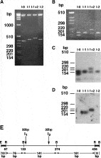
Fig. 3. Characterization of mRNA isoforms of the DNA-binding domain of SmHSF. RT-PCR products, prepared from adult worms RNA serving as template and primers flanking the DBD, were cloned into the KS Bluescript and characterized by restriction enzyme digestions and hybridizations. Ethidium bromide-stained agarose gels (A, B) of clones digested by EcoR1/XbaI (A) to release the intact DBD insert, and by EcoR1/BamH1 (B) to release fragmented insert cleaved at internal BamH1 sites. (C) Autoradiogram of (B) reacted with 32P-labelled probe of SmHSF cDNA (2·3 kb) hybridizing with all fragments of all DBD isoforms. (D) After removal of radioactivity from (C) the blot was rehybridized with a 32P-labelled intron I1 probe hybridizing with a single fragment only in DBD isofroms containing intron I1. Representative clones corresponding to the 4 DBD isoforms are shown. (1) I-0, product identical to cDNA composed of exons only and no introns, (2) I-1, product contains intron I1, (3) I-2, product contains intron I2, (4) I-1+2, product contains introns I1 and I2. Positions of DNA size markers (bp) are given on the left. (E) Scheme of cloned cDNA encoding DBD I-0. RT-PCR product digested by Rsa1 was cloned into Sma1 cleaved pJC40 expression vector (R, cloning site). Numbering of nucleotides of DBD is according to the SmHSF cDNA sequence deposited in GenBank (Acc. no. AF043418). BamH1 (B) sites in DBD, locations of introns I1 and I2 in DBD, positions of EcoR1 (E) and BamH1 sites of the vector, short DNA segments of the vector flanking DBD (black bar), and sizes of DNA fragments (bp) between restriction sites, are given.

Table 1. Clones of SmHSF mRNA isoforms containing introns
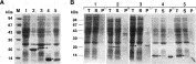
Fig. 4. Expression of protein isoforms of the DNA-binding domain of SmHSF. Coomassie blue staining of SDS–PAGE of lysates from PlysS bacteria transfected with the pJC40 vector (1), and with pJC40 constructs expressing DBD isoforms with His-tails: DBD I-0 (2), DBD I-1 (3), DBD I-1+2 (4) and DBD I-2 (5). (A) Total bacterial lysate. (B) Total bacterial lysate (T), supernatant (S) and insoluble pellet (P) of the lysate. Positions of the protein size markers (kDa) are indicated.
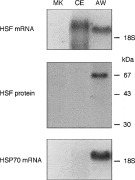
Fig. 5. Expression of HSF mRNA and protein, and of HSP70 mRNA, in cercariae and adult worms. Northern blots (10 μg RNA/lane) hybridized with 32P-labelled 2·3 kb SmHSF cDNA (top) or 1 kb HSP70 cDNA of Schistosoma mansoni (bottom). Western blot (50 μg protein/lane) developed with rabbit purified antibodies to SmHSF protein (2 μg/ml) followed by 125I-labelled goat purified antibodies to rabbit Ig. Total RNA and proteins were prepared from cercariae (CE), adult worms (AW), and mouse kidney (MK) serving as control. Positions of the 18S rRNA and protein size markers (kDa) are indicated.
 You have
Access
- 12
- Cited by