Introduction
Pinworms are nematodes from the family Oxyuridae that occupy the large intestine of many species of Old and New World primates (Hugot, Reference Hugot1985; Hugot et al., Reference Hugot, Gardner and Morand1996). There are peculiar morphological characteristics in each group that may be explained by specific host–parasite co-evolution (Cameron, Reference Cameron1929; Hugot, Reference Hugot1999; Sorci et al., Reference Sorci, Skarstein, Morand and Hugot2003). The subgenus Trypanoxyuris (Vevers, Reference Vevers1923) is one of four subgenera of pinworms found in Neotropical primates and has been reported in the primate families Cebidae, Pitheciidae and Atelidae (Artigas, Reference Artigas1936; Inglis & Dunn, Reference Inglis and Dunn1964). Trypanoxyuris infections have been also reported in captive primates, and in some cases are difficult to eradicate, causing different degrees of pathology and even mortality (Amato et al., Reference Amato, Amato, Calegaro–Marques and Bicca–Marques2002; Monteiro et al., Reference Monteiro, Coutinho, Araujo, Monteiro, Castro, Silva, Benigno and Vicente2009).
The close evolutionary relationship between humans and other primates means that their parasite faunas are also similar and the possibility of cross-infection makes the identification of new species relevant to human medicine. In recent years the number of known species in the genus Trypanoxyuris has increased, adding Trypanoxyuris (P.) lagothricis and Trypanoxyuris (T.) minutus (Souza et al., Reference Souza, Magalhães, Vieira and Souzalima2010; Trejo-Macías et al., Reference Trejo-Macías, Mosqueda-Cabrera, García-Prieto and Estrada2011; Pinto et al., Reference Pinto, Ferreira, Mati and de Melo2013), which show interesting morphological variation and different geographical ranges compared to previously known species, such as Trypanoxyuris (T.) croizati and T. (T.) callicebi (Hugot et al., Reference Hugot, Morand and Guerrero1994).
The Amazon region is highly diverse in megafauna, and in consequence harbours an even richer microfauna within animal hosts (Gonçalves et al., Reference Gonçalves, Vicente and Pinto2002; Muniz-Pereira et al., Reference Muniz-Pereira, Vieira and Luque2009). However, this high biodiversity, and the remoteness of the many parts of the Amazon, mean that the biology and geographical distributions of many potential hosts are poorly known. One such host is the Peruvian red uakari monkey (Cacajao calvus ucayalii), a subspecies endemic to the Peruvian Amazon that occurs in patchy and sometimes isolated populations in north-eastern Peru (Bowler et al. Reference Bowler, Noriega Murrieta, Recharte, Puertas and Bodmer2009). The species is included in Appendix II of CITES (the Convention on International Trade in Endangered Species of Wild Fauna and Flora) and is described as Vulnerable by IUCN (International Union for Conservation of Nature), mainly due to habitat loss and hunting (Peres, Reference Peres2001; Veiga & Bowler, Reference Veiga and Bowler2013). This rareness makes the collection of data on red uakari biology difficult and, until now, parasitological studies have been restricted to faecal samples (Conga et al., Reference Conga, Bowler, Tantaleán, Montes, Serra-Freire and Mayor2014). We report on the nematodes found in the large intestine of Peruvian red uakari monkeys on the Yavari-Mirín River and describe new species.
Materials and methods
Study site
This study was conducted in the Yavari–Mirín River basin, in the north-eastern Peruvian Amazon (04°19’53’’S, 71°57’33’’W; UT-5:00), Loreto region. The study area spans 107,000 ha of continuous, predominantly non-flooding terra firma, forest. The climate in the region is typically equatorial with an annual temperature ranging from 22 to 36°C, a relative humidity from 80 to 100%, and an annual rainfall from 1500 to 3000 mm. Seasons are defined as dry (January–February and July–September) and wet (March–June and October–December).
Collection and examination of nematodes
During 2010 and 2011, hunters living in the study area collected gastrointestinal organs from five adult Peruvian red uakari monkeys (hosts numbers 1 and 3, females; hosts numbers 2, 4 and 5, males), as part of a participatory conservation programme that involves local hunters in implementing community-based wildlife management. The hunters were trained to remove all the abdominal and pelvic organs intact, and store them in 10% formalin solution.
Researchers were responsible for the dissection of the gastrointestinal organs and collection of the dead parasites using a stereomicroscope. We washed specimens in saline and stored them in 70% ethanol (Gardner, Reference Gardner, Wilson, Cole, Nichols, Rudran and Foster1996). We made temporary study slides of nematodes in lactophenol for morphological and morphometric studies. Cross-sections of the bodies were hand-cut using a razor blade and manipulated using a small brush. We made diagrams with the aid of a drawing tube coupled to the light microscope (Olympus BX41; Melville, New York, USA) at LBCH–ICB, UFPA (Laboratório de Biologia Celular e Helmintologia Profa. Dra. Reinalda Marisa Lanfredi – Instituto de Ciěncias Biológicas, Universidade Federal do Pará). For scanning electron microscopy (SEM), we post-fixed the specimens in 2% osmium tetroxide, dehydrated them in a graded series of alcohol, dried them to ‘critical point’ using liquid CO2, coated them in gold and examined them using a scanning electron microscope (LEO 1450 VP; Clifton Road, Bristol, UK) in the Museu Paraense Emilio Goeldii – Belém, PA, Brazil (MPEG). Morphometric analysis was based on ten mature male and ten mature female specimens for each species, except for the measurements of the distance between amphids, for which we used five males and five females. All measurements are given in micrometres, with the mean and standard deviation, followed by a range in parentheses.
Results
Oxyuridae Cobbold, 1864; Trypanoxyuris Hugot, 1984; Trypanoxyuris (Trypanoxyuris) cacajao n. sp.
Description
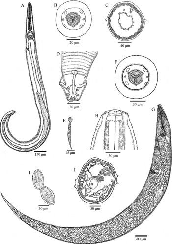
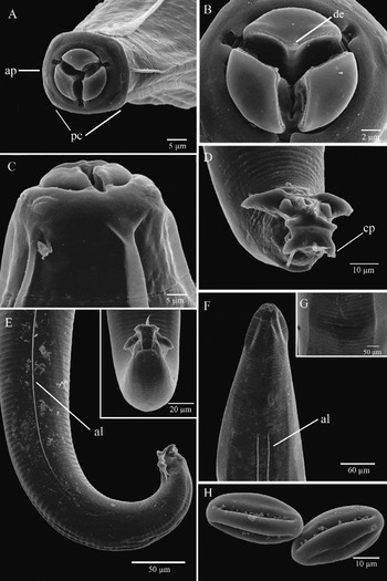
Small white worm. Cephalic structure or cephalic-tray presents a rectangular form, buccal opening is framed by three lips, each of which is separated by a groove (figs 1B, 1F, 2A). Each lip covers partially correspondent oesophageal triangular-shaped teeth clearly visible in male and female (fig. 2B). Four cephalic papillae, two ventral and two dorsal, present laterally a pair of amphidial pores, both clearly visible (fig. 2A). Cuticle with transverse striations (figs 1H, 2C) extends the full length of the worm, ending in females before the anal opening as a transverse fissure (fig. 2G) and in males before the caudal alae. Lateral alae are present in both sexes. The oesophagus is composed of a short isthmus with a bulb and valve. The lumen of the oesophagus is triradiate and the nerve ring is localized around the oesophagus in the first anterior third. Excretory pore located after oesophageal bulb with a small vestibule and aperture in the ventral part of the worm (fig. 1A). All these structures are easily visible, with no major difference in anterior extremity, apart from the size of the cephalic structure of the male and female (fig. 1A, G).
Morphology of Trypanoxyuris (Trypanoxyuris) cacajao n. sp. Male: (A) entire worm, right lateral view; (B) cephalic end, apical view; (C) cross-section of lateral alae in middle body; (D) posterior end, ventral view; (E) spicule, left lateral view. Female: (F) cephalic end, apical view; (G) entire worm, right lateral view; (H) cephalic end, left lateral view; (I) cross-section of lateral alae in middle body; and (J) eggs.

Ultrastructure of Trypanoxyuris (Trypanoxyuris) cacajao n. sp. Male: (A) cephalic end, apical view with detail of papillae (pc) and amphids (ap); (B) oesophageal teeth, apical view showing subteeth (de); (C) cephalic end, lateral view; (D) posterior end, ventral view with detail of caudal papillae (cp); (E) posterior end, lateral view with detail of end lateral alae (al). Female: (F) anterior extremity, lateral view with detail of lateral alae (al); (G) vulvar aperture, right ventral view; and (H) eggs.

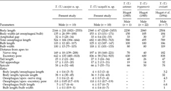
Males (based on ten mature specimens; table 1). Total worm length is less than half the length of the female (fig. 1A). Lateral alae commencing posterior to oesophageal bulb, bilaterally single crested, ending anterior to anus (figs 1C, 2E). Posterior extremity of body bent ventrally, four pairs of caudal papillae (figs 1D, 2D), with a unique spicule without gubernaculum (fig. 1E), with an anal aperture and the tip of the tail appendage clearly visible. Of the four pairs of caudal papillae, the first and the fourth pairs are peduncles and the second and the third pairs are sessile; each of the second and third pairs of papillae is surrounded by a strong ring-shaped thickening (fig. 2D, E).
Morphometric data of male Trypanoxyuris (Trypanoxyuris) cacajao n. sp. and Trypanoxyuris (Trypanoxyuris) ucayalii n. sp. in Cacajao calvus ucayalii from the Peruvian Amazon, and measurements (in μm) reported by other authors are given for comparison with Trypanoxyuris (Trypanoxyuris) sp. of Pitheciidae hosts; n= number of specimens examined; ranges of measurements are given in parentheses.

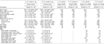
Females (based on ten mature specimens; table 2). Total length greater than that of the male (fig. 1G). Lateral alae commencing posterior to oesophageal isthmus or bulb level, becoming parallel bilaterally double-crested, extending posteriorly, and ending at level of anal aperture (figs 1I, 2F). Excretory pore located after oesophageal bulb, with a small vestibule and aperture in the ventral part of the worm. Vulvar aperture is located in the ventral anterior third of the worm. Tail long, conical, from the anus to the posterior end (fig. 1G). Eggs (obtained from an average of ten eggs measured per female): thin shell, oval format, symmetrical or asymmetrical, finely granulated, three equidistant crests are present around the eggs (figs 1J, 2H).
Morphometric data of female Trypanoxyuris (Trypanoxyuris) cacajao n. sp. and Trypanoxyuris (Trypanoxyuris) ucayalii n. sp. in Cacajao calvus ucayalii from the Peruvian Amazon, and measurements (in μm) reported by other authors are given for comparison with Trypanoxyuris (Trypanoxyuris) sp. of Pitheciidae hosts; n= number of specimens examined; ranges of measurements are given in parentheses.

Taxonomic summary
-
Type host. Cacajao calvus ucayalii Thomas, 1948 (Primate: Pitheciidae).
-
Site of infection. Large intestine.
-
Locality. Yavari-Mirim River, Peruvian Amazon (S0 04°27.5′; W0 71°45.9′).
-
Date. August 2010.
-
Prevalence. 100% (five hosts examined).
-
Intensity of infection. Forty-nine (11 males and 38 females) in host no. 1; 81 (27 males and 54 females) in host no. 2; 107 (23 males and 84 females) in host no. 3; 36 (5 males and 31 females) in host no. 4; 50 (9 males and 41 females) in host no. 5. Average: 64.6 ± 28.9 (15.0 ± 9.5 males and 49.6 ± 20.9 females).
-
Specimens deposited. The holotype (No. MPEG 0059), allotype (No. MPEG 0060) and one paratype (No. MPEG 0061) were deposited in the Coleção de Nematoda do Museu Paraense Emílio Goeldi (MPEG).
Oxyuridae Cobbold, 1864; Trypanoxyuris Hugot, 1984; Trypanoxyuris (Trypanoxyuris) ucayalii n. sp.
Description
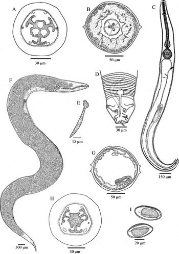
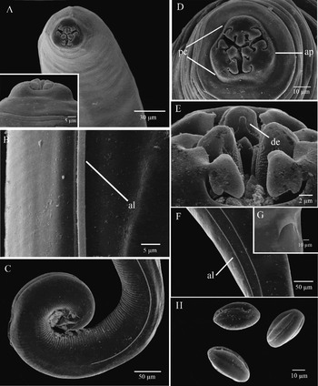
Small white worm. Cephalic structure or cephalic-tray presents a clearly hexagonal form, buccal opening is framed by three lips, and each is divided into two lobes, also called bilobate lips (figs 3A, 3H, 4A, 4D). Each lip partially covering one of three oesophageal teeth, with triangular format clearly visible (fig. 4E). Four cephalic papillae – two ventral and two dorsal – with a pair of amphidial pores present laterally (fig. 4D). Cuticle with transverse striations. Lateral alae present in both sexes. Oesophagus composed of a short isthmus with bulb and valve. The lumen of the oesophagus is triradiate and the nerve ring is localized around the oesophagus in the first anterior third. Excretory pore located after oesophageal bulb, with a small vestibule and aperture in the ventral part of the worm. All these structures are easily visible, with no major difference in anterior extremity, apart from the size of the cephalic structure of the male and female (fig. 3C, F).
Morphology of Trypanoxyuris (Trypanoxyuris) ucayalii n. sp. Male: (A) cephalic end, apical view; (B) cross-section of lateral alae in middle oesophagus; (C) entire worm, left lateral view; (D) posterior end, ventral view; (E) spicule, right lateral view. Female: (F) entire worm, right lateral view; (G) cross-section of lateral alae in middle body; (H) cephalic end, apical view; and (I) eggs.

Ultrastructure of Trypanoxyuris (Trypanoxyuris) ucayalii n. sp. Male: (A) anterior extremity, apical and lateral view; (B) anterior extremity, lateral view with detail of lateral alae (al); (C) posterior extremity, lateral view with detail of end lateral alae. Female: (D) cephalic end, apical view with detail of papillae (pc) and amphids (ap); (E) oesophageal teeth, apical view showing subteeth (de); (F) posterior extremity, lateral view with detail of lateral alae (al); (G) anal aperture, ventral view; and (H) eggs.

Males (based on ten mature specimens; table 1). Total worm length is four times smaller than the length of the female (figs 3C, 4A). Lateral alae commencing posterior to oesophageal bulb, bilateral with a single crest, ending anterior to anus (figs 3B, 4B). Posterior extremity of body bent ventrally, four pairs of caudal papillae (fig. 3D), with a unique spicule without gubernaculum (fig. 3E) and tail finalized with a tail appendage. Of the four pairs of caudal papillae, the first and the fourth pair are peduncles and the second and third pairs are sessile, each papilla of the second and third pairs is surrounded by a strong ring-shaped thickening (figs 3D, 4C).
Females (based on ten mature specimens; table 2). Total length greater than that of the male (fig. 3F). Lateral alae commencing posterior to oesophageal isthmus or bulb level, becoming parallel bilaterally double-crested, clearly visible, extending posteriorly, and ending at level of anal aperture (fig. 4F, G). Excretory pore located after oesophageal bulb, with a small vestibule and aperture in the ventral part of the worm. Vulva aperture is located in the ventral anterior third of the worm. Tail long, conical, from the anus to the posterior end (fig. 3F). Eggs (obtained from an average of ten eggs measured per female): thin shell, oval format, symmetrical or asymmetrical, finely granulated, three equidistant crests are present around the eggs (figs 3I, 4H).
Taxonomic summary
-
Type host. Cacajao calvus ucayalii Thomas, 1948 (Primate: Pitheciidae).
-
Site of infection. Large intestine.
-
Locality. Yavari-Mirim River, Peruvian Amazon (S0 04°27.5′; W0 71°45.9′).
-
Date. August 2010.
-
Prevalence. 100% (five hosts examined).
-
Intensity of infection. Thirty-eight (13 males and 25 females) in host no. 1; 85 (17 male and 68 female) in host no. 2; 35 (6 males and 29 females) in host no. 3; 18 (3 males and 15 females) in host no. 4; 29 (9 males and 20 females) in host no. 5. Average: 41.0 ± 25.8 (7.2 ± 6.3 males and 38.6 ± 26.6 females).
-
Specimens deposited. The holotype (No. MPEG 0056), allotype (No. MPEG 0057) and one paratype (No. MPEG 0058) were deposited in the Coleção de Nematoda do Museu Paraense Emílio Goeldi (MPEG).
Discussion
In our samples, the salient characteristics on the lips (Vevers, Reference Vevers1923) and the specimen proportions – oesophagus and bulb length (Hugot et al., Reference Hugot, Gardner and Morand1996) – allowed us to include them in this genus and subgenus Trypanoxyuris. Thus far, in Neotropical forest regions, eight species of the subgenus Trypanoxyuris have been described in non-human primates, including T. (T.) minutus (Schneider, 1866); Trypanoxyuris (Trypanoxyuris) microon (Linstow, 1907); Trypanoxyuris (Trypanoxyuris) sceleratus (Travassos, 1925); Trypanoxyuris (Trypanoxyuris) clementinae (Hugot, Reference Hugot1985); T. (T.) trypanuris (Vevers, Reference Vevers1923); T. (T.) satanas (Hugot, Reference Hugot1985); T (T.) callicebi (Hugot & Vaucher, Reference Hugot and Vaucher1985) and T (T.) croizati (Hugot et al., Reference Hugot, Morand and Guerrero1994). Of the eight species listed, the last four have been recorded parasitizing monkeys of the Pitheciidae (genera: Callicebus, Chiropotes and Pithecia) and are morphologically closer species to those described in this study. The new species, T. (T.) cacajao n. sp. and T. (T.) ucayalii n. sp., also live in this family of hosts, and share morphological characteristics. However, there are important details that distinguish these new species from the others in this subgenus.
The cephalic structure of T. (T.) cacajao n. sp. is almost quadrangular, with three simple lips surrounding the three oesophageal teeth; this format is distinct from that of the four closest species. The new species differs from T. (T.) trypanuris and T. (T.) satanas by having an hexagonal cephalic-tray and by the arrangement of each lip, divided into three lobes each. Trypanoxyuris (T.) cacajao n. sp. can be distinguished easily from T. (T.) callicebi and T. (T.) croizati, which have a hexagonal shaped cephalic-tray and a mouth opening bounded by three lips that partially cover the teeth. We also emphasize a small ornate subtooth in the ventral part of each oesophageal tooth, as reported in the studies by Hugot et al. (Reference Hugot, Gardner and Morand1996) and Pinto et al. (Reference Pinto, Ferreira, Mati and de Melo2013), and now observed in detail through scanning microscopy of our specimens.
Another important distinguishing feature is that of the lateral alae, which are easily visible in both sexes through scanning electron microscopy: one pair in the male and two pairs in the female. This feature is similar in females of the species T. (T.) callicebi and T. (T.) croizati, but differs in male T. (T.) croizati in having a lateral thickening. The lateral alae of T. (T.) satanas are similar in males, but differ in females. In T. (T.) trypanuris differences in lateral alae between the sexes are clearly evident, as in T. (T.) cacajao. Thus either males or females differ in this aspect in each of the four related species.
In the female T. (T.) cacajao n. sp. general measurements are smaller than those reported in the literature for the other eight species in the subgenus Trypanoxyuris (Inglis & Diaz-Ungría, Reference Inglis and Diaz-Ungría1960), which may be useful in identifying this species. Sexual characteristics of males and females conform to the patterns described by Hugot et al. (Reference Hugot, Gardner and Morand1996): the male has four caudal papillae, with a single spicule and no gubernaculum. Females have a robust vulvar aperture with a divided uterus and eggs with three equidistant crests, as described in studies by Hugot et al. (Reference Hugot, Morand and Guerrero1994). Eggs are longer than those of T. (T.) callicebi, T. (T.) croizati, T. (T) trypanuris, and similar to those of T. (T.) satanas.
Trypanoxyuris (Trypanoxyuris) ucayalii n. sp. is morphologically closest to T. (T.) satanas, which was described from a holotype male and an allotype female parasitizing Chiropotes satanas (Hoffmannsegg, 1807) captured in Venezuela (Hugot, Reference Hugot1985). Morphological characteristics and measurements of the specimens studied, as well as the cephalic-tray, shape of the lips and the presence of lateral alae agree with morphological studies on T. (T). satanas (Hugot, Reference Hugot1985), except for the detail of the lateral alae in the female. According to the previous work, this is described as a marked lateral protuberance, whereas we observed two pairs of lateral alae as two parallel crests, very evident by light microscopy or scanning electron microscopy. An important feature to highlight is the presence of subteeth, clearly visible here but absent in T. (T.) satanas.
General measurements in females are similar, except the total body length, which is slightly larger than that described in T. (T.) satanas by Hugot (Reference Hugot1985). Trypanoxyuris (T.) trypanuris (Vevers, Reference Vevers1923) is another similar species, with similar cephalic structure, but is sharply differentiated by the lateral alae, described as a ‘thickening’ in males and females (Hugot, Reference Hugot1985), unlike the evident lateral alae shown in our specimens. Since differences are marked in such important characteristics as lateral alae of the female, the oesophageal subteeth and the worm length, we propose that this species is separated from the rest of congener pinworms previously observed in Pitheciidae hosts, and may be a competitor of T. (T.) cacajao n. sp. in the uakari host. The other four species of the subgenus Trypanoxyuris within the Pitheciidae hosts are distributed in the eastern Amazon: T. (T.) satanas in Chiropotes satanas and T. (T.) croizati in Callicebus torquatus from Venezuela; T. (T.) trypanuris in Pithecia pithecia from British Guyana and T. (T.) callicebi in Callicebus moloch from Paraguay. The present study could be the westernmost record of the Trypanoxyuris subgenus in the Amazon.
The increasing reduction and fragmentation of threatened populations, such as the uakari, might alter the host–helminth equilibrium in the wild, by increasing the parasite prevalence and altering parasitic wealth in the host's restricted area of distribution (Wenz et al., Reference Wenz, Heymann, Petney and Taraschewsk2009; Bordes et al., Reference Bordes, Morand, Pilosof, Claude, Krasnov, Cosson, Chaval, Ribas, Chaisiri, Blasdell, Herbreteau, Dupuy and Tran2015). Additionally, some helminth infections, such as Trypanoxyuris, may be difficult to eradicate, causing pathologies and even death in captive and wild primates (Amato et al., Reference Amato, Amato, Calegaro–Marques and Bicca–Marques2002; Nunn et al., Reference Nunn, Altizer, Jones and Sechrest2003). These factors may act as a feedback mechanism to further reduce the density of threatened populations (Medina-Vogel, Reference Medina-Vogel2010), increasing the risk of extinction. We described two species of the genus and subgenus Trypanoxyuris co-habiting the large intestine of C. c. ucayalii, showing significant morphological characteristics. Similar examples of helminths inhabiting the same niche were described in Old World primates, such as lemurs and colobus, with particular eating habits, as in uakari, that could explain the presence of sister species (Wahid, Reference Wahid1961; Chabaud et al., Reference Chabaud, Brygoo and Petter1965; Hugot, Reference Hugot1987). This is the first record of two species within a platyrrhine primate in the Peruvian Amazon, under natural conditions of infection. The description of pinworms in this study reinforces the need for further parasitological studies in free-range wildlife, especially in threatened species.
Acknowledgements
The authors thank all the people from Nueva Esperanza, who participated actively in data collection, showing that communal participation is an important step in the development of wildlife management, and the institutional support provided by Servicio Nacional de Sanidad Agraria (SENASA) and Dirección General de Flora y Fauna Silvestre – Ministerio de Agricultura of Peru (no. 0229–2011–DGFFS–DGEFFS). We are especially thankful to Dra. Jeannie Nascimento dos Santos (head professor of Laboratório de Biologia Celular e Helmintologia ‘Profa. Dra. Reinalda Marisa Lanfredi’, Universidade Federal do Pará, Belém-Pará) for her support with the analyses.
This paper is dedicated to the memory of the parasitologist Nicolau Maués da Serra Freire, professor of Programa de pós-graduação em Saúde e Produção Animal na Amazônia of the Universidade Federal Rural da Amazônia and researcher of the Instituto Oswaldo Cruz, who died 24 June 2015. Our eternal gratitude.
Financial support
This work was possible thanks to institutional and financial support of the Fundació Autònoma Solidària from the Universidad Autónoma de Barcelona. The scholarship for D.F.C. was supported by the Coordination for the Improvement of Higher Level Personnel (CAPES, Foundation – Ministry of Education of Brazil) and the Programma de pós-graduação em Saúde e Produção Animal na Amazônia – Universidade Federal Rural da Amazonia (UFRA-Belém/Pará).
Conflict of interest
None.
Ethical standards
The research protocol was approved by the Dirección General de Flora y Fauna Silvestre from Peru (Research Ethics Committee for Experimentation in Wildlife no. 0229–2011–DGFFS–DGEFFS and Export Authorization no. 02309-MINAGRI-DGFFS). Hunters preserved all species hunted, and were not paid for the sample collection, so no animals were killed specifically for the research.