Introduction
Astronotus ocellatus, popularly known as Oscar, is a cichlid fish from South America that lives in the Amazon river of Brazil and Peru. This species has great potential for fish farming, and is cultivated around the world as an ornamental fish (Machado, Reference Machado1983), as well as being extremely intelligent, and suitable for sport fishing (Fury & Morelo, Reference Fury and Morello1994) and also for human nutrition, because their meat is tasty, firm and there is absence of intramuscular bones (Fontenele e Nepomuceno, Reference Fontenele and Nepomuceno1983). Adult specimens may reach 33 cm in length and weigh up to 1.5 kg (Machado, Reference Machado1983). Their first gonadal maturation is reached at around 10 to 12 months of age, and an average about 1500 to 2000 eggs per clutch is obtained (Braga, 1962). It is a species of partitioned spawning that exhibits parental care behaviour and has three to four reproductive cycles per year (Silva et al., Reference Silva, Regis and Bezerra1993).
Despite the economic importance of this species, there are scarce data in the current literature, with information mainly about its reproductive biology and the early development of eggs and larvae.
Little is known about the early development of native species with partitioned spawning. Knowledge about embryos and larvae of any species is very important because it represents a useful tool for localization of spawning sites and studies on growth in a natural environment (Reynalte-Tataje et al., Reference Ribeiro, Leme dos Santos and Bolsan2001).
Data about the early developmental stages in native species are essential for research and fish farming (Ribeiro et al., Reference Reynalte-Tataje, Zaniboni-Filho and Muelbert1995). Most research with fish larvae has been focused on systematics and phylogeny, but lacks particular attention to morphological development in relation to function. The study of fish larvae is necessary to understand the biological role of larval characteristics in survival, mainly involving those functional mechanisms related to vital areas such as locomotion, feeding and breathing (Osse, Reference Osse1990).
Studies using stereomicroscopy and scanning electron microscopy have provided three-dimensional images of the surface, very important for knowledge of external morphology and identification of eggs and larvae.
Given the biological and economical importance of A. ocellatus for fish farming, the aim of this present research was to analyse its embryonic and larval development using stereomicroscopy and scanning electron microscopy.
Material and methods
Two sets of 150 specimens of A. ocellatus from 1 to 2 years old were kept in separated 50 m3 masonry tanks and fed twice a day with commercial food containing 40% raw crude protein. Two plastic cylinders and ceramic bricks were placed in the tanks to provide both shelter and an area for spawning. Natural spawn were then observed from October 2007 to February 2008.
As soon as the spawn in the tanks was observed, they were removed and transported to plastic incubators that had a continuous water flow. Sample collection was performed according to the following procedure: initial collection at the moment the spawn in the tank was observed, every 6 h up to larval mouth opening, then every 12 h up to 161 h post-hatching (hPH) and every 24 h up to 383 hPH.
Samples were fixed in Karnovsky's solution for 24 h, washed up in phosphate-buffered saline, pH 7.4 and stored in 70% ethanol in a refrigerator. Afterwards, the samples were observed and the images were captured under a stereomicroscope, model MZ8 equipped with LEICA DFC 280 automatic photomicrography. For scanning electronic microscopy analysis, the samples were post-fixed in 1% osmium tetroxide for 2 h and washed again in the same buffer. Then, the samples were dehydrated in a graded series of ethanol at 30, 50, 70, 80, 90 and 95% concentrations plus three washes at 100% (15 min each) and dried to the critical point in a liquid CO2 drier, mounted in a copper grid, coated with gold–palladium ions and observed and photographed under a scanning electron microscope (JEOL-JSM 5410).
The average physico-chemical water parameters monitored in the incubators were as follows: temperature 27.45°C; pH 4.83; dissolved oxygen 5.52 mg/l; alkalinity 52 mg/l; ammonia 117.45 μg/l and conductivity 35.51 μS/cm.
Results
Astronotus ocellatus exhibits external fertilization and parental care. The eggs adhere to a substrate and spread in a single layer. They are demersal, adherent and fragile to touch, with a slight ovoid shape, a large yolk sphere and a small perivitelline space. The fertilized eggs have a yellowish colouration and, when unfertilized, they are opaque white.
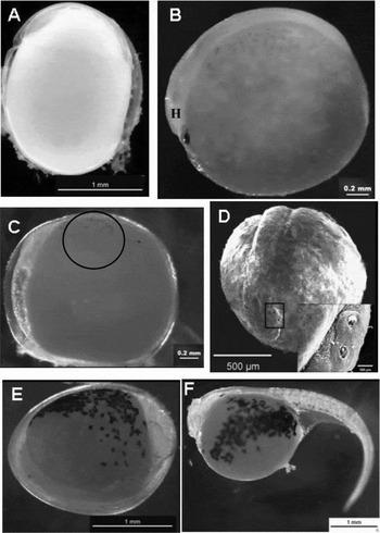
The first egg collection was performed immediately after their removal from the tank (initial collection – IC). At this moment, most of eggs were in the initial gastrula stage, with about 30% epiboly (Fig. 1A) and covered with a thick jelly that made the eggs adherent. After 6 h, the eggs presented 50% epiboly, and at 12 h after IC, the formation of the embryo had already begun.
Photomicrographs and electron micrographs of Astronotus ocellatus (A) Stage gastrula. (B) Differentiation of the head (H) and tail. (C) Yolk pigmentation initial (circle). (D) Formation of the cephalic gland. (E) Prelarva shortly before the outbreak. (F) Newly hatched larva.

The beginning of embryo organogenesis is characterized by the formation of the embryonic axis and the appearance of somites followed by tail and head differentiation (Fig. 1B). From 15 h after IC we observed the tail extension, the beginning of yolk pigmentation (melanophores) (Fig. 1C) and the formation of adhesive glands on the head (Fig. 1D), and optic and otic vesicles. Prior to hatching, the prelarvae (Fig. 1E) showed strong and continuous tail movements to break the chorion.
Hatching
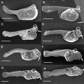
Hatching took place between 46 to 58 h after the initial collection. The recently hatched larvae were transparent, with pigmentation restricted to the yolk (Fig. 1E). The finfold was very large, the mouth was closed and the adhesive glands were highly developed, comprising two pairs in the upper part and one pair in the lower part of the head (Fig. 2A).
Electron micrographs of Astronotus ocellatus showing their development. (A) Newly hatched larva; (B) 30 hPH; (C) 65 hPH; (D) 77 hPH; (E) 101 hPH; (F) 149 hPH; (G) 185 hPH; (H) 209 hPH. hPH, hours post-hatching. FF, finfold; Y, yolk; white arrow, mouth; circle, adhesiveness glands; black arrow, pectoral fin.

6 hPH
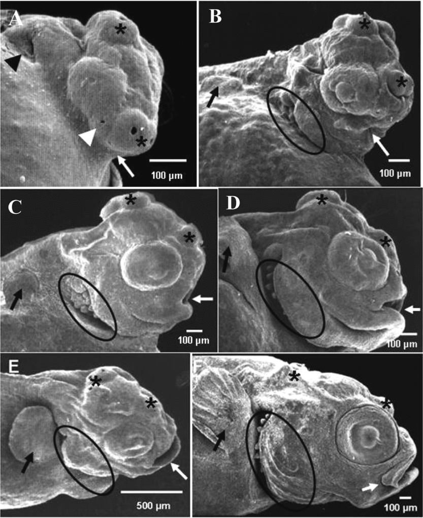
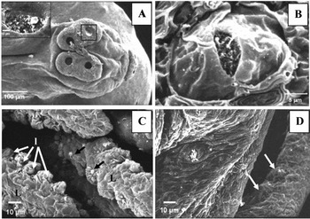
A primordial heart started beating in the inner part of the embryo. By scanning electronic microscopy, it was possible to observe the cephalic groove, the conspicuous adhesive glands and both otic vesicle and nasal orifices (Figs. 4A and 5A). Due to the absence of fins and swim bladder and the large amount of yolk, the larvae remained grouped in the bottom of incubators, moving with rapid tail movements.
12–30 hPH
The body pigmentation of larvae increased and the pectoral fin bud appeared; the head was already detached from the yolk; the heart was pumping blood throughout the entire larval extension; the yolk was highly vascularized and the eyes were clearly pigmented. At this stage, the larvae tried to escape the pipette aspiration and proved to be negative photosensitively when exposed to the stereomicroscope light.
41–77 hPH
There was a deep fold in the epithelium covering the mouth entrance and the formation of branchial arches had started (Fig. 4B). At 53 hPH, the mouth was open and occupied the anterior head portion, below the eye line. As well as the head pigmentation, the pectoral fin was already noticeable; the gill lamellae were formed and the opercle was starting to cover them (Figs. 2C and 4C). At 77 hPH, the larvae presented free pectoral fins, the embryonic finfold was reduced, the opercle covered nearly the entire well developed gills and the lower jaw was completely formed (Figs. 2D and 4D) and constantly moving.
89–113 hPH
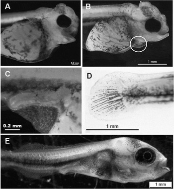
The larvae presented a formed branchial apparatus. There was evidence of the main lamellar of the gill arches and of the intense blood vascularization in the yolk and head (Fig. 3A). The adhesive glands were now not as evident, the pectoral fins moved strongly and the opercle covered the gills slit at 101 hPH (Fig. 2E, 3B and 4E), with the annus opened (Fig. 3C).
Photomicrographs of Astronotus ocellatus. (A) 89 hPH larva beginning on the pigmentation of the head, yolk sac is richly vascularized, training and irrigation of the branchial apparatus. (B) 101 hPH larva with pectoral fins free, heart (circle) and vascularization of the yolk. (C) 113 hPH opened anus. (D) 137 hPH Pigmentation of caudal fin rays. (E) 383 hPH larva with yolk completely absorbed, well trained eyes, heart, staying under the cap, body pigmentation, digestive tract formed, inflated swim bladder and early onset ray of the dorsal and anal. hPH: hours post-hatching.

Electron micrographs of Astronotus ocellatus showing the development head structures. (A) 6 hPH; (B) 41 hPH; (C) 65 hPH; (D) 77 hPH; (E) 101 hPH; (F) 149 hPH. White arrowhead: nasal orifice; black asterisks: adhesive glands; black arrowhead: otic vesicle; black arrow: pectoral fin; black circle: gill formation; white arrow: mouth. hPH: hours post-hatching.

125 hPH
Exogenous feeding was initiated. Taste buds could be noticed only on the lip surface (Fig. 5D). The olfactory epithelium had many cilia; larvae already presented vertical swimming and chased brine shrimp napuli.
Electron micrographs of Astronotus ocellatus. (A) 6 hPH adhesive glands present on the head. (B) 185 hPH neuromast around the eyes. (C) 383 hPH gill filaments showing the main (L) and secondary (l) lamellae that compose the gill with the presence of mucus released by the secretory cells (black arrows). (D) 383 hPH taste buds in the mouth (arrows). hPH: hours post-hatching.

137–383 hPH
The first rays of the caudal fin appeared (Fig. 3D); only one pair of adhesive glands was present in the upper region of the head and the other were absorbed. The heart was already located between the bones of the opercle. At 149 hPH, the formation of dorsal and anal fins was initiated; the opercle was completely formed and rays appeared in the pectoral fins, the mouth assumed a terminal position (Fig. 2F) and the neuromasts appeared around the eyes (Figs. 2G and 5B) and bile excretion through the anus could also be observed. With 209 hPH the larva is well formed but still has a yolk (Fig. 2H). At 257 hPH, the yolk was completely absorbed. From 383 hPH on, six branchial arches with mucus-secreting cells were detected (Fig. 5C), similar to those found in the skin. The pectoral fins were completely formed; the dorsal and anal fins were starting to develop; the adhesive glands were degenerated; the eyes were completely formed, with intense pigmentation and definitive coloration of retina (Fig. 3E).
Discussion
Oscar's eggs are ovoid shaped, demersal, adhesive, fragile to the touch, with a large yolk sphere and a small perivitelline space. The characteristics of Oscar's eggs confirmed the description presented by Perini et al. (Reference Perini, Sato, Rizzo and Bazzoli2009) who asserted that eggs of non-migratory or sedentary species usually exhibit some degree of adhesiveness, a small perivitelline space, are less numerous and may be subject to parental care.
The first stage observed in the studied spawnings was the gastrula phase. Chacon (Reference Chacon1959) analysed the development of eggs of A. ocellatus by stereomicroscopy and reported that the gastrula phase occurs approximately 5 h after the first cleavage. However the environmental conditions of each experiment must be taken into account, as several factors might influence development time, in particular temperature (Osman et al., Reference Osman, Wuertz, Mekkawy, Verreth and Kirschbaum2008).
In this present research, somitogenesis in A. ocellatus occurred before the end of epiboly, similar results were obtained by Morrison et al. (Reference Morrison, Miyake and Wright2001) for Nile tilapia. This occurrence is due to the large size of the eggs, a common characteristic of species with parental care (Suzuki, Reference Suzuki1992).
The larvae hached with closed mouth and anus, were transparent, with a large yolk sac and less eye pigmentation, similar to most of the larvae of freshwater fish (Nakatani et al. Reference Nakatani, Agostinho, Baumgartner, Bialetzki, Sanches and Cavicchioli2001). However, A. ocellatus had glands used for adhesion present in the head that appeared even before hatching and developed mainly in the newly hatched larvae. These glands have been described in some freshwater species (Hoplias malabaricus, Araújo-Lima & Bittencourt, Reference Araujo-Lima and Bittencourt2002; Symphysodon spp., Morais, Reference Morais2005; Paracheirodon axelrodi, Anjos & Anjos, Reference Anjos and Anjos2006). The adhesive glands in fish are composed of a group of mucous-producing cells and serve to prevent the larvae dispersing from the nest, which would reduce the efficiency of parental care (Araújo-Lima & Bittencourt, Reference Araujo-Lima and Bittencourt2002).
The development of many structures and organs, such as, eyes, taste buds, fins, neuromasts, heart, opening and position of the mouth, branchial apparatus, etc., is closely related to survival and adaptation of the larvae. The eyes of Oscars develop very quickly, even before the mouth opening, representing the first sensitive organ to be effectively functional.
The larvae of A. ocellatus, which had constituted and pigmented eyes, escaped easily from pipette suction during sample collection. These data reveal typical characteristics of a species whose escape from predators and feeding is mainly dependent on their sight (Osman et al., Reference Osman, Wuertz, Mekkawy, Verreth and Kirschbaum2008).
Taste buds are also important structures involved in the capture of food. According to Matsuoka (Reference Matsuoka2001) the olfactory organs and taste buds act as chemoreceptors, whose function is to capture the smell and the taste of saturated substances present in the water. The number of taste buds would also be related in the ability of fish to effectively locate a food source (Caprio, Reference Caprio, Atema, Fay, Popper and Tavolga1988). Species that inhabit deep waters and/or turbid water, for example, Pseudoplatystoma corruscans, have large numbers of chemical receptors, not only around the eyes, but also in the barbels and on the body surface and have poorly developed eyes. In contrast, the A. ocellatus, which inhabits clear, calm and shallow water (http://fishbase.org, 2010) was noted to have few taste buds, free neuromasts were located in the head and big eyes developed.
The development of the circulatory system begins with the appearance of the heart in the embryo during somitogenesis, and it continues during larval development (Hu et al., Reference Hu, Sedmera, Yost and Clark2000). The heart is a structure that develops rapidly, being the first functional organ in the larvae. Osman et al. (Reference Osman, Wuertz, Mekkawy, Verreth and Kirschbaum2008) argued that in Clarias gariepinus, the rapid formation of the heart indicates an adaptation of native species to warm waters, the increased metabolic rate being a result of oxygen demand and nutrients under elevated temperatures.
The Oscar has a large yolk sac that within 257 h of hatching (HPH) was completely absorbed. According to Nakatani (Reference Nakatani, Agostinho, Baumgartner, Bialetzki, Sanches and Cavicchioli2001) species with larger yolk sacs are better prepared to survive for longer periods without relying on exogenous feeding. Tengjaroenkul et al. (Reference Tengjaroenkul, Smith, Smith and Chatreewogsin2002) argued that to prevent larvae mortality and feed waste, exogenous feeding must begin when the yolk is completely absorbed. However, in this research exogenous feeding was offered at 125 hPH and the larval survival index from then until the end of the experiment was of an order of 100%. The size of the yolk and its absorption time are also species specific. Larvae with higher amounts of endogenous nutrition have a longer period to adapt to capturing external foods items during the period that they are maintained by yolk sac reserves (Woynarovich & Horváth, 1983; Bonislawska et al., Reference Bonislawska, Formicki and Winnicki2000; Gisbert et al., Reference Gisbert, Williot and Castelló-Orvay2000).
A. ocellatus development is considered slow when compared with species that migrate, but it is in line with the development of the most lentic species. The occurrence of morphological structures that allow the capture of food even before the exhaustion of endogenous energy reserves provides for an increased chance of survival during the larval stage.
The simultaneous differentiation of structures such as pectoral fins, heart and gills was remarkable, as well as the rapid formation of the eyes and reveals that this fish is a typical tropical species and can be characterized as a visual predator.
The results presented here provide information that will be useful for the biology, reproduction and management of rearing A. ocellatus.
Acknowledgements
The authors are grateful to FAPESP (process 2006/54048–0) and CNPq (process 134265/2006–8) for financial support.