Introduction
Since its introduction in 1994, the Hunsaker Mon-Jet tube for subglottic jet ventilation has revolutionised microlaryngeal surgery. This system is widely used, and its safety and efficacy are well established.Reference Barakate, Maver, Wotherspoon and Havas1–Reference Orloff, Parhizkar and Ortiz3 The Hunsaker Mon-Jet tube has a positioning device which keeps the distal tip of the tube in a central position within the airway. The system allows adequate ventilation together with control of the airway, and has been shown to have few complications.Reference Davies, Hillel, Maronian and Posner2 From the surgeon's perspective, the Hunsaker Mon-Jet tube provides an excellent visual field along with complete and unimpeded access for microlaryngeal surgery, while providing the patient with a safe and effective airway.
Case report
A 46-year-old Caucasian woman was scheduled for microlaryngoscopy and laser removal of a vocal cord polyp. After intravenous induction of anaesthesia, the vocal cord polyp was visualised on direct laryngoscopy.
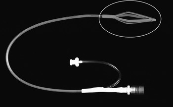
A Hunsaker Mon-Jet tube (Xomed Surgical Products, Jacksonville, Florida, USA) was passed through the vocal cords to permit jet ventilation (using a Manujet 3 injector; VBM GmbH, Sulz, Germany). However, introduction of the Hunsaker tube was blocked at the vocal cords, and it could not be advanced or withdrawn. It was noticed that the polyp had slipped into the distal positioning cage of the tube. However, it was still possible to inflate the lungs using the jetting device (Figure 1).
Hunsaker Mon-Jet ventilation tube showing the distal positioning basket within which the vocal fold polyp became trapped during intubation.

A cuffed microlaryngeal tube was inserted alongside the Hunsaker tube and positioned in the trachea beyond the vocal cords, permitting conventional ventilation. Using a microlaryngoscope, the surgeon stabilised the polyp with microlaryngeal forceps, released the polyp from the Hunsaker tube cage, and removed the Hunsaker tube safely. The vocal cord polyp was excised using a ‘cold steel’ technique.
Discussion
Jet ventilation was first introduced by Saunders in 1967 for use during routine bronchoscopy. This technology has since been adapted to provide supraglottic, subglottic and transtracheal jet ventilation for microlaryngeal surgery. Subglottic ventilation has been shown to be more efficient than supraglottic ventilation as it provides ventilation below the vocal cords, propels any blood and debris upwards and outwards with the expiratory flow of air, and causes minimal displacement of the vocal folds during ventilation (thereby facilitating visualisation and operative access).
In 1994, Hunsaker and colleagues introduced a fluoroplastic, laser-safe, self-centring, subglottic jet ventilation device, the Hunsaker Mon-Jet tube.Reference Hunsaker4 This tube is 35.5 cm long with a maximum outer diameter of 4.3 mm and an inner diameter of 2.7 mm. A removable metal stylet provides the rigidity, yet flexibility, required to facilitate tracheal intubation. The basket at the distal end positions the tube centrally within the trachea, preventing the jet port from coming into contact with the mucosa. The larger diameter lumen allows jet ventilation, usually with oxygen. A secondary lumen allows monitoring of airway pressures and sampling of expired CO2.
Compared to other techniques and devices used for subglottic ventilation for microlaryngeal surgery, the Hunsaker Mon-Jet tube has been shown to be safe, with few complications, and efficacious for use in both complicated and uncomplicated airways.Reference Davies, Hillel, Maronian and Posner2 Barotrauma associated with Hunsaker Mon-Jet tube subglottic ventilation is greatly minimised by the use of automatic jet ventilation with a preset switch-off facility, which activates when airway pressures exceed certain limits.
• The Hunsaker Mon-Jet tube is commonly used for jet ventilation during microlaryngeal surgery, with few complications
• In this case, a vocal fold lesion became trapped within the Hunsaker tube basket, causing potential airway compromise
• Anaesthetists and surgeons should be aware of this complication, and its appropriate management
Since 2004, subglottic ventilation with the Hunsaker Mon-Jet tube has been our department's preferred technique for routine, uncomplicated microlaryngeal surgery. We have used this technique successfully for treating laryngeal lesions (including polyps, vocal nodules, granulomas and Reinke's oedema) and for obtaining vocal fold biopsies. Prior to the presented case, we had not encountered any previously reported complications associated with its use, such as hypoxia, hypercapnia or barotrauma.Reference Cook and Alexander5 However, the presented case highlights the rare but potential complication of entanglement of the Hunsaker tube basket when dealing with large laryngeal lesions, especially pedunculated polyps.
We recommend caution and careful placement of the Hunsaker tube in cases of large laryngeal polyps. If such a problem is encountered, we recommend leaving the Hunsaker tube in situ, securing the airway using a standard endotracheal tube, and then commencing traditional ventilation. We do not recommend attempting to withdraw or advance the Hunsaker Mon-Jet tube, as this would risk bleeding and airway obstruction. Direct microlaryngoscopy is recommended, allowing the polyp to be disengaged from the basket under direct vision. The laryngeal lesion can then be dealt with as appropriate.
Conclusion
This case highlights the potential complication of entrapment of a laryngeal growth (e.g. a pedunculated polyp) in the positioning cage of the Hunsaker Mon-Jet tube, while intubating a patient for microlaryngeal surgery.