Introduction
Radiotherapy to the whole breast is frequently offered as adjuvant treatment to breast cancer patients following breast conserving surgery. Patients with one or more factors that indicate a high risk of local recurrence are treated with an additional radiotherapy breast boost dose to the former tumour bed/surgical excision site. 1
Studies have been conducted which clearly tested and demonstrated the benefits and risks associated with the accepted standard radiation doses prescribed for whole breast radiotherapy across the world.Reference Whelan, Mackenzie and Julian 2 , Reference Haviland, Owen and Dewar 3
Currently across the United Kingdom, oncology consultants prescribe any prescription from a range of breast boost doses. It has been reported that almost 75% of the eventual local recurrence risk occurs during the first 5 years post diagnosis. Variations in local treatment that seriously affect 5-year local recurrence rates could reduce 15-year overall mortality.Reference Clarke, Collins and Darby 4 Research showed that by reducing the incidence of local recurrence post-surgery through optimising all treatment options including radiotherapy would prevent one breast cancer death for every four cases of local recurrence over a 15-year period.Reference Clarke, Collins and Darby 4
There is a paucity of published clinical evidence, which shows which or why a particular breast boost radiotherapy dose is appropriate for different patients.Reference Bartelink, Horiot and Poortmans 5 – Reference Guenzi, Vagge and Azinwi 8 Despite this weakness, a retrospective study conducted using patient data from 18 institutions based in Australia, Europe, Israel and the United States showed the use of three different dose regimes 10 Gy (65%), >10 Gy (27%) and <10Gy (8%).Reference Omlin, Amichetti and Azria 9 As a result of reading this literature the following questions arose. Which one of these boost doses is best for a range of patients with different recurrence risk factors? Are any of these doses much more appropriate for patients with invasive ductal carcinoma, invasive lobular cancer or invasive tubular cancer?
These observations and failure to identify clinical guidelines about the optimal breast boost dose for patients with different histopathological characteristics initiated the development of a survey to determine current clinical practice in the United Kingdom. The nature of the condition in this group of patients means that the conduction of prospective randomised trials may be costly, due to the long recruitment and follow-up periods (~8–10 years) required before the treatment outcomes are determined. Therapeutic Radiographers were chosen to be the survey participants because they have the experience and technical expertise on all aspects of breast boost patient treatment pathways.
Literature Review
The European organisation for research and treatment of cancer (EORTC) 22881–10882 trial recruited over 5,000 patients to investigate the impact of prescribing a breast boost radiation dose to Stage I and II breast cancer patients. Patients were allocated a boost dose of 16 Gy in 8 fractions or iridium 192 implants or no boost dose randomly.Reference Bartelink, Horiot and Poortmans 5 The 10-year cumulative incidence of local recurrence was 6·2% in the patients prescribed a boost versus 10·2% in those not treated with a boost dose. The 10-year severe fibrosis rate was statistically significantly higher (p<0·0001): 4·4% in the boost group and 1·6% in the no boost group. Although there was no difference in survival the use of a 16 Gy boost dose improved local control and reduced the number of salvage mastectomies. The 5-year rate of local recurrence was 5·4%. Women aged <50 years had a ≥1% risk of local relapse after radiotherapy to the whole breast plus a tumour bed boost.Reference Bartelink, Horiot and Poortmans 5
A substudy of the same EORTC trial was designed to assess the effect of two boost doses in patients with involved surgical margins. Patients were randomly allocated a low boost dose of 10 Gy or a high boost dose of 26 Gy. Unfortunately, only 251 patients were recruited instead of the intended 660 patients.Reference Bartelink, Horiot and Poortmans 6 After 10 years, there was difference in survival between the two groups, but the cumulative incidence of local recurrence was 17·5% (95% CI: 10·4–24·6%) in the low-dose group and 10·8% (95% CI: 5·2–16·4%) in the high-dose group patients. They did not find a significant difference in local control, but severe fibrosis in the boost area was 3 and 13% in the low-dose and high-dose groups, respectively.Reference Bartelink, Horiot and Poortmans 6
The majority of local recurrences apparently occur in the area of the primary tumour, which has led to justification of the concept of administering a higher radiation dose to the original tumour bed; in the EORTC 22881–10882 trial 47% of the local recurrences occurred at the site of the primary tumour and 9% occurred at the site of the scar.Reference Bartelink, Horiot and Poortmans 5 It is important to note that for older patients, the benefits of improved local control attributed to the higher radiation dose must be weighed against the increase in treatment adverse effects, in particular breast fibrosis. The researchers’ analysis showed that moderate or severe fibrosis increased from 13 to 28% when a boost was added.Reference Bartelink, Horiot and Poortmans 5
A retrospective study reported a 5-year local relapse rate of 15% in breast cancer aged ≤45 years at the time of diagnosis.Reference Omlin, Amichetti and Azria 9 There is limited published data about clinical trials comparing the different breast radiotherapy boost doses in current use.Reference Omlin, Amichetti and Azria 9 Radiotherapy localisation treatment and verification techniques used nationally and internationally are also variable.Reference Hurkmans, Meijer, van Vliet-Vroegindeweij, van der Sangen and Cassee 10 , Reference Benda, Yasuda, Sethi, Gabram, Hinerman and Mendenhall 11 The introduction of surgical clips several years ago has improved the identification of the tumour bed, but is unknown whether better planning target volume localisation has had an effect on reported short-term (5 years) recurrence rates.Reference Benda, Yasuda, Sethi, Gabram, Hinerman and Mendenhall 11
According to the Royal College of Radiologists, Radiotherapy Dose Fractionation publication in 2006 they acknowledged the use of multiple fractionation regimes across the United Kingdom as a topic that required further research. 12 They stated that it would be unlikely for further randomised trials of sequential boost therapy to be undertaken. 12 No reasons have been provided for this.
The purpose of this research is to identify variation in breast boost radiotherapy across the country in order to determine whether the differences in practice merit the need for further research that may quantify patients clinical outcomes based on individual breast boost doses or standardisation of radiotherapy practice.
Methodology
Design
A cross-sectional study design was utilised. Categorical data were collected using a questionnaire consisting of closed ended questions and likert scales.
Survey development
The researcher developed a questionnaire which was reviewed, amended and tested on a group of Therapeutic Radiographers at Imperial College Healthcare NHS Trust who provided feedback about the format, questions and response options. The final approved version was distributed electronically from June 2014 until April 2015, requesting Therapeutic Radiographer responses to questions about equipment, localisation, doses and decision making about radiotherapy breast boost practice at their institution. Therapeutic Radiographers involved in research or advanced practice were targeted. The respondents were permitted to provide anonymised their cancer centre, but regional locations were provided. Responses ranged between one and nine participants from the 12 regions of the United Kingdom.
The survey was distributed by an e-mail by the researcher. It was also promoted in the Society and College of Radiographers (SCoR) Synergy News publication and on the SCoR website which included a link to the online questionnaire.
After the survey closed, e-mail reminders to encourage participation were sent to Research Radiographers or Specialist Radiographers based in regions which lacked contributions.
Statistical analysis
The information collected will be analysed using descriptive statistics.
Results
Regional profile of Therapeutic Radiographers
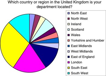
Responses were collected from June 2014 until April 2015. In all, 32 out of 34 radiographers responded to the question, ‘Which country or region in the United Kingdom is your department collected?’ The response rates can be viewed in Figure 1. Only one radiographer responded from each of the following regions: North East, North West, Ireland, Wales, Yorkshire and Humber, East Midlands and the West Midlands. The region which provided the highest radiographer response was the South East with a total of nine radiographers.
Regional distribution of respondents.

Electrons versus megavoltage (MV) photons
Most radiotherapy departments use both MV photons and electrons to deliver breast boost treatment. Therapeutic Radiographers were asked which modality was used most frequently at their centre for the delivery of breast boost treatment. The results showed electrons were used most commonly by 77·4% of the survey participants. The centres that used MV photons as the main treatment modality were based in the East of England, North West England, West Midlands and Scotland. There was an even split in Scotland. Only 5·9% of respondents did not use electrons at all in their department and they were based in East of England. Although 8·8% based in the South West and London reported that they did not use MV photons for this patient group.
A relatively large proportion amounting to 93·94% of respondents use electrons and MV photons as the main method for the delivery of radiotherapy breast boost. Currently, only 6·06% of respondents stated that brachytherapy (multichannel interstitial planar brachytherapy) was an option offered in their departments. The centres offering brachytherapy are based in the North West of England and South East England only. One Therapeutic Radiographer commented that although brachytherapy was available, it was not utilised very often.
Radiation doses
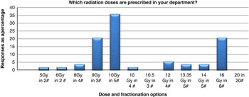
There is a broad range of radiotherapy boost doses prescribed across the United Kingdom with a total of 10 different fractionation regimes available. This diversity of doses is also evident within single departments where a choice of one of two or three different dose regimes are offered. Figure 2 summarises the distribution of boost doses routinely prescribed in different departments.
Dose and fractionation range.

Planning target volume delineation
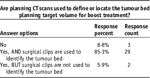
A significant percentage of respondents indicated that planning computed tomography (CT) scans are used to define the tumour bed planning target volume, where 85·3% indicated that surgical clips were used to identify the tumour bed for radiotherapy planning purposes (Table 1).
Surgical clip usage (planning target volume delineation)

Factors that influence dose prescription
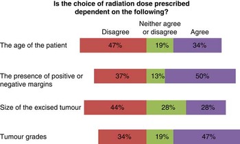
There is limited consensus among Therapeutic Radiographers regarding the factors that influence breast boost dose prescription. Figure 3 shows that radiographers do not agree or agree on which clinical factors (patient age, tumour size, tumour grade or positive or negative margins) influenced the choice of breast boost dose.
Clinical factors and prescription choice.

The study also sought to explore how a patient’s age might affect treatment. Exactly 25% of respondents (eight out of 32) agreed that younger patients were more likely to be treated with more fractions than older patients.
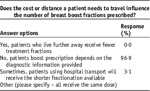
In order to explore whether financial resources affected the treatment patients were offered the following question was asked: ‘Does the cost or distance a patient needs to travel influence the number of breast boost fractions prescribed’. The responses are shown in Table 2.
Decision making based on travel costs

No respondents believed that the costs associated with long travel journeys influenced the choice of treatment given. Only one centre located in Wales which routinely prescribes one standard dose for all patients permits an exception for patients using hospital transport occasionally.
Discussion
This survey demonstrates the great variation in boost dose practice across the United Kingdom and the differences in opinions about which factors are important in the determination of prescription choice. Therapeutic Radiographers were considered to be appropriate respondents as they have broad experience on detailed aspects of the entire patients’ treatment pathway. It would have been beneficial to obtain summarised data from a national database or audit, but during the set-up of this project access was quite restricted.
A larger proportion of respondents were acquired from the regions of London and the South West, possibly reflecting the distribution of radiotherapy centres nationally or the professional networks of the researchers. The number of radiotherapy centres in certain parts of the country is related to the total population resident in that region. One response from each radiotherapy department was preferable, but as participation was voluntary and anonymous the results suggest that this was not a major limitation.
Ten different doses were identified as being routinely administered nationally. Two of the three most frequently used doses 10 Gy in 5 fractions and 16 Gy in 8 fractions and can be attributed to previous breast radiotherapy trials, which reported good outcome data. Romestaing et al.Reference Romestaing, Lehingue and Carrie 13 reported 5-year results for patients treated with a boost dose of 10 Gy which showed as 3·6% Kaplan–Meier estimate of local relapse rate with acceptable cosmetic results. The EORTC 22881–10882 used 16 Gy.Reference Bartelink, Horiot and Poortmans 5 It is unknown whether these doses have been adopted as a result of these or more current clinical trials, or whether they reflect prior local practice.
Results from this study indicate that the boost technique of choice across the United Kingdom is electrons 76% versus photons 24%. The EORTC Trial 22881/10882 recruited 2,661 patients, and clinical oncologists were permitted to select their preferred boost technique. The results from the EORTC study showed a distribution of electrons 63%, photon beams 28% and brachytherapy 9%.Reference Poortmans, Bartelink and Horiot 14 Therefore, this study’s results are broadly similar to practice across Europe despite the rare use of brachytherapy in the treatment of this patient group nationally.
A significantly high percentage of centres (85·3%) utilise surgical clips to identify the tumour bed for planning purposes. The use of surgical clips was found to improve clinical delineation of the tumour bed in other studies.Reference Benda, Yasuda, Sethi, Gabram, Hinerman and Mendenhall 11 , Reference Krawczyk and Engel 15 It was beyond the scope of this study to explore the underlying reasons a few centres did not use surgical clips for planning.
The results indicate that Therapeutic Radiographers have different opinions about which clinical factors (such as age, positive or negative excision margins, tumour grade, tumour size) underpin the choice of boost doses prescribed. The highest agreement relates to the presence of positive or negative margins, where 50% of Therapeutic Radiographers agreed that this factor influenced the choice of dose. The absence of a broad consensus may be attributed to the absence of research-based evidence in this area which would provide robust clinical guidelines for radiotherapy practitioners. This is in agreement with a study carried out in 2003, which indicated that although breast boosts are routine practice no standard technique has been established.Reference Omlin, Amichetti and Azria 9
There were limited differences in practice identified which could be attributed to clinical decisions being altered based on access to financial resources. Exceptions were made to reduce the total number of fractions for patients who needed hospital transport in the region of Wales where patients have to travel long distances to attend appointments. Significant regional agreement was indicated by the results which showed respective access to electrons 96·9%, photons 90·6%, planning CT scans and the insertion of surgical clips 84·4%.
Conclusion
The purpose of this project was to identify the range of breast boost radiotherapy doses and treatment techniques offered in the United Kingdom. Analysis showed substantial variation in boost dose practice and the absence of consensus about which clinical factors determined the treatment of patients with different clinical features (grade, histology, positive or negative margins). Initial examination of Therapeutic Radiographers responses shows that the topics of highest agreement, such as the use of surgical clips and presence or absence of positive margins, exist where such clinical practice is underpinned by medical literature.
It was quite positive to observe that financial costs did not override clinical needs, therefore radiotherapy departments do not reduce the number of breast boost fractions in order to minimise the financial costs associated with a longer treatment. However, it was observed that the absence of financial and/or technical resources restrict access of most radiotherapy centres in the United Kingdom from providing brachytherapy equipment as an option for breast boost cancer treatment.
Further review of these results in comparison with data collected by national cancer statistics institutions would confirm national practice on this subject. Overall, the results of this study identify the need for further research or audit in order to establish guidelines or decision making tools to aid practitioners on the allocation of breast boost radiotherapy doses.
Acknowledgements
The author thanks the Imperial College Healthcare NHS Trust: Camarie Welgemoed and Society of Radiographers: Rachel Harris and Therapeutic Radiographers who participated in this study.
Financial support
This research received no specific grant from any funding agency, commercial or not-for-profit sectors.
Conflicts of Interest
None.