I. INTRODUCTION
Highly complex and hierarchically structured materials, together with abilities to spontaneously repair with minimal treatment or scarring are often needed for medical applications such as bone replacement and repair.Reference Logeart-Avramoglou, Anagnostou, Bizios and Petite1 While allowing for the bone’s geometry to adapt to the new loading conditions, the replacement material should also enhance the osteogenic healing process, when the defects are too large. Autograft, allograft, and xenograft techniques are common bone grafting methods, with specific shortcomings in each case, such as limited amount of extraction from the iliac crest, additional trauma and possible morbidity,Reference Ben-Nissan2–Reference Pelker and Friedlaender4 immune rejection and inflammation possibilities,Reference Mankin, Gebhardt, Jennings, Springfield and Tomford5–Reference Simonds, Holmberg, Hurwitz, Coleman, Bottenfield, Conley, Kohlenberg, Castro, Dahan, Schable, Rayfield and Rogers7 and high residual risk of disease transmission and immune rejection.Reference Jensen, Aarboe, Pinholt, Hjorting-Hansen, Melsen and Ruyter8 Bone replacement materials based on natural coral derived hydroxyapatite (HA)/calcium carbonate compositesReference Moore, Graves and Bain9 and synthetic calcium phosphatesReference Albee and Morrison10 offer to resolve some or all of these issues and attract wider medical applications despite limitations such as shrinkage.Reference Goodridge, Dalgarno and Wood11
Biomedical polymers such as polyethylene (PE), polyamide, poly(ε-caprolactone), poly(lactic acid), poly(glycolic acid), poly(lactic-co-glycolic acid), and poly(methyl methacrylate) (PMMA) were identified to offer mechanical strengths suitable to be considered for permanent and bioresorbable implants.Reference Asti and Gioglio12,Reference Vroman and Tighzert13 HA and tricalcium phosphate (TCP) composites prepared by precipitation and sintering with a Ca to P ratio of 1.5–1.6 have been used as bone replacement materials.Reference Taş, Korkusuz, Timuçin and Akkaş14 The biphasic composites of TCP and HA with varying compositions have also been used in dental applications.Reference Gauthier, Goyenvalle, Bouler, Guicheux, Pilet, Weiss and Daculsi15 While neat biopolymer options lack bioactivity,Reference Baxter, Macdonald, Kurtz and Steinbeck16 bioceramics are weak and brittle; consequently, combinations of natural and synthetic polymer-ceramic composites became choice-materials, targeting both strength and bioactivity responses.Reference Kim, Kim, Jang, Kim, Kim, Lee and Khang17 By combining biodegradable polymers and bioceramics such as HA and β-TCP, it is possible to achieve biodegradable, osteoconductive, and bioactive materials.Reference Seunarine, Gadegaard, Tormen, Meredith, Riehle and Wilkinson18 Suitable manufacturing methods are then needed to achieve highly porous and interconnected structures with suitable surface chemistry and topography, for cell attachment, proliferation, and differentiation, together with sufficient mechanical properties.Reference Agrawal and Ray19,Reference Mooney, Langer and Bronzino20 The material and process combinations should also enable achieving controlled volume fractions and appropriate distribution of the bioceramic phases within the polymer/ceramic system to meet the mechanical and physiological requirements when used as implants.Reference Bonfield, Wang and Tanner21
The conventional processing methods often used for the fabrication of scaffolds for tissue engineering with these biopolymer composites include solvent casting/salt leaching, phase separation, foaming, gas saturation, and use of textile meshes. Drying and sol–gel methods often result in shrinkage problems as noted by Goodridge et al.Reference Goodridge, Dalgarno and Wood11 The powder material form was identified to be a better option to develop the bone replacement solutions, but the lower mechanical strength and stiffness were limitations as also the low fracture toughness of ceramics. The other issues with these methods are the difficulties associated with controlling the pore size, geometry, and the spatial distribution. Furthermore, complex re-entrant sections are difficult to achieve, and toxic organic solvents are often used, apart from long fabrication times.Reference Thomson, Shung, Yaszemski, Mikos, Lanza, Langer and Vacanti22–Reference Niklason and Langer24 Most processes also depend largely on individual skills and pose serious limitations toward achieving consistent results and complex shapes.Reference Leong, Cheah and Chua25
In recent years, rapid prototyping methods such as 3D printing, Fused Deposition Modeling (FDM), Selective Laser Sintering (SLS), and inkjet printing came up as possible solutions to achieving complex shapes.Reference Chua and Leong26 In biomedical research in particular, SLS has proved to play a greater role as controlled microporosity could be achieved through varying process parameters; laser power, scan speed, and part bed temperature considering composites of HA and different other biopolymers. Naing et al. evaluated the surface morphologies of sintered HA/PEEK composites and also fabricated scaffolds for evaluating the porosity levels and the effects of balling,Reference Naing, Chua and Leong27 while high density PE (HDPE), with HA as the filler, was evaluated for laser sintering by Hao et al. and Savalani et al.Reference Hao, Savalani, Zhang, Tanner and Harris28,Reference Savalani, Hao and Harris29 In terms of developing strong bonds with the host bone, tricalcium phosphate Ca3(PO4)2, and in particular, the β form has been the choice as a bioceramic reinforcement for both organic and inorganic composites.Reference Jarcho, Kay and Gumaer30 Kalita et al. developed polypropylene (PP) and TCP composites as filaments for extrusion printing based on FDM.Reference Kalita, Bose, Hosick and Bandyopadhyay31 Scaffolds with different geometries were fabricated and the average pore size was noted to be in the range 150–200 µm while the compressive strength was comparable to that of a cancellous bone. Duan et al.Reference Duan, Wang, Zhou, Cheung, Li and Lu32 processed Ca–P and poly hydroxybutyrate-co-hydroxyvalerate nanocomposites for bone tissue engineering via SLS and achieved controlled material microstructures, totally interconnected porous structures and high porosities.
The progress is evident combining layered manufacturing and biopolymer composite processing; however, biological responses and the biocompatibility of the materials after processing would be the key factor for end use. Correspondingly, the PP and tricalcium phosphate FDM samples evaluated by Kalita et al. proved the matrices to be nontoxic and promoting cell attachment based on in vitro characterization using immortalised human preosteoblastic cells (OPCI).Reference Kalita, Bose, Hosick and Bandyopadhyay31 Similar investigations on the laser sintered polymer nanocomposites showed improved cell proliferation and alkaline phosphate activity in the presence of Ca–PReference Duan, Wang, Zhou, Cheung, Li and Lu32 While these preliminary investigations indicate no obvious losses due to processing by additive methods, the choice of the material components could vary, as also the process and the parameters.
Poly(methyl methacrylate) [(PMMA)–(C5O2H8)] has been a popular choice for biomedical applications offering potential compatibilities with human tissue. The interconnected porosity characteristics allow PMMA to be used as a bone cement to fix implants and remodel lost bones, though the lack of bioactivity is the main drawback, limiting its use in bone repair applications.Reference Lewis33,Reference Puska, Kokkari, Närhi and Vallittu34 As TCP is a bioactive element, the polymer composite based on PMMA and TCP is an option to achieve both biocompatibility and activity attributes. Descamps et al.Reference Descamps, Richart, Hardouin, Hornez and Leriche35 developed PMMA and β-TCP composites by means of thermal forming techniques and evaluated the resulting macroporous structures. The scaffold properties matched with the native bone tissue and also allowed the growth of the neo-tissue as the scaffold degrades with time. Velu and SingamneniReference Velu and Singamneni36 evaluated the possible roles of laser process parameters on the sintered structures, surface morphologies, and mechanical properties of thin film samples produced by SLS PMMA/β-TCP composites.
Evidently, the PMMA/β-TCP polymer composites are potential candidates for medical applications such as bone repair and replacement. SLS is also proved to be applicable to processing these polymer composites targeting application-specific micro- and macrostructures and overall forms. However, the polymer degradation possible due to the laser interaction and the variations in the biological responses are not ascertained sufficiently yet. This is the focus of the experimental research presented in this paper. The optimum parameters for processing selected grades of PMMA and β-TCP powders mixed in different compositions are established first beginning with a thermal characterization. Possible polymer degradation aspects due to photonic interactions are evaluated by means of the FTIR analysis. The in vitro osteoblast functions of the sintered samples are studied by detecting attachment, spreading, proliferation, and osteogenic differentiation of the MG-63 cells.
II. MATERIALS AND METHODS
Both PMMA (C5O2H8) and β-TCP (Ca3(PO4)2) powders are sourced from Aldrich chemicals. The average PMMA particle size is around 75 µm, the melting and glass transition temperatures are 160 and 106 °C, and the theoretical density is 1180 kg/m3. The β-TCP (Ca3(PO4)2) powder particles are at an average diameter of around 5 µm and the molecular weight is 310.18 g/mol. Different blend compositions were developed combining 5, 10, 15, and 20% by weight of β-TCP with PMMA, physically mixing the two together. The β-TCP particles being small, usually agglomerateReference Lohfeld, Cahill, Barron, McHugh, Dürselen, Kreja, Bausewein and Ignatius37 and entangle around the relatively large PMMA particles and disperse easily within the powder matrix.
SLS was applied to PMMA powders earlierReference Velu and Singamneni38 but the process parameter combinations needed further adjustments based on the thermodynamic responses of the current powders. Differential scanning calorimetry (DSC) is performed using the Simultaneous Thermal Analyzer-STA 449 F5 Jupiter DSC system (NETZSCH Australia Pty Ltd., Kings Park, Australia). The DSC data generated are processed using the NETZSCH Proteus software (NETZSCH Australia Pty Ltd., Kings Park, Australia) and the thermodynamic and kinetic parameters; heat capacity (C p) and glass transition temperature (T g) are derived. The activation energy is obtained evaluating the slope of the Ozawa plot. To generate the Ozawa kinetics plot, the nonisothermal DSC values are measured at different heating rates with the same mass of samples. The heating rate varies from 2 to 10 °C/min. Various heating rates and constant sample masses generate different peak temperatures. The Ozawa plot, i.e., the heating rate β in logarithmic scale (ln(β)) against the reciprocal of the peak temperature 1/T p curve. From the slope (−E a/R) of the line, the activation energy E a can be obtained using the following equation:
 $${E_{\rm{a}}} = {{dR} \over {1.052}}\quad ,$$
where d represents the trend line slope. The glass transition temperature value from the DSC curve is used in energy density equation, together with a selected laser scan velocity to establish the energy density required to achieve sufficient melting and interparticle coalescence.
A make-shift SLS system is designed and built in-house, to serve as an SLS test bed for the experimental investigations planned and conducted in the current research. The design is centered around an off-the shelf 60 W CO2 laser that can provide an average power output ranging from 0.6 to 60 W and scan speeds up to 5000 mm/s. A suitable powder bed system is also developed with manual powder dispersion and overall, this system allowed economical use of the expensive materials for experimental investigation with a better variation and control of the laser sintering parameters. The CO2 laser has a wave length of 10.6 µm, suitability for sintering PMMA powders in terms of the absorptivity, a spot size of 540 µm, and a focal length of 370 mm. A Pulse Width Modulation (PWM) system allows controlling the laser at frequencies 5, 10, and 20 kHz. The current experimental trials are conducted with the PWM frequency set at 5 kHz, as this is the common standard and works well in most laser applications. A computer controlled optical deflection system together with a software control allowed to program the raster scans while sintering the powder bed surface.Reference Velu and Singamneni36 Single layer samples of 10 × 70 mm are printed using pure PMMA and the four combinations of PMMA and β-TCP composites using process parameter settings as identified best from preliminary trials. Each trial is repeated for three times, making it a total of 12 printed samples. Tests are conducted based on these samples for different responses as detailed in the following sections.
A. SEM for microstructural characterisation
The Scanning Electron Microscopy (SEM) image is captured for different compositions of PMMA/β-TCP composites to analyze the porosity level as mentioned below. A Hitachi SU-70 (HITACHI Australia Pty Ltd., Macquarie Park, Australia) field emission SEM is used at a voltage of 5–15 kV, and with magnifications up to 800,000 times. The SEM images are obtained with a very small electron probe (or electron spot), scanned over the surface of the specimens, and by mapping the electron signals detected from each specimen 73 pixel onto the corresponding pixel of the cathode ray tube, or charge coupled device, i.e., the screen. All samples are coated with platinum to provide a conductive surface for imaging.
B. Polymer degradation analysis based on FTIR
The Thermo-Scientific (Thermo Fisher, Waltham, Massachusetts) FTIR spectrometer at the School of Applied Sciences of the Auckland University of Technology is used for establishing the consequences of laser sintering on the basic chemistry of the polymers under investigation. When the sample segment is in place, the IR beam is reflected off the surface of the sample, while specific frequencies of energy, characteristics of the sample are absorbed. The beam energy finally falling on the detector allows developing the special interferogram signal. The digitised signal is then processed on a computer for Fourier transformation using OMNIC software and the infrared spectrum is developed to interpret the molecular composition based on standard commercial tables.
C. In vitro studies
All biological tests are conducted at the Biotechnology Lab of the PSG College of Technology in India, where the samples are trimmed down to 10 × 10 mm size for the in vitro studies. Procedures followed for specific tests are as follows:
1. Cell culture
The human osteosarcoma cell line, MG-63, obtained from the National Center for Cell Science NCCS, Pune, India is cultured in Dulbecco’s Modified Eagle’s Medium (DMEM-AL007S, HiMedia, Mumbai, India), a modified basal medium for cellular proliferation. The DMEM is supplemented with 200 mM L-glutamine, 10% fetal bovine serum, and 1× of antibiotic antimycotic solution (A002 from HiMedia, India). The cells are incubated with 5% CO2 at 37 °C and trypsinized with trypsin–EDTA after attaining 80–90% confluency.
2. Cell proliferation studies
The 3-(4,5-dimethylthiazol-2-yl)-2,5-diphenyltetrazolium bromide (MTT) colorimetric assay measuring the reduction of yellow 3-(4,5-dimethythiazol-2-yl)-2,5-diphenyl tetrazolium bromide (MTT) by mitochondrial succinate dehydrogenase is used to assess the cell viability and proliferation after interacting with the laser sintered materials. The MTT enters the cells and passes into the mitochondria where it gets reduced to an insoluble, colored (dark purple) formazan product. The insoluble product is then solubilized with an organic solvent (dimethylsulphoxide), and the released solubilized purple colored formazan is measured spectrophotometrically at 570 nm. Since the reduction of MTT can only occur in metabolically active cells, the level of activity is a measure of the viability of the cells. The intensity of the purple color is proportional to the number of live cells.
Each well containing a UV sterilized scaffold is seeded with 1000 µL of a medium containing approximately 1 × 10⁴ cells in a 48-well plate and incubated for 1, 3, and 5 days. MG 63 cells cultured on the 48-well plate without any scaffold are used as the control. After the incubation, 5 mg/mL of freshly prepared MTT (HiMedia, India) solution is added to each well and incubated for 3.5 h at 37 °C. After the incubation time, the solution is discarded, and the samples are air dried, followed by the addition of 200 µL of dimethyl sulphoxide (SD Fine Chemicals, Coimbatore, India) to dissolve the insoluble formazan crystals formed. The plate is kept in shaking condition for 10–15 min in dark. The absorbance is then read at 570 nm in a multiwell plate reader (Thermo Multiscan Labsystems, Illinois, Model 352).Reference Jang, Nguyen, Sarkar and Lee41 The spectrophotometric values of the MTT assay were used in Microsoft Excel sheets for plotting the bar graphs. Statistical analyses were performed using one-way ANOVA, and paired t-test in the MINITAB 15 Software, trial version. 95% confidence interval and P < 0.005 was considered as statistically significant. To denote the statistical significance, symbols such as *, **, #, ##, and & were inserted manually in the Microsoft Excel sheet.
3. Alkaline phosphatase assay
The alkaline phosphatase assay is carried out using the p-nitrophenol pyrophosphate (N7653 Sigma Aldrich, Bengaluru, India) as the substrate. According to the protocol specified by the manufacturer, ALP catalyzes the hydrolysis of the colorless ρ-nitrophenyl phosphate to a colored ρ-nitrophenol. This product has a strong absorbance at 407 nm, which is a measure of the level of the enzyme activity. Each well containing a UV sterilized scaffold is seeded with 1000 µL of medium containing approximately 1 × 10⁴ cells in a 48-well plate, and incubated for 4, 7, and 10 days. MG 63 cells cultured on the 48-well plate without any scaffold are used as the control. After incubation, 200 µL of p-nitophenol pyrophosphate solution is added to each well. The plates are then incubated in the dark for 30 min at room temperature. After the incubation, 50 µL of 3 M NaOH solution is added per 200 µL of the reaction mixture to stop the reaction. The absorbance is then read at 407 nm in a multiwell plate reader (Thermo Multiscan Lab systems, Model 352). The absorbance values were normalized to the number of viable cellsReference Tsai, Liou, Lin, Kuo, Hung, Weng and Hsu44 and the graph was plotted using MS Excel. The data were analyzed for statistical significance using one-way ANOVA and paired t-test using MINITAB 15 software. P < 0.005 is considered to be statistically significant.
4. Alizarin red S staining
Alizarin red S, an anthraquinone derivative, is used to detect calcium depositions on the sample. The reaction is not strictly specific for calcium since magnesium, manganese, barium, strontium, and iron may interfere, but these elements usually do not occur in sufficient concentrations to interfere with the staining. Calcium forms an alizarin red S-calcium complex in a chelation process. The test is carried out according to the protocol specified by Gregory et al.Reference Gregory, Gunn, Peister and Prockop42 Alizarin red S powder (A5533 Sigma Aldrich, Bengaluru, India) is used to prepare the fresh solution (pH 4.1–4.2) each time as per the protocol given by the manufacturer.Reference Ma, Tang, Tan, Lin, Wang, Wei, Zhao, Zhao and Preparation43
Each well containing a UV sterilized scaffold is seeded with 1000 µL of medium containing approximately 1 × 10⁴ cells in a 48-well plate and incubated for 25 and 28 days. The media in the 48-well plate are removed, and the wells are rinsed gently with 1 mL PBS pH 7.4. The cells are then fixed with 1.0 mL of 10% formalin in each well for 15 min at room temperature. Formalin solution is then removed, and the wells are washed twice gently with double distilled water. To each well, 500 µL of 40 mM alizarin red S solution is added and left at room temperature for 20 min with gentle shaking. After incubation, the dye solution is removed; the wells are washed 4 times with 1.0 mL distilled water, replacing the water at each 5-min’ interval. The specimens are then air dried, and images are taken as mineralized areas appear red in color. The images of the stained samples were captured using a SONY CYBER-SHOT (Model no. DSC-WX50; Sony, San Mateo, California) 16.2 MP camera.
D. Quantitative real-time PCR
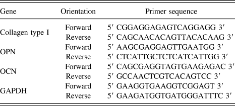
Each well containing a UV sterilized scaffold was seeded with 6000 µL of medium containing approximately 1 × 10⁴ cells in a 6-well plate and incubated for 7 days at 37 °C with 5% CO2. MG 63 cells cultured without scaffold were used as the control to compare the gene expression profiling in those cells cultured with the scaffold. After 7 days, RNA was isolated using TRI reagent (Cat. No. 93289, Sigma-Aldrich, Bengaluru, India), as per the manufacturer’s protocol. In this study, collagen type I, osteopontin (OPN), osteocalcin (OCN), and GAPDH are considered and analyzed in real-time PCR (CFX 96, Bio-Rad, Hercules, California). To calculate the relative gene expression, a constitutive gene (GAPDH) is used as the internal control. The sequences reported by Tsai et al. are adopted for the oligonucleotide primer synthesis.Reference Tsai, Liou, Lin, Kuo, Hung, Weng and Hsu44 The primer sequences are presented in Table I.
Genes and their primer sequences used for RT-PCR.

The reaction volume (25 µL) included 12.5 µL SYBR Premix Ex Taq II (TliRnaseH Plus) (2×), 2 µL diluted cDNA (<100 ng), 8.5 µL sterile distilled water, and 1 µL each of forward and reverse primers (10 µM). Reactions are performed in triplicate, and the values are analyzed for calculating the fold change. After initial denaturation at 95 °C for 30 s, the target genes are amplified with 40 cycles of denaturation at 95 °C for 5 s and annealed at 62.5 °C for 60 s. Real-time PCR reactions are carried out in a CFX96 real-time PCR system (Bio-Rad, Hercules, California). The levels of RNA expression are determined according to the relative gene expression method. The expression levels of the target genes are calculated by normalizing the mRNA level for a particular gene against that of GAPDH mRNA, which are used as a constitutive control according to the procedure described by Schmittgen and Livak.Reference Schmittgen and Livak45 While presenting the relative gene expression profile, the gene of interest has to be normalized with the internal control. Relative gene expression is expressed as (−∆C T), where ∆C T = (C T gene of interest − C T internal control).Reference Schmittgen and Livak45 GAPDH is used as the internal control for this study. The normalized data were analyzed for statistical significance one-way ANOVA and paired t-test in MINITAB 15 software. P < 0.005 is considered to be statistically significant.
III. RESULTS AND DISCUSSION
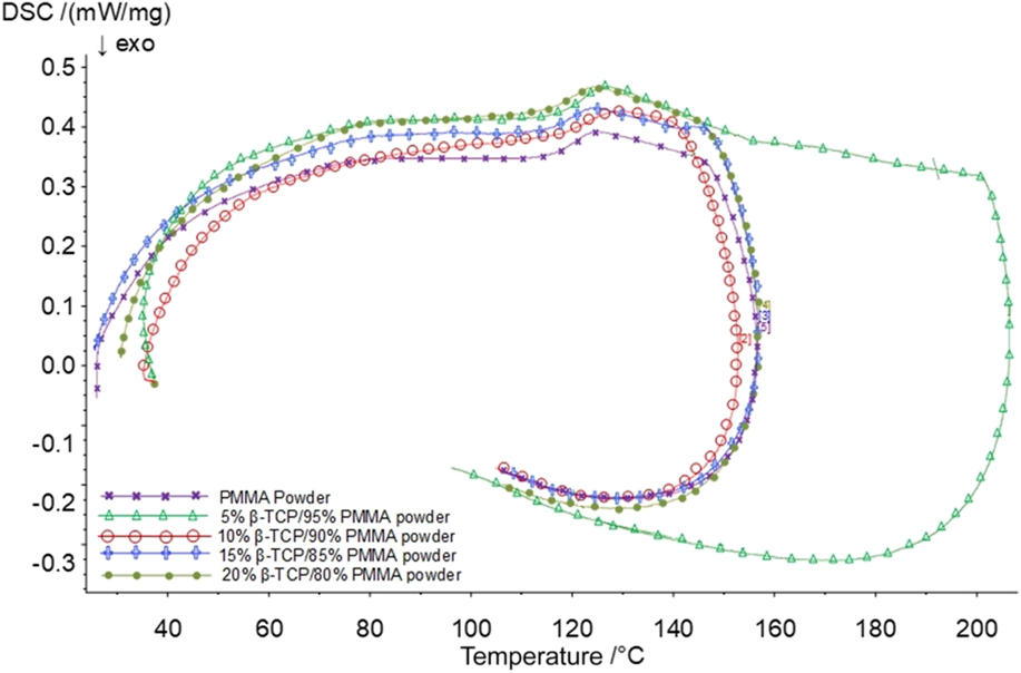
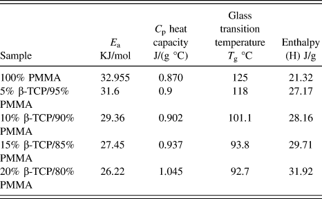
Critical glass transition and melting temperatures are established based on the DSC curves presented in Fig. 1. Evidently, the DSC curves did not show any sharp variations in the differential energy absorbed, ascertaining the amorphous nature of the polymer.Reference Rudin and Choi39 The kinetic and thermodynamic parametersReference Rantuch, Kačíková and Nagypál40 established for the polymer composites of varying compositions are listed in Table II. The glass transition temperature values are used together with a selected laser power to establish the minimum energy density required for sufficient melting and interparticle coalescence. Based on the 5% β-TCP case, the energy density essential is identified as 0.05 J/mm2, which is used as the a priori data to establish the optimum process conditions as follows.
DSC curves.

Kinematic parameters of PMMA and PMMA/β-TCP composites derived from the DSC curves.

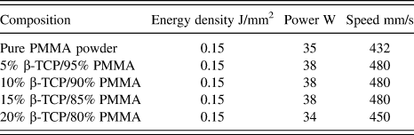
Starting with the energy density established with the DSC results, a series of experimental trials involving sintering single layer samples are carried out for each composition of the polymer composite. Based on the morphologies of the SEM images and the porosity levels, the optimum energy density level for all compositions is established to be at 0.15 J/mm2. A detailed description of this process was presented earlier with a different grade of the same material combinations by Velu and Singamneni.Reference Velu and Singamneni36 All the samples for the current investigations other than the 20% β-TCP case are produced at the optimum energy density of 0.15 J/mm2. Slightly lower energy densities became necessary to be used with the 20% β-TCP case, due to the decomposition and charring of the specimens resulting possibly from the higher energy absorption in the presence of excessive β-TCP. Considering the confounding effects of laser power and scan velocities, each combination of the polymer composites is sintered at three different power settings with corresponding changes in the scan velocities while maintaining the energy density at the optimum level.
Based on SEM photomicrographs of sintered samples at different laser parameter combinations, the porosity levels are identified to be higher at the two extremes of the power settings in the first three cases. Better coalescence and lesser porosities are noted with the middle level of laser power and speed settings at 38 W and 480 mm/s. However, the 20% β-TCP case indicated that the best sintered sample can be obtained with the lowest power and speed setting at 34 W and 450 mm/s. Based on these results, the laser power and scan speed settings as listed in Table III are finally used for the four compositions of the composite material to build specimens for further analyses. While ensuring sufficient coalescence and mechanical strengths, these settings are also expected to provide the necessary porosity and pore sizes for the cells to adhere and proliferate during biological testing.
Material and process parameter combinations used for the printed test pieces.

A. Porosity
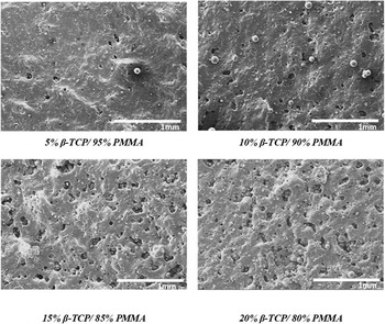
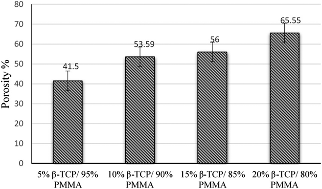
The actual mechanisms and mechanics of interparticle coalescence and consolidation of layers were discussed in greater detail in an earlier publication.Reference Velu and Singamneni38 While the current focus is on the after effects of laser sintering, some attention is paid first to the porosity levels and their variation with process conditions considering the significance of achieving controlled porosities in the target applications. SEM photomicrographs of single layer specimens of different compositions printed as per the conditions are set in Table III and presented in Fig. 2 are used as the basis. The formation of a continuous sintered layer is evident from these photomicrographs, while the porosity levels varied with varying compositions. The theoretical density is calculated based on the weight percentage and the density of the composite powder while the density of the sintered specimen is established by weighing the sample. Porosity levels of different samples are then estimated using Eq. (1) and plotted in Fig. 3. Evidently, the higher the β-TCP, the higher is the porosity.
 $${\rm{Porosity}} = {{\rho \left( {{\rm{theoretical}}} \right) - \rho \left( {{\rm{sintered}}} \right)} \over {\rho \left( {{\rm{sintered}}} \right)}} \times 100\quad .$$
SEM photomicrographs of single layer sintered specimens (accelerating voltage 15 kV).

Percent porosity with varying PMMA/β-TCP compositions.

The pore size and the extent of porosity have significant roles to play in controlling the bioactivity of the sintered samples, possibly leading to increased cell proliferation and bone ingrowth in specific applications.Reference Yokozeki, Hayashi, Nakagawa, Kurosawa, Shibuya and Ioku46 In general, the pore sizes may be observed to vary from 5 to 100 µm which are similar to the pores in natural bone.Reference Hao, Lawrence and Chian47 Presumably, this porosity allows cells to attach and initiate growth forming a local micro-environment. This environment in turn presents potential biochemical, cellular, and physical stimuli orchestrating cellular processes such as proliferation, differentiation, migration, and apoptosis.Reference Tsang and Bhatia48
B. Polymer degradation
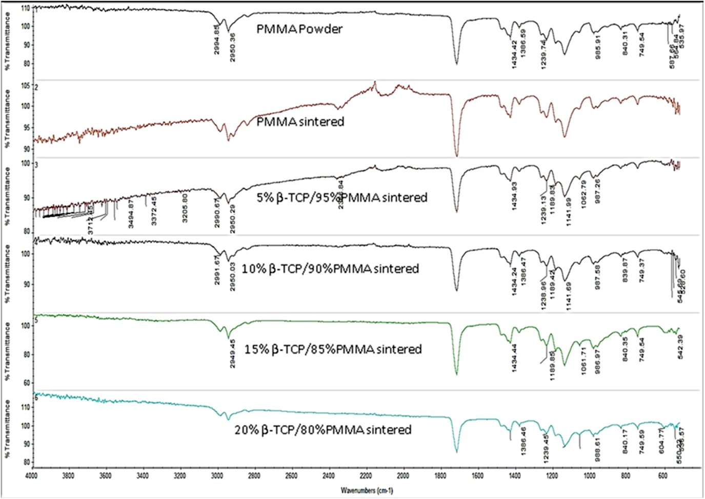
Comparatively, the infrared spectrum obtained based on the pure PMMA powder as shown in Fig. 4 is exactly similar to the patterns reported in the literature.Reference Balamurugan, Kannan, Selvaraj and Rajeswari58 The C–O bonds dominate the majority of the vibrational peaks and the presence of C=O ester carbonyl group stretching vibration appeared at the 1723 cm−1 peak. The broad peak ranging from 3000 to 2900 cm−1 is due to the presence of C–H stretch vibration and the 1385–1450 cm−1 band can be assigned to the CH3 and CH2 deformation vibration.Reference Duan, Zhang, Li, Yang, Lu and Wang49 The spectrum thus established for the pure PMMA powder sample is then used to compare the spectra obtained with the sintered samples of other compositions presented in Fig. 4.
FTIR results based on neat PMMA powder and sintered samples of PMMA and β-TCP varying compositions.

It may be noted that the spectra in all the cases have similar patterns and peaks with the raw PMMA powder. This indicates that the polymer chemistry has not changed due to the interaction with the laser-induced energy. Furthermore, the presence of β-TCP particles also did not have any significant effects on the spectral results other than very minor variations. Based on these observations, it is inferred that the laser sintering at 0.15 J/mm2 energy density and the selected combinations of laser power and scan speeds do not alter the basic chemistry of the polymer.
C. In vitro responses
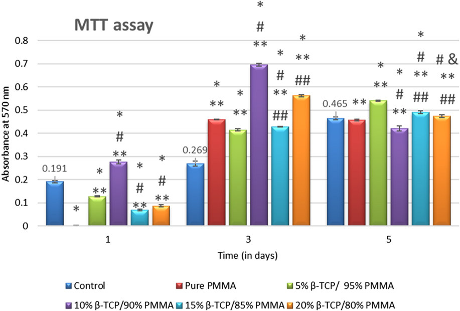
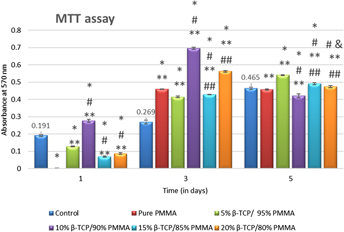
Cell proliferation results using sintered specimens of pure PMMA, 5% β-TCP/95% PMMA, 10% β-TCP/90% PMMA, 15% β-TCP/85% PMMA, and 20% β-TCP/80% PMMA composites, quantified based on the MTT assay for 1, 3, and 5 days are presented in Fig. 5. All sintered specimens showed cytocompatibility and facilitated the growth and proliferation of MG 63 cells throughout the 10-day culture period. One-way ANOVA and paired t-test analyses indicate the differences to be statistically significant (P < 0.005) with the 5% β-TCP/95% PMMA and 15% β-TCP/85% PMMA samples. It may be observed that on the first day, there is apparently no cell growth in the pure PMMA sample, while significant cell proliferation is noted with both 5 and 10% β-TCP samples from Fig. 5. Evidently, the sintered PMMA surfaces promote cell viability and proliferation almost at the same level as the control by the fifth day. However, it appears that the sintered surface needs an initial incubation period of about two days to be able to promote cell attachment and proliferation, probably due to the nature of the surface direct from sintering. Beyond this, the cells multiply quickly due to the affinity toward PMMA and a sudden growth is observed as evidenced in Fig. 5 after 3 days. The sudden increased activity apparently shows the cell absorbance of the sintered samples to be higher than that of the control, which is eventually balanced by the fifth day.
Results of the MTT assay with varying compositions. “*”—significant difference (P < 0.005) compared to the control; “**”—significant difference (P < 0.005) compared to PMMA; “#”—significant difference (P < 0.005) compared to 5% β-TCP/95% PMMA; “##”—significant difference (P < 0.005) compared to 10% β-TCP/90% PMMA; “&”—significant difference (P < 0.005) compared to 15% β-TCP/85% PMMA. The 20% β-TCP/80% PMMA has no statistical significance compared to the remaining samples.

The fifth day saw stabilization in most cases, while the 5% β-TCP case still shows an upward trend. Based on the statistical significance (P < 0.005), it may be noted that both 5 and 15% β-TCP samples scored higher than the normal expected levels in terms of cell proliferation. Besides β-TCP being osteoconductive, increasing its volume percentage in the composite did not show a linear increase in cellular proliferation and differentiation. These results are in similar lines to the observations made by Di Silvio et al.Reference Di Silvio, Dalby and Bonfield50 Measuring the cell proliferation with varying compositions of HA within the HDPE matrix, they noted that the cellular proliferation and differentiation did not linearly increase with the increase in the volume percentage of bioactive HA in the HA/HDPE composite. All the graphs under cell line testing were constructed using Microsoft Excel, and the symbols denoting significance were inserted manually.
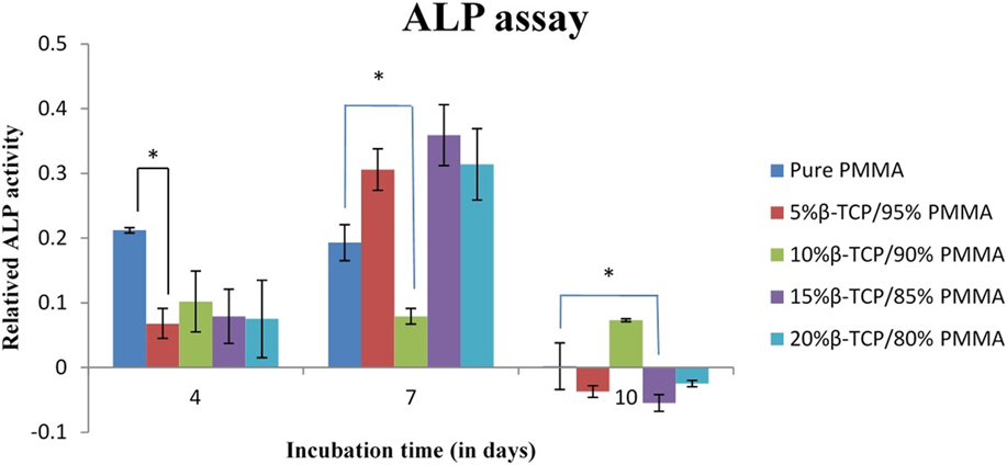
The ALP assay results of MG 63 cells cultured on PMMA/β-TCP specimens for 4, 7, and 10 days are presented in Fig. 6. The ALP enzyme is present in higher amounts in the actively bone forming cells, where it can catalyze the hydrolysis of the phosphate esters at alkaline pH and play an important role in the bone matrix mineralization process. Again, based on ANOVA and t-test, the difference observed for 5% β-TCP/95% PMMA and 15% β-TCP/85% PMMA are noted to be statistically significant (P < 0.05). The 5% β-TCP/95% PMMA, 15% β-TCP/85% PMMA, and 20% β-TCP/80% PMMA demonstrated increased ALP activity from day 4 to day 7. There is a relative decrease observed in the ALP activity on day 10. This could be due to the formation of the extracellular matrix (ECM) and the consequent masking of the cells present. Muhammad et al. observed the formation of Extra Cellular Matrix (ECM) after five days of culturing in the healthiest cases. On culturing the bone marrow stromal cells for 14 days, the cells found in the porous holes are not visible on the surface.Reference Muhammad, Abas, Kim, Pingguan-Murphy, Zain and Akram51 Since ALP is an enzymatic activity, only the cells interacting with the substrate will contribute to the color formation, indicating the initiation of the mineralization process.Reference Kim, Park, Jeon, Choi and Kim52 Samples with 15% β-TCP/85% PMMA and 20% β-TCP/80% PMMA did not show differences in statistical significance levels (P < 0.005) compared to the remaining samples. On day 4, the β-TCP has supported cellular attachment and proliferation (Fig. 5) but could less likely support cellular differentiation, whilst the differentiation is observed on day 7. Surprising results were observed on day 10. Probable masking of cells by the formation of ECM is a hypothesis. This activity of the 10% β-TCP sample has to be further elucidated from the biological perspective.
Results of the ALP assay. Bars represent standard deviation. Statistical significance (P < 0.005) is represented using *.

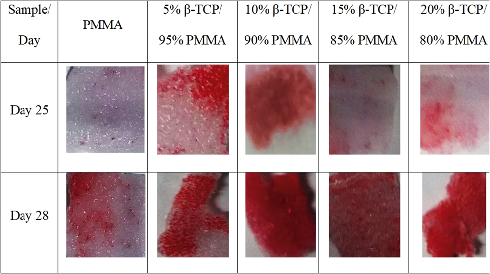
The alizarin red S staining test results with samples of varying compositions are presented in Fig. 7. The staining in samples made of the composites is evidently much greater than that of pure PMMA. Also, the higher the β-TCP content, the better is the staining. These results clearly indicate that the cellular attachment, proliferation, and calcium mineralization on the composite samples are significantly higher compared to those on pure PMMA samples after 25 and 28 days. This means better mineralization with increasing β-TCP in laser sintered PMMA-based composites. Since the alizarin red S stains the mineralized areas on the scaffold, no control was used.
Photographic images of alizarin red S staining results with varying composition.

D. Quantitative real-time PCR
Collagen type I, OPN, OCN, and reference gene GAPDH are chosen for the qPCR studies. The bone’s ECM comprises two types of proteins: collagen type I (90% of the bone matrix) and noncollagenous proteins (OPN and OCN). Collagen type I is the structural portion of the bone. This is also found in other fibrous tissues such as tendons, ligaments, cartilages, intervertebral disc, and skin. OPN is a highly sulphated, phosphorylated, and glycosylated protein, that is expressed in the preliminary stages of matrix mineralization. This allows for open and flexible structures that facilitate strong binding to calcium ions. In addition, this also enables anchoring of osteoclasts to the mineral matrix of bones during bone remodeling. OCN, expressed at the later stage of osteoblastic differentiation binds to calcium and plays a significant role in matrix mineralization. In the absence of OCN, OPN would contribute to the bone formation.
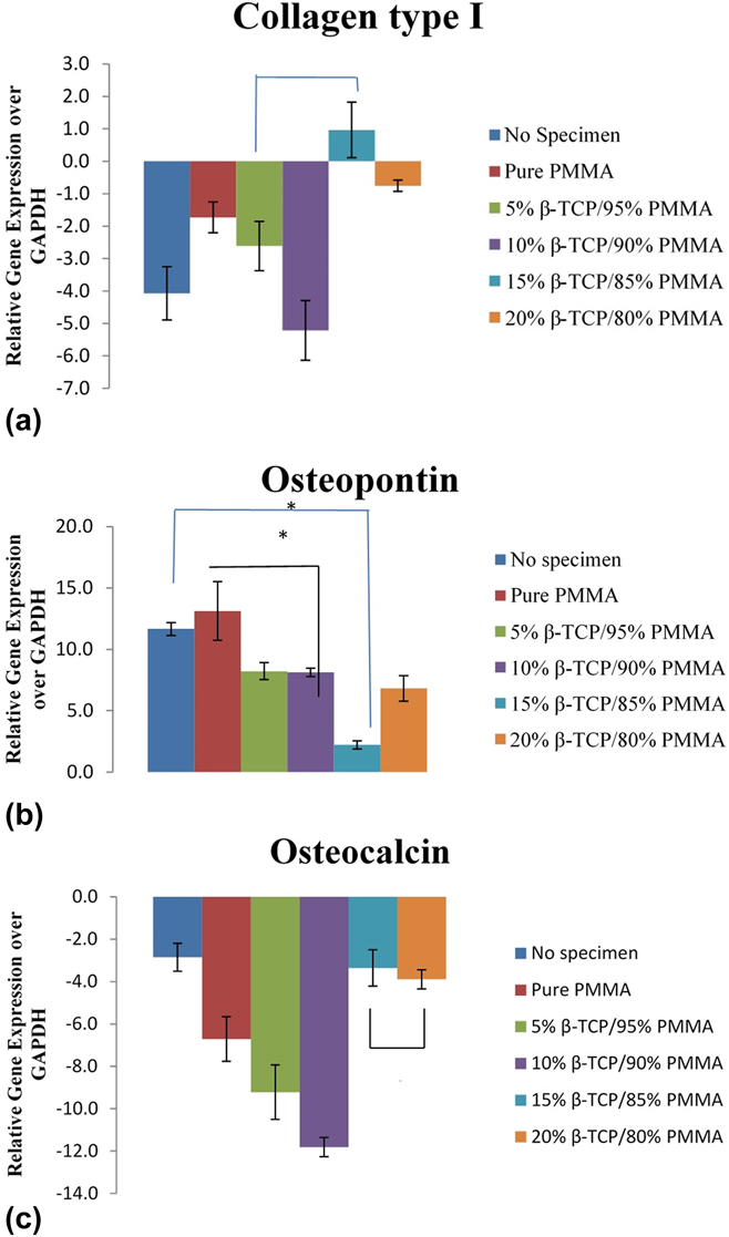
The real time PCR results are presented in Fig. 8, where the statistical significance (P < 0.005) is represented by *. From Fig. 8(a), it can be observed that 10% β-TCP/90% PMMA expressed lesser collagen type I, while 15% β-TCP/85% PMMA expressed the highest collagen type I expression. In Fig. 8(b), except pure PMMA, all other β-TCP/PMMA composites displayed lower expression of OPN compared to the “No Specimen” control. Other than the 15% β-TCP/85% PMMA sample, remaining samples show lower expression of collagen type I compared to OPN. A similar observation was reported on osteoblast-like cell line, MC3T3-E1 on day 7.Reference Sun, Gan, Fan, Zhuang and Hu53 Contradictorily, the 15% β-TCP/85% PMMA sample showed higher expression of collagen type I, and lower expression of OPN. This pattern was also observed and reported by Sulaiman et al. when testing HA/TCP scaffold.Reference Sulaiman, Keong, Cheng, Saim and Idrus54
Representative graphical images of relative gene expression of (a) collagen type I, (b) OPN, and (c) OCN with GAPDH as the reference in the MG 63 cell line. Statistical significance (P < 0.005) is represented using *.

In Fig. 8(c), it is observed that all the sample types (including the “No specimen” control) have lower expression of OCN compared to the reference gene, GAPDH. However, the OCN expressed by 15% β-TCP/85% PMMA and 20% β-TCP/80% PMMA are on par with the “No specimen” control. This trend of OCN < Collagen type I < OPN relative gene expression was observed by MC3T3-E1 cells when tested on a polymeric bone scaffold on day 7.Reference Gumusderelioglu, Tuncay, Kaynak, Demirtas, Aydin and Hakki55 Yamano et al. also reported a down regulation of OCN when testing on a collagen membrane, with GAPDH as the reference gene.Reference Yamano, Lin, Dai, Fabella and Moursi56 More interestingly, Xi et al. reported that collagen type I, OPN, and OCN expressions remained constant from day 4 through day 14, when testing β-TCP scaffolds.Reference Xu, Lv, Zhang, Zhang, Jiang and Zhang57
The in vitro test results are based on the levels of cellular interaction with PMMA/β-TCP components in the sintered material matrices further to surface exposure. Evidently, increasing concentration of β-TCP in the composite did not proportionately increase the bioactivity of the laser sintered materials. However, it may be observed based on photomicrographs such as the ones in Fig. 2 that the surface exposure of β-TCP has not been uniform and proportional to the level of the bioceramic component. This is due to the limitations of the manual mixing of the powder composites. Presumably, this nonuniform presence of the β-TCP phase on the surfaces of the samples caused the loss of bio-activity in some specimens. Furthermore, the 15% β-TCP/85% PMMA polymer composite sample of Fig. 2 has relatively optimum porosity level, better surface exposure of β-TCP, and notable surface roughness patterns compared to the other cases. These factors could have significantly contributed for the relatively higher bioactivity of 15% β-TCP/85% PMMA observed from the in vitro results.
IV. CONCLUSIONS
The post-sintering attributes of PMMA and PMMA plus β-TCP polymer composites are evaluated. The FTIR results ascertained the polymer chemistry to be intact even after the interaction with the laser energy. The in vitro results established no loss of the biological responses in the sintered specimens, though the trends are slightly different to the normal expectations due to the issues with the filler particle dispersion and the porosity arising with higher ceramic contents.
Though the chemical compositions are intact in the sintered states, the process parameters greatly influenced critical responses such as porosity, surface exposure of β-TCP, and surface roughness. Besides the chemical nature, surface characteristics of the sintered specimens greatly affected the cellular responses. Both 5% β-TCP/95% PMMA and 15% β-TCP/85% PMMA composites performed consistently well in the MTT and ALP assays, alizarin red S staining, and real-time PCR. Overall, the 15% β-TCP/85% PMMA composite scored relatively better in terms of bioactivity.