As a highly rare congenital defect, cor triatriatum sinister represents only 0.1% of congenital cardiac anomalies.Reference Van Praagh and Corsini 1 , Reference Talner 2 The clinical presentation can mimic those of mitral stenosis, supravalvular mitral ring, or pulmonary venous stenosis. However, features of systematic embolisation, heart failure, atrial fibrillation, cyanosis, cardiac asthma, syncope, and sudden cardiac arrest have also been reported in the literature.Reference Gheissari, Malm, Bowman and Bierman 3 In the following, we present a rare case of a cor triatriatum sinister with almost complete obstructed flow between pulmonary veins, mitral valve, and left ventricle through only 3 mm perforation on the fibromuscular membrane as well as left upper and lower anomalous pulmonary venous return to the innominate vein through the vertical vein.
Case report
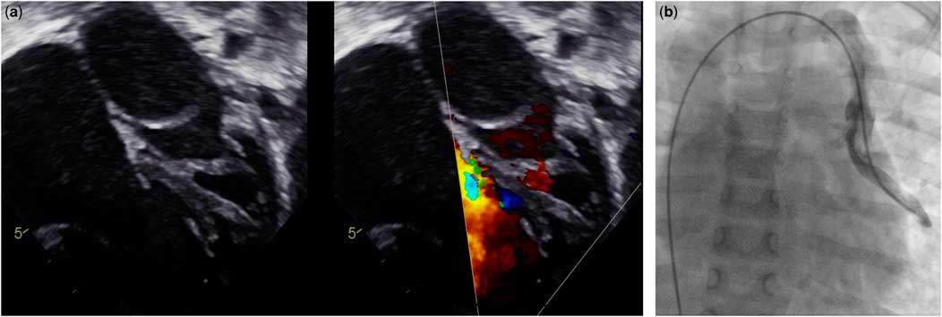
A 3-year-old girl with a heart murmur was admitted to our hospital with recent onset of dyspnoea, recurrent respiratory tract infections, appetite loss, and fatigue since 1 week. Physical examination revealed tachypnoea, tachycardia with a heart rate of 140/minute, and a diastolic blowing murmur with a loud P2. Her body weight was less than that of normal child of her age. Electrocardiography indicated sinus tachycardia, right-axis deviation, and right ventricular hypertrophy. A chest X-ray showed a moderate-to-large right pleural effusion and cardiomegaly. Following transthoracic echocardiography and cardiac catheterisation revealed the presence of an obstructing membrane separating the left atrium into proximal and distal chambers with small patent foramen ovale (type A1 of Lam classification),Reference Talner 4 left upper and lower anomalous pulmonary venous return to the innominate vein through the vertical vein (16 mm), right ventricular enlargement and hypertrophy, as well as reduced left ventricular pumping function (Fig 1a and b).
(a) Transthoracic echocardiography showing the presence of cor triatriatum sinister. (b) Cardiac catheterisation confirming the vertical vein.

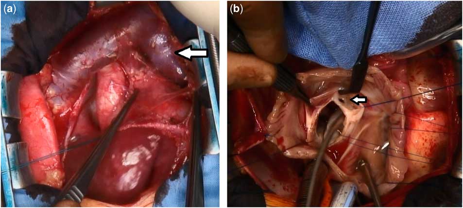
The usual approach through median sternotomy, standard cardiopulmonary bypass with aorto-bicaval cannulation, mild hypothermia, and cardioplegic arrest was performed. The left upper and lower pulmonary veins as well as vertical vein with drainage to innominate vein were exposed (Fig 2a). Then, the vertical vein was severed without stenosis from the innominate vein and connected with the left atrium.
(a) Vertical vein (arrow) with drainaige to innominate vein. (b) The obstructing membrane (arrow) that was excised through a right atriotomy incision.

The left atrium was accessed via a right atrium-transseptal approach. The fibromuscular membrane with 3–4 mm central perforation is visualised (Fig 2b), while the pulmonary veins can be identified as not obstructed. The mitral valve is hidden below the membrane. The abnormal tissue was very carefully resected in toto. The atrial septum defect was closed during the process, as was the 2 mm patent foramen ovale. Intra-operative transoesophageal echocardiography showed significant improvement of the left ventricular pumping function.
Duration of intraoperative cardiopulmonary bypass was 77 minutes, and duration of aortic cross-clamping was 39 minutes. The child remained in the ICU for 10 days after surgery. Ventilation with NO was discontinued 3 days after operation. There were no post-operative complications. The control echocardiographic investigations demonstrated significant improvement of biventricular pumping function and radical resection of membrane without any mitral valve insufficiency. The child was discharged 16 days after surgery for rehabilitation in good condition.
Discussion
The three main embryological theories explaining the development of cor triatriatum sinister are malseptation, malincorporation, and entrapment. Typically, the wall between the two chambers is bulky and contains muscular tissue and has one or multiple openings in it.Reference Aliter, Youssef, Al-Murayeh and Gallo 5 The atrial septal defect is usually present and may be in the septum between the right atrium and either the proximal or distal left atrial chamber.
Cor triatriatum sinister is classified, according to Lam (1962), as type A, without associations, as in the present case; A1, in which the atrial septal defect occurs in the proximal chamber (50%); A2, in which the atrial septal defect occurs in the distal chamber (10%); B, in which the pulmonary veins drain into the coronary sinus (1%); and C, when there is total anomalous pulmonary venous drainage (5%).Reference Lam, Green and Drake 4 , Reference Atik and Tavares 6
Since the first surgical approach in 1956 by Lewis et al, surgery is the treatment of choice in symptomatic patients with cor triatriatum sinister. This case highlights the association between cor triatriatum sinister, left upper and lower anomalous pulmonary venous return to the innominate vein through the vertical vein and patent foramen ovale. The child underwent a successful surgical repair, with rapid normalisation of both ventricular function and pulmonary artery pressure. It remains unclear why the patient could survive so long with severe and oligosymptomatic pulmonary hypertension; specific rescue-mechanisms were not observed.
In conclusion, the principles for surgical management of cor triatriatum can be summarised as radical resection of the membrane to allow adequate confluence of the genuine and the auxiliary atrium, maintenance of a sufficient volume of the left atrium, and radical correction of other intracardiac anomalies.
Acknowledgements
None.
Financial Support
This research received no specific grant from any funding agency, or from commercial or not-for-profit sectors.
Conflicts of Interest
None.
Ethical Standards
The authors assert that all procedures contributing to this work comply with the ethical standards of the relevant national guidelines on human experimentation and with the Helsinki Declaration of 1975, as revised in 2008.