Introduction
Dermatofibrosarcoma protuberans (DFSP) is a rare, infiltrative, skin soft tissue tumour of intermediate malignancy. It is locally aggressive tumour with lesser propensity to spread to regional nodes or distant organs.Reference Malkud and Dyavannanavar 1 , Reference Lal, Goel and Mandal 2 It is known to have male preponderance and can occur in any age group but more common in the third and fourth decades. It has an indolent nature and presents as a painless skin nodule that gradually progresses in size initially and then gains a spurt of growth into many nodules and ulceration.Reference Cai, Wang, Wu and Shi 3 Pathologically, they are commonly low grade and are CD34 positive on immunohistochemistry (IHC). A few of them (5–10%) are high grade with fibrosarcomatous component and behave aggressive with greater metastatic potential.Reference Hiraki-Hotokebuchi, Yamada and Kohashi 4 Surgery is the mainstay of treatment but adequate margins of 3 cm or more is needed to cure them. With inadequate margins, the recurrence rate with surgery alone ranges from 20 to 60%. Better surgical techniques such as Moh’s micrographic surgery can reduce the recurrence rates to less than 6%.Reference Acosta and Vélez 5 , Reference Veronese, Boggio and Tiberio 6 In recent years, there is increasing research on systemic therapy after the discovery of abnormality of rearrangement of chromosomes 17 and 22 [t(17;22)(q22;q13)] leading to fusion of the platelet-derived growth factor B (PDGFB) to collagen 1 alpha 1 (COL1A1) in DFSP. This results in response to therapy with tyrosine kinase inhibitors. Imatinib has shown to be promising in advanced and metastatic disease.Reference Hiraki-Hotokebuchi, Yamada and Kohashi 4 , Reference Oh, Jeong and Kwon 7 , Reference Rutkowski and Debiec-Rychter 8 In disease that is locally aggressive, radiation therapy (RT) has been used to treat for positive margins and recurrent tumours when further surgery is not feasible. The evidence for benefit of RT in DFSP is scarce.Reference Ballo, Zagars, Pisters and Pollack 9 – Reference Haas, Keus, Loftus, Rutgers, van Coevorden and Bartelink 11 We aim to determine the evidence to support the efficacy of RT in local control and in terms of recurrence-free survival (RFS) in patients treated for DFSP in our institution.
Materials and Methods
In total, 45 patients who had been diagnosed with DFSP in our institution between January 2004 and December 2013 were studied retrospectively after obtaining Institutional Review Board (IRB) approval to estimate RFS. This time frame was chosen in order to obtain at least 5-year follow-up data. The data of patients’ demographics, their clinical details including tumour size, site, previous treatment details, histopathology findings including margins, fibrosarcomatous changes, IHC, treatment details – surgery and RT details including dose, and follow-up details were obtained from institutional medical records. The patients who had no recent follow-up were contacted by phone.
Tumour size documented for this study was the maximum size in the transverse direction obtained either from clinical findings, cross-sectional imaging or pathological data. Pathological margins were noted from histopathological findings and the least of all margins were recorded for margin status. Margin was considered to be positive if the resection margin was involved. Close margin was defined to be ≤5 mm for this study. Recurrence-free interval was determined from date of histopathological diagnosis at the institution to the date of recurrence.
Treatment protocol for DFSP at our institution
All patients who presented with a primary or recurrent swelling underwent wide local excision surgery for a suspicious or proven DFSP. After obtaining histopathology and margin status, the patients who could undergo further surgery aiming to obtain adequate margins were re-excised. Adjuvant RT was offered for the following indications: positive or less than 5 mm margins and if further surgery would be mutilating or not feasible; if patient was operated for recurrent tumour irrespective of margins and high-grade tumours with sarcomatous changes. Chemotherapy with either single agent Doxorubicin or combination with Ifosfamide was used for metastatic disease.
Statistical analysis
Categorical variables were summarised using counts and percentages. Quantitative variables were summarised using mean and standard deviation or median and interquartile range. χ 2 Test was used to compare the proportions between categorical variables. Since the event was the time to recurrence, the Kaplan–Meier analysis and survival curve were given to determine the mean time to recurrence. Log-rank test was used to compare the equity of the survivors among categories. For all the analysis, 5% level of significance was considered to be significant.
Results
The patients’ characteristics
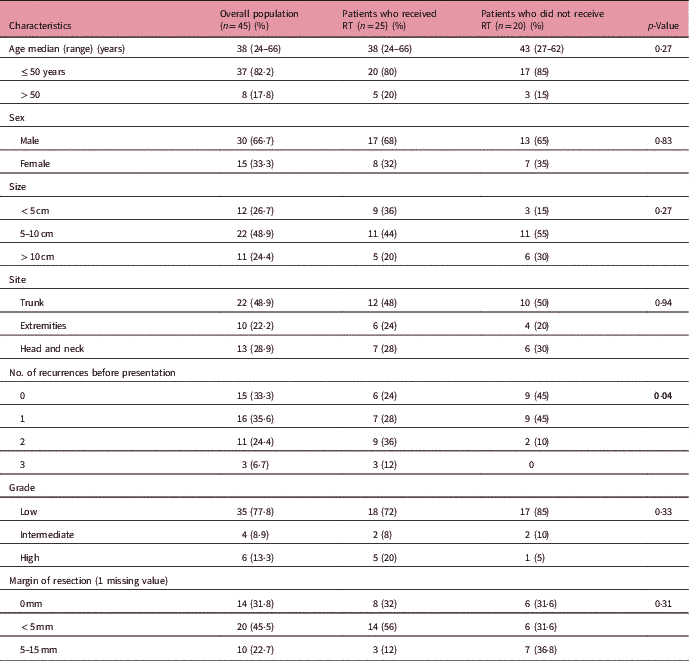
As described in Table 1, the most common age group was third and fourth decade. Males were twice in number compared with females. The most common site was trunk followed by head and neck followed by extremities. Anterior abdominal wall was the most common site in our cohort. Nearly half of the tumours were between 5 and 10 cm in size. The majority (66·7%) of patients had at least one recurrence before presenting to our institution and the reason for repeated recurrences appears to be inadequate surgery.
Patient characteristics

Abbreviation: RT, radiation therapy.
DFSP is known to be a low-grade tumour and 77·8% (n=35) of patients in this study were of low grade, 8·9% (n=4) were intermediate grade and 13·3% (n=6) were high grade. Four out of 6 patients with high-grade histology had sarcomatous changes. The IHC status was available for 26 patients and all were positive for CD34. All patients underwent wide local excision and plastic surgeons were involved when needed to cover the surgical defect with a skin flap. There was no gross residual tumour for any of them. Acquiring adequate margins was a difficult task mainly because of the location of the tumour. When least margin of all margins for the patient was taken into account, 31·8% (n=14) had positive margins, 45·5% (n=20) had margins <5 mm.
The patients in the RT group were found to have aggressive tumours with an increased risk of recurrence. The known poor prognostic factors were found in the group though statistically not significant such as: males were more in RT group compared with surgery alone group (17 versus 13 patients); and presented with high-grade tumours (5 versus 1 patients), multiple recurrences before presentation (25 versus 20 patients) and positive or <5 mm margins (22 versus 12 patients).
p-Value (0·04) was significant only for number of recurrences before presentation in our cohort between adjuvant RT and surgery alone group.
Radiotherapy
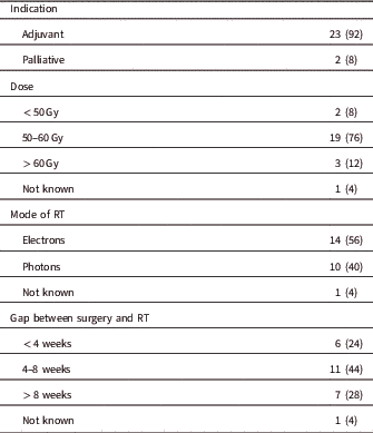
The radiotherapy details are given in Table 2. Radiation was given as adjuvant to surgery in 92% (n=23) and 8% (n=2) received RT with palliative intent. The dose given was more than 50 Gy in adjuvant setting. Treatment with electrons therapy was sufficient in 56% (n=14). Among the 40% (n=10) patients who had photon treatment, 8 of them were treated with conventional technique, one patient with three-dimensional conformal radiation therapy and another patient with intensity-modulated radiation therapy (IMRT). In conventional technique, CT scan with reference markers was not done; the patient has planned photon fields either single field to depth or parallel opposed fields. The regions treated by conventional technique were extremities or chest wall. There was more than 8 weeks gap from surgery to RT in 28% (n=7) and the common reason was delayed wound healing and graft acceptance. There was skin flap cover over the defect after excision which needed extra time for healing. There were no post-RT grade 3 or 4 adverse effects in this cohort.
Radiotherapy details

Abbreviation: RT, radiation therapy.
Details of outcome
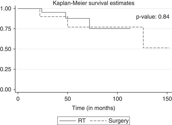
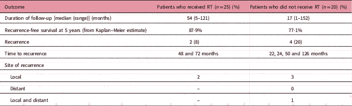
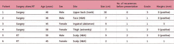
The patients in our cohort had a median follow-up of 39 months ranging from 1 to 152 months. Table 3 gives account of outcomes of the two groups: surgery alone and postoperative RT. Among the patients who received surgery alone, there were four (20%) recurrences. One of them had both local recurrence and distant metastases in the lungs, bone and in the abdomen (paraaortic nodes, peritoneal deposit). This patient had sarcomatous component in the initial histology. Those who had only local recurrence were treated with reexcision and reirradiation. There were two (8%) recurrences among those who received RT in adjunct to surgery. They had local recurrences that could be treated with reexcision and reirradiation. One of them had local recurrence again in the scalp after 48 months after reirradiation and died due to disease progression 60 months after the last recurrence. There were no patients with distant recurrence in RT group. Table 4 describes the characteristics of the patients who had disease recurrence. Inadequate margins after excision was common to all six patients. Figure 1 estimates the recurrence-free survival for patients with DFSP with surgery alone and surgery followed by postoperative RT. The difference between both groups was not statistically significant.
Recurrence-free survival. Abbreviation: RT, radiation therapy.

Details of outcome

Abbreviation: RT, radiation therapy.
Characteristics of patients who had recurrence

Abbreviation: RT, radiation therapy; H&N, head and neck.
Discussion
Clinicopathologic features
DFSP are slow growing tumours initially but once they begin to recur, they begin to grow and recur aggressively. This was noticed in our study too as reported by Li et al. in their series of recurrent DFSP.Reference Li, Wang, Xiang, Chen, Li and Ji 12 The previous recurrences before presentation to our institution showed that the duration between 1st and 2rd, 3rd recurrences was much shorter compared to that between 0th and 1st recurrence. The reasons include inadequate surgery. Prognostic factors for this disease from the US database have been found to be male, older age, margin positivity, higher grade, head and neck location.Reference Trofymenko, Bordeaux and Zeitouni 13 – Reference Kreicher, Kurlander, Gittleman, Barnholtz-Sloan and Bordeaux 15 Size was not found to be a prognostic factor revealing that wide local excision with adequate margins is curative for patients. Better surgical techniques are warranted for these tumours and Veronese et al. have shown that Moh’s micrographic surgery (8·3% recurrence rate) and Moh Tübingen technique (5·5%) reduces the chances of recurrence compared with wide local excision (41%). High-grade tumours and tumours with fibrosarcomatous component did not differ in the clinical features and prognostic factors but did behave aggressively.Reference Li, Wang, Xiang, Chen, Li and Ji 12 CD34 IHC helps in diagnosis of DFSP. The presence of specific COL1A1–PDGFB fusion transcript is a recent finding that has helped in differentiating high grade from low grade DFSP.Reference Hiraki-Hotokebuchi, Yamada and Kohashi 4 , Reference Oh, Jeong and Kwon 7 , Reference Rutkowski and Debiec-Rychter 8 As a consequence, the era of systemic therapy has begun for DFSP with Imatinib being the first successful systemic therapy.Reference Rutkowski and Debiec-Rychter 8 , Reference Ugurel and Becker 16 – Reference Wang, Luo and Chen 19
Radiotherapy details
The role of RT was defined in the late 1990s when Ballo et al.,Reference Ballo, Zagars, Pisters and Pollack 9 Haas et al.Reference Haas, Keus, Loftus, Rutgers, van Coevorden and Bartelink 11 and Suit et al.Reference Suit, Spiro, Mankin, Efird and Rosenberg 10 along with the soft tissue tumours working group demonstrated the control of gross tumours of DFSP with RT along with when being used as adjuvant to surgery.Reference Dagan, Morris, Zlotecki, Scarborough and Mendenhall 20 Ballo et al. reported 6-year progression-free survival of gross tumour with RT alone. Haas et al. demonstrated 67% control probability with surgery alone as compared with 82% when combined with RT. Suit et al. reported 9-year progression-free survival with RT alone for gross DFSP. Yet, the level of evidence is low for RT in DFSP. Our study demonstrated 92% control rate with a median follow-up of 54 months. RT is indicated for recurrent lesions and for positive or close margins (<3 cm).Reference Malkud and Dyavannanavar 1 , Reference Rutkowski and Debiec-Rychter 8 , Reference Trofymenko, Bordeaux and Zeitouni 13 Some surgeons suggest the use of adjuvant RT even in negative margins.Reference Farma, Ammori and Zager 21 , Reference Chen, Tu, Lee and Huang 22 Dose of RT is used in the range of 50–66 Gy.Reference Castle, Guadagnolo, Tsai, Feig and Zagars 23 , Reference Uysal, Sager and Gamsiz 24 Though there are no consensus guidelines available for RT, RT is unanimously offered for recurrent tumours or in cases with positive margins where further surgery is not feasible and in difficult sites such as head and neck where wider margin excision is not feasible.Reference Trofymenko, Bordeaux and Zeitouni 13 , Reference Angouridakis, Kafas and Jerjes 25 The volumes to be treated include tumour bed, postoperative scar with 3–5 cm margin.Reference Sun, Wang and Huang 26 , Reference Ugurel, Kortmann, Mohr, Mentzel, Garbe and Breuninger 27 The role of neoadjuvant RT is unknown and there are few reports indicating an increased risk of wound complication but in the era of accurate delineation and dose sculpting, this is an area of research that should be explored further.Reference Sun, Wang and Huang 26
Outcome and benefit of RT
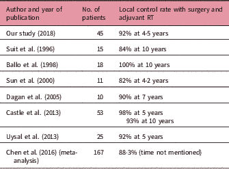
The median time to recurrence for these tumours has been reported to be 2–3 years.Reference McPeak, Cruz and Nicastri 28 , Reference Liang, Jambusaria-Pahlajani, Karia, Elenitsas, Zhang and Schmults 29 In our study, we found it to be 49 months (4 years). This difference is probably due to the increase use of RT for our patients. In our cohort, 55·6% of patients were treated with RT in comparison with the US database wherein only 8% of patients had RT.Reference Trofymenko, Bordeaux and Zeitouni 13 Table 5 compares our study outcome with the published series. DFSP is found to be radiosensitive tumour.Reference Castle, Guadagnolo, Tsai, Feig and Zagars 23 , Reference Angouridakis, Kafas and Jerjes 25 The patients who received RT in our cohort had aggressive tumours. They were tumours with known increased risk of recurrence compared with those in the surgery alone group. There were more males in the RT group compared with surgery alone group; higher grade tumours received RT; number of recurrences before treatment was higher in RT group; margins were positive or closer (<5 mm) in the RT group compared with surgery alone group. Both groups are not comparable. This indirectly shows that RT has benefit in DFSP in recurrence-free survival. The overall survival benefit is unknown.
Benefit with radiation therapy (RT) in dermatofibrosarcoma protuberans (DFSP) from literature review – selected references

Limitation of this study
Though there were double the recurrences in the surgery group compared with postoperative RT group in our cohort, statistical significance could not be tested as the sample size was less and the median survival was not reached for both groups. The sample size required for a comparative study of postoperative RT versus no RT in DFSP for recurrence-free survival with 80% power and to show a difference in outcome of 10% between RT and surgery, the sample size calculated would be 300, with 150 in each group.
Conclusion
RT delays the time to recurrence in DFSP. RT does improve the outcome of recurrence-free survival of DFSP with recurrent tumours and with positive margins.
Acknowledgements
None.
Financial support
This research received no specific grant from any funding agency, commercial or not-for-profit sectors.
Conflicts of interest
None.
Ethical standards
The authors assert that all procedures contributing to this work comply with the ethical standards of the relevant national guidelines on human experimentation (The Indian Council of Medical Research) and with the Helsinki Declaration of 1975, as revised in 2008, and has been approved by the institutional committees (Institutional Review Board, Christian Medical College).