Introduction
In vitro culture of primordial follicles enclosed in ovarian tissues is a crucial method for studying follicular development and generating a large population of in vitro grown oocytes, which may be utilized in future assisted reproduction technologies (Silva and Silva, Reference Silva and Silva2023). Ovarian stromal cells represent around 80% of the cell population in ovarian tissues (Shahri et al., Reference Shahri, Chiti and Amorim2019; Wagner et al., Reference Wagner, Yoshihara, Douagi, Damdimopoulos, Panula, Petropoulos, Lu, Pettersson, Palm, Katayama, Hovatta, Kere, Lanner and Damdimopoulou2020; Asadi et al., Reference Asadi, Najafi and Benson2022), which, in association with granulosa and theca cells (Pei et al., Reference Pei, Xiong, Guo, Wang, La, Chu, Liang, Yan and Guo2023), play a key regulatory role in the regulation of follicle development (Telfer and Zelinski, Reference Telfer and Zelinski2013; Najafi et al., Reference Najafi, Asadi and Benson2023). The density of stromal cells also reflects tissue integrity and its mechanical responsiveness (Grosbois et al., Reference Grosbois, Bailie, Kelsey, Anderson and Telfer2023). Similarly, the ovarian extracellular matrix (ECM) interacts with the surface of ovarian cells, forming a network of macromolecules that surrounds follicles and stromal cells, along with other constituents such as fibrous proteins, water and proteoglycans (Theocharis et al., Reference Theocharis, Skandalis, Gialeli and Karamanos2016; Grosbois et al., Reference Grosbois, Bailie, Kelsey, Anderson and Telfer2023).
Considering the large population of cells and follicles within ovarian tissues, in vitro culture systems must manage oxidative stress, which has been associated with a high rate of follicular degeneration in cultured tissues (Barbato et al., Reference Barbato, Genovese, De Gregorio, Di Nardo, Travaglione, De Napoli, Fragomeni, Zanetti, Adiga, Mondrone, D’Hooghe, Zheng, Longobardi, Catapano, Gualtieri and Talevi2023; Silva et al., Reference Silva, Barberino and Matos2023). High levels of reactive oxygen species (ROS) impair mitochondrial activity and chromosome configuration, leading to loss of membrane integrity and reduced ATP production (He et al., Reference He, Leong, Zhuo, Majeska, Cardoso, Spray, Goldring, Cobelli and Sun2016; Sreedhar et al., Reference Sreedhar, Wiese and Hitosugi2020). Excessive levels of ROS also damage ovarian stromal cells and the ECM (Grosbois et al., Reference Grosbois, Bailie, Kelsey, Anderson and Telfer2023). It can also impact endogenous antioxidant enzymes, such as superoxide dismutase (SOD), catalase (CAT), glutathione peroxidase (GPX) and peroxiredoxin (PRDX) (Hori et al., Reference Hori, Kuno, Hosoda and Horio2013; Chen et al., Reference Chen, Gao, Du, Yao, He, Niu, Wang and Zhang2020). In this context, the addition of naturally derived antioxidant substances to culture media has gained emphasis (Kaderides et al., Reference Kaderides, Kyriakoudi, Mourtzinos and Goula2021). Acetyl-L-carnitine is a short-chain ester derived from L-carnitine, and it is more bioavailable than L-carnitine when used endogenously (Placidi et al., Reference Placidi, Di Emidio, Virmani, D’Alfonso, Artini, D’Alessandro and Tatone2022; Hsu et al., Reference Hsu, Fatuzzo, Weng, Michno, Dong, Kienle, Dai, Pasca, Abu-Remaileh, Rasgon, Bigio, Nasca and Khosla2023). Acetyl-L-carnitine participates in lipid biosynthesis within mitochondria and can protect against oxidative stress (Wang et al., Reference Wang, Xu, Zheng, Zhang, Shao, Zhao and Hao2020). The acetyl and carnitine compounds play a crucial role in maintaining acetyl-coenzyme A levels (Adeva-Andany et al., Reference Adeva-Andany, Calvo-Castro, Fernández-Fernández, Donapetry-García and Pedre-Piñeiro2017; Albogami, Reference Albogami2023) during the transport of fatty acids to mitochondria for energy production through β-oxidation (Tauqir et al., Reference Tauqir, Israr, Rauf, Malik, Habib, Shah, Usman, Raza, Shah, Badshah, Ehtesham and Shah2021). Modak et al. (Reference Modak, Alam, Islam, Paul, Akter, Hashem, Kabir and Moniruzzaman2022) reported that L-carnitine enhanced β-oxidation, provided energy to support both cytoplasmic and nuclear maturation and significantly increased the maturation rate of oocytes from early antral follicles cultured in vitro. As an antioxidant, L-carnitine combats free radicals, particularly superoxide ions, protecting cells from oxidative damage that can lead to apoptosis (Shahreza et al., Reference Shahreza, Ahmadpour, Almasi, Seyyed Hosseini, Akhavan Taheri and Moshkdanian2023). Several studies have highlighted that acetyl-L-carnitine reduces oxidative stress in various cell types (oocytes: Dunning et al., Reference Dunning, Russell and Robker2014; stem cells: Qian et al., Reference Qian, Wang, Ji, Chen, He, Lv, Zhang, Li, Pan, Li and Lin2021; granulosa cells: Li et al., Reference Li, Wu, Ma, Zhang, Sun, Qi and Ma2023; HepG2 cell lines: Albogami, Reference Albogami2023). In cryopreserved ovarian tissue from mice, L-carnitine reduced follicular apoptosis and inhibited oxidative stress, thereby improving follicular survival in grafts (Zhang et al., Reference Zhang, Wang, Yao, Zhang, Zhang, Chen, Fu and Zhang2015). Thus, it is hypothesized that acetyl-L-carnitine may enhance the control of oxidative stress during in vitro culture of bovine ovarian tissues.
This study aims to evaluate the effects of different concentrations of acetyl-L-carnitine on primordial follicle activation, survival and development, as well as on ovarian stromal cell density and collagen distribution in the ECM of in vitro cultured ovarian tissues. In addition, the effects of acetyl-L-carnitine on mRNA expression for NRF2, SOD, CAT, GPX and PRDX6 were investigated.
Material and methods
Chemical products
The culture media and other chemicals used in this study were purchased from Sigma Chemical Co. (St Louis, MO, USA), unless otherwise indicated in the text. This study was approved by the Ethics and Animal Welfare Committee of the Federal University of Ceará (Nº 03/2022).
Ovaries and in vitro culture of cortical tissues
Ovaries from cows (n = 10) were collected at a local slaughterhouse (Sobral, Ceará, Brazil). Immediately after slaughter, the ovaries were washed with 70% ethanol for approximately 10 s, followed by two washes in α-MEM supplemented with penicillin (100 µg/ml) and streptomycin (100 µg/ml) and buffered with HEPES. The ovaries were transported to the laboratory within 1 h in α-MEM at 4°C (Costa et al., Reference Costa, Vasconcelos, Azevedo, De Assis, Paulino, Silva, Silva and Batista2021a).
In the laboratory, the ovarian cortex from each pair of ovaries was divided into 18 fragments of approximately 3 × 3 × 1 mm in a dissection medium composed of α-MEM supplemented with penicillin (100 μg/ml) and streptomycin (100 μg/ml). The culture medium was α-MEM (pH 7.2–7.4) supplemented with 50 μg/ml ascorbic acid, 2 mM glutamine, 2 mM hypoxanthine, 100 μg/ml penicillin, 100 μg/ml streptomycin, 1.25 mg/ml bovine serum albumin, 10 μg/ml insulin, 5.5 μg/ml transferrin and 10 μg /ml selenium (ITS). This control medium was designated as α-MEM+. The cortical tissues were cultured in α-MEM+ alone or supplemented with 10, 50 or 100 μM of acetyl-L-carnitine (Altun et al., Reference Altun, Olgun, Ercetin, Aktas, Kirkim, Serbetcioglu, Olgun and Guneri2014; Ishii et al., Reference Ishii, Shimpo, Atsuoka and Kinoshita2000) at 38.5°C with 5% CO2 in humidified air. The culture medium was partially replaced (60%) every 2 days (Costa et al., Reference Costa, Vasconcelos, Azevedo, Paulino, Soares, Silva, Silva and Souza2021b). Before (uncultured control) or after in vitro culture, the ovarian tissues were fixed in 4% paraformaldehyde for 12 h for histological analysis or stored at –80oC for mRNA analysis of NRF2, SOD, CAT, PRDX6 and GPX1. This experiment was repeated eight times.
Morphological analyses of ovarian follicles
Histological analysis was performed following the established methodology of Bizarro-Silva et al. (Reference Bizarro-Silva, Santos, Gerez, González, Lisboa and Seneda2018). The ovarian tissues were fixed in 4% paraformaldehyde for 12 h, dehydrated in ethanol, clarified with xylene and embedded in histological paraffin. The tissues were serially sectioned at a thickness of 7 µm, and every fifth section was mounted on slides and stained with haematoxylin-eosin. Only the follicles in which the oocytes had a clearly visible nucleus were analyzed to avoid double counting. The slides were identified by anonymous codes to avoid subjective analysis and were examined under a microscope (Nikon, TS 100, Japan) at 100× and 400× magnifications. Based on the morphology of the oocyte and granulosa cells, the follicles were classified as primordial or developing follicles (primary and secondary stages of development) (De Figueiredo and De Lima, Reference De Figueiredo and De Lima2017). These follicles were classified as morphologically normal if they contained an intact oocyte, granulosa cells well-organized in one or more layers and no pyknotic nucleus. Degenerated follicles exhibited a retracted oocyte, pyknotic nucleus and/or disorganized granulosa cells (Telfer et al., Reference Telfer, McLaughlin, Ding and Thong2008). The percentages of healthy primordial and developing follicles were calculated both before and after culture.
Analysis of ovarian stromal cell density and collagen fibres
Ovarian stromal cell density was evaluated in both uncultured tissues and tissues cultured for 6 days under the different treatments. The number of stromal cells in a 100 μm2 area was counted in five fields from different histological sections of five different animals. The number of stromal cells per field was counted as described previously by Cavalcante et al. (Reference Cavalcante, Matos-Brito, Paulino, Silva, Aguiar, DE Almeida, Souza, Vasconcelos, De Assis, Silva and Silva2019).
Collagen fibres in the ECM were evaluated after staining the sections with Picrosirius Red (Abcam Kit), following the methodology described by Rittié (Reference Rittié2017). Picrosirius red selectively stains collagen fibres, leaving all other ECM components colourless. To this end, the sections were clarified in xylene and incubated in a 0.1% Sirius Red solution at room temperature for 1 h. For each treatment, the percentage of area occupied by collagen fibres in five different fields was measured using a camera attached to a microscope (Nikon, Eclipse, TS 100, Japan), and the images were analyzed using ImageJ software (version 1.451p, 2017) with constant exposure and brightness parameters. To quantify the area occupied by collagen, the images were sequentially converted to 8-bit greyscale using ImageJ software. The analysis software automatically excluded the circumference of unstained follicles from the total area marked in red. In this format, the software analyzes and measures the fraction of the area stained by collagen fibres in the tissue sections. The sample size used for the analysis was based on a previous study (Silva et al., Reference Silva, Costa, De Lima Neto, Caetano Filho, de Assis, Aguiar, Silva, Martins, Araújo, Matos, Costa and Silva2024). The results are expressed as the percentage of the total area stained in each treatment.
Expression of mRNA for NRF2, SOD, CAT, PRDX6 and GPX1 in ovarian tissues
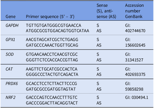
Ovarian tissues were stored at –80°C immediately after the end of the culture period until extraction of total RNA. Tissues cultured in control medium (α-MEM+) alone or supplemented with 100 μM aceyl-L-carnitine were selected for mRNA expression analysis. After maceration of ovarian tissues, total RNA was extracted using a TRIzol® purification kit (Invitrogen, São Paulo, Brazil) according to the manufacturer’s instructions. The SYBR Green was used to quantify the mRNA levels. The polymerase chain reaction (PCR) consisted of 1 μl cDNA as template in 7.5 μl of SYBR Green Master Mix (PE Applied Biosystems, Foster City, CA, USA), 0.5 μM of each primer and 5.5 μl of ultrapure water, performed on a Step One Plus instrument (Applied Biosystems, Foster City, CA, USA). The primers were designed to amplify GAPDH, SOD, CAT, PRDX6, GPX1 and NRF2 (Table 1). The thermal cycling profile for the initial PCR cycle consisted of denaturation and polymerase activation at 95°C for 10 min, followed by 40 cycles of 15 s at 95°C, 30 s at 58°C and 30 s at 72°C. The final extension was carried out at 72°C for 10 min. The 2^ΔΔCt method was used to convert the Ct values into normalized relative expression levels (Livak and Schmittgen, Reference Livak and Schmittgen2001).
Primer sequence

Statistical analysis
Statistical analysis was performed using the chi-square test to evaluate the percentages of normal, primordial and developing follicles (GraphPad Prism software 9.0). Data of collagen fibres, stromal cell number and mRNA levels for NRF2, SOD, CAT, PRDX6 and GPX1 were analyzed using the Kruskal–Wallis test, followed by Dunn’s post hoc comparison. Results were expressed as the mean ± standard deviation (mean ± SD). Differences were considered significant when P < 0.05.
Results
Effect of acetyl-L-carnitine on follicular morphology
Morphologically normal follicles displayed oocytes with a central nucleus and intact, well-organized granulosa cells, while degenerated follicles showed nuclear pyknosis, cytoplasmic retraction or disorganization of granulosa cells (Figure 1). After 6 days of culture, a significant decrease in the percentage of morphologically healthy follicles was observed in all treatments compared with the non-cultured tissues (Figure 2). Among the treatments, ovarian tissues cultured in the presence of 100 µM acetyl-L-carnitine exhibited higher percentages of normal follicles compared with those cultured in control medium (α-MEM+) alone or supplemented with 50 µM acetyl-L-carnitine. In addition, tissue cultured in the presence of 10 µM or 50 µM acetyl-L-carnitine showed similar percentages of normal follicles (Figure 2).
Bovine ovarian tissues showing morphologically normal (A–C) and degenerated (D–F) follicles at different stages of development. (A) Normal and (D) degenerated primordial follicles; (B) normal and (E) degenerated primary follicles; (C) normal and (F) degenerated secondary follicles. Granulosa cells (GC); oocyte (O); oocyte nucleus (N). Scale bar: 100 μm.

Percentage of normal follicles in uncultured ovarian tissues and in tissues cultured in α-MEM+ alone or supplemented with 10, 50 or 100 µM acetyl-L-carnitine for 6 days. *Represents a significant difference compared with uncultured tissues. a,b Different letters represent statistically significant differences between uncultured tissues and tissues cultured in different treatments (P < 0.05).

Effect of acetyl-L-carnitine on primordial follicle activation and development
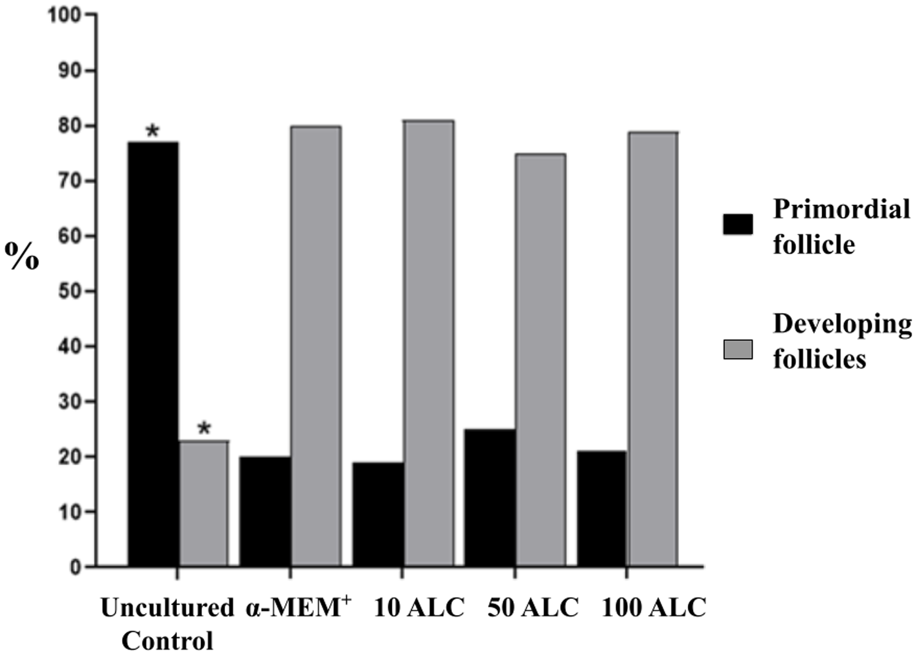
Regardless of the treatment, ovarian tissues cultured in vitro showed reduced percentages of primordial and an increased rate of developing follicles compared with uncultured tissues. However, the presence of acetyl-L-carnitine in the culture medium did not affect the percentages of primordial follicle activation and development after 6 days in vitro culture (Figure 3).
Percentage of primordial and developing follicles in uncultured tissues and in tissues cultured in α-MEM+ alone or supplemented with 10, 50 or 100 µM acetyl-L-carnitine for 6 days. *Represents a statistically significant difference between uncultured tissues and tissues cultured in different treatments (P < 0.05).

Effect of acetyl-L-carnitine on stromal cell density and collagen fibres after in vitro culture
The number of stromal cells in ovarian tissues cultured for 6 days in α-MEM+ alone or supplemented with 10 µM or 50 µM acetyl-L-carnitine did not differ from that observed in uncultured tissues. In contrast, the presence of 100 µM acetyl-L-carnitine increased the number of stromal cells in ovarian tissues compared with uncultured tissues or those cultured in α-MEM+ alone or supplemented with 10 µM or 50 µM acetyl-L-carnitine (Figure 4). Cultured tissues had a reduced percentage of area occupied by collagen fibres compared with non-cultured tissues. The presence of acetyl-L-carnitine in the culture medium, however, did not influence the percentage of collagen in the ovarian tissue after 6 days of culture (Figure 5).
Stromal cell density in uncultured ovarian tissues and in tissues cultured for 6 days (A). Representative images of ovarian stromal cell density in (B) fresh tissues and in tissues cultured with (C) α-MEM+, (D) 1, (E) 50 or (F) 100 µM acetyl-L-carnitine. (400×, scale bar: 100 μm). *Represents a statistically significant difference between uncultured tissues and tissues cultured with 100 µM acetyl-L-carnitine (P < 0.05).

Collagen fibre levels in uncultured ovarian tissues and in tissues cultured for 6 days (A). Representative images of ovarian stromal cell density in (B) fresh tissues and in tissues cultured with (C) α-MEM+, (D) 10, (E) 50 or (F) 100 µM acetyl-L-carnitine. (400×, scale bar: 100 μm). *Represents a statistically significant difference between uncultured tissues and tissues cultured in different treatments (P < 0.05).

Expression of mRNA for NRF2, SOD, CAT, PRDX6 and GPX1 in ovarian tissues
Relative mRNA levels for NRF2, CAT, SOD, GPX1 and PRDX-6 are shown in Figure 6A–E. The presence of Acetyl-L-carnitine in the culture medium did not influence the expression of these transcripts (P > 0.05).
Levels of mRNA for (A) CAT, (B) SOD, (C) GPX1, (D) PRDX6 and (C) NRF2 in ovarian tissue cultured in α-MEM+ alone or with 100 μM acetyl-L-carnitine for 6 days.

Discussion
To the best of our knowledge, this is the first study to demonstrate that 100 μM acetyl-L-carnitine increases the percentage of morphologically normal follicles and stromal cell density in cultured bovine ovarian tissues. Acetyl-L-carnitine plays a crucial role in maintaining bioenergetic homeostasis during in vitro cellular culture (Qian et al., Reference Qian, Wang, Ji, Chen, He, Lv, Zhang, Li, Pan, Li and Lin2021). Li et al. (Reference Li, Wu, Ma, Zhang, Sun, Qi and Ma2023) showed that 80 µM L-carnitine reduced apoptosis in human ovarian granulosa cells, thereby exerting a protective effect on ovarian follicles. In a recent study, acetyl-L-carnitine also reduced ROS; increased the expression of proliferating cell nuclear antigen, B-cell lymphoma 2 (BCL-2), cyclin B1 and cyclin D1; and decreased the levels of BCL-2-associated X (BAX), Caspase 3 and tumour protein P53 in granulosa cells (Jiang et al., Reference Jiang, Liu, Wu, Gong, Ma and Zi2023). The mechanisms through which carnitines exert positive effects on follicles, oocytes and embryos include β-oxidation, antioxidant response and protection against apoptosis (Placidi et al., Reference Placidi, Di Emidio, Virmani, D’Alfonso, Artini, D’Alessandro and Tatone2022). Xu et al. (Reference Xu, Yang, Lu, Liang, Lu, Zhang and Lu2018) reported that acetyl-L-carnitine improves mitochondrial function, regulates oocyte-derived paracrine factors and increases steroid hormone production, thereby enhancing the quality of mature oocytes and promoting in vitro embryonic development in buffaloes. Thus, acetyl-L-carnitine plays an important role in regulating mitochondrial metabolic activity and promoting antioxidant activity – key factors for maintaining bioenergetic homeostasis during in vitro culture. These functions may have played a key role in the survival of bovine ovarian follicles.
This study shows that acetyl-L-carnitine did not influence the expression of mRNAs for NRF2, SOD, CAT, PRDX6 and GPX1, emphasizing that this substance uses different pathways to protect the cells. In vitro studies have demonstrated that acetyl-L-carnitine increases energy production and protects mitochondria against oxidative stress by playing a crucial role in transporting acetyl-CoA into mitochondria and facilitating fatty acid oxidation to generate ATP and to reduce the levels of ROS) and, consequently, reducing the damaging effects of oxidative stress (Shenk et al., Reference Shenk, Liu, Fischbach, Xu, Puchowicz, Obrenovich, Gasimov, Alvarez, Ames, Lamanna and Aliev2009; Pancotto et al., Reference Pancotto, Mocelin, Marcon, Herrmann and Piato2018). In the ovarian tissue context, the influence of Acetyl-L-Carnitine on mitochondria may contribute to the maintenance of follicular and ovarian stroma survival (Di Emidio et al., Reference Di Emidio, Rea, Placidi, Rossi, Cocciolone, Virmani, Macchiarelli, Palmerini, D’Alessandro, Artini and Tatone2020). Mitochondria play a key role in cellular energy metabolism and apoptosis and are involved in signal transduction for cell proliferation (Zhang et al., Reference Zhang, Bao, Zhou and Zheng2019). Furthermore, mitochondria are well known as the source of ROS production. Thus, mitochondrial protection may be a good approach to increase ovarian stromal cell survival (Abdolmaleki et al., Reference Abdolmaleki, Ghayour and Behnam-Rassouli2020). Thus, we believe that maintaining cell survival through energy preservation is positively associated with stromal cell density and follicle morphological integrity during culture (Kinnear et al., Reference Kinnear, Tomaszewski, Chang, Moravek, Xu, Padmanabhan and Shikanov2020).
In the current study, 100 µM acetyl-L-carnitine increased the stromal cells density after 6 days of culture. Ovarian stromal cells are responsible for producing growth factors and peptides essential for follicular development (Kinnear et al., Reference Kinnear, Tomaszewski, Chang, Moravek, Xu, Padmanabhan and Shikanov2020). According to Barbato et al. (Reference Barbato, Genovese, De Gregorio, Di Nardo, Travaglione, De Napoli, Fragomeni, Zanetti, Adiga, Mondrone, D’Hooghe, Zheng, Longobardi, Catapano, Gualtieri and Talevi2023), the in vitro culture environment must maintain the health of both follicular and stromal cells, preserving the relationship between follicles and ovarian stromal cells, as well as growth factors and hormones that influence this follicular development. Stromal cells are associated with theca and granulosa cells (Pei et al., Reference Pei, Xiong, Guo, Wang, La, Chu, Liang, Yan and Guo2023), establishing a connection between the follicles and surrounding cells, thereby influencing follicular development (Dolmans et al., Reference Dolmans, Donnez and Cacciottola2021). Hartanti et al. (Reference Hartanti, Hummitzsch, Irving-Rodgers, Bonner, Copping, Anderson, McMillen, Perry and Rodgers2019) reported that stromal cells are primarily responsible for stromal expansion during ovarian development. Some studies have reported that stromal cells can enhance granulosa cell proliferation and hormone production (Qiu et al., Reference Qiu, Liu, Han, Wu, Yang, Su, Quan and Zhang2014; Asadi et al., Reference Asadi, Najafi and Benson2022). Therefore, our findings corroborate previous data showing a positive relationship between follicular morphology and stromal cell density after ovarian tissue culture (Cavalcante et al., Reference Cavalcante, Matos-Brito, Paulino, Silva, Aguiar, DE Almeida, Souza, Vasconcelos, De Assis, Silva and Silva2019). Thus, ovarian stromal cell density may play an important role in both follicle growth and survival, as well as in the regulation of follicular growth in vitro (Telfer and Zelenski, Reference Telfer and Zelinski2013; Asadi et al., Reference Asadi, Najafi and Benson2022; Najafi et al., Reference Najafi, Asadi and Benson2023).
Devos et al. (Reference Devos, Grosbois and Demeestere2020) showed that the intense follicular activation and accelerated growth observed in vitro are explained by the fragmentation of ovarian tissue, which interrupts the Hippo signalling pathway. To corroborate this, we observed high rates of follicular activation in cultured ovarian tissue fragments, regardless of the treatment. The ECM present in the ovary promotes contact between the follicles and stromal cells, causing natural stiffening and loosening processes, and is rich in substances such as collagen, glycoproteins and elastin (Tang, Reference Tang2020; Grosbois et al., Reference Grosbois, Bailie, Kelsey, Anderson and Telfer2023). However, acetyl-L-carnitine did not influence collagen fibres.
In conclusion, 100 μM acetyl-L-carnitine increases follicle survival and stromal cell density in cultured bovine ovarian tissues, but does not affect collagen fibre distribution or the expression of mRNAs for NRF2, SOD, CAT, PRDX6 and GPX1. The maintenance of follicular and stromal cell survival is a key step to develop new alternatives to have competent oocytes from in vitro grown follicles. This technology has great potential to restore fertility in humans or to produce large numbers of embryos from livestock animals.
Acknowledgements
The authors thank the National Council for Scientific and Technological Development (CNPq, Brazil, Process No. 407992/2021-9).
Competing interests
The authors declare no conflict of interest.