INTRODUCTION
The use of radiation in diagnostic radiology, nuclear medicine and radiation oncology is associated with its hazards and risk.Reference Broadbent and Hubbard1–Reference Gough4 It is becoming more apparent that the quantitative knowledge of radiation exposure measurement in radiology is essential. For radiation exposure measurements in radiology and radiation therapy different type of ionization chambers are available in various sizes and shapes. However, the fundamental principle of operation is the same for all chambers, and the mechanism involved in the measurement of radiation is well known.Reference Attix5–Reference Johns and Cunningham7
Exposure measurements with ionization chambers are dependent on the correction factor related to beam energy, temperature and pressure, high voltage placed across the chamber, ionization recombination and polarity effect. These factors and the construction of the chambers are directly related to sensitive volume of the chamber. The sensitive volume is the volume of gas (air) inside the chamber that gives maximum response to the radiation, and all these factors affect the response of the chamber.Reference Das and Akber8–Reference Kofler, Gray and Daly10
With the introduction and the use of conventional simulator, cone beam CT and CT simulator and 4DCT in the radiotherapy, the radiation survey of the facility and the exposure measurements are performed with the diagnostic chambers. It is therefore, important that not only the diagnostic physicists, but also the therapy physicists should be aware of the chamber characteristics.
In this work, partial volume (spatial) response of ionization chambers is evaluated.
MATERIALS AND METHODS
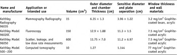
Four ionization chambers of 10 cm3, 15 cm3, 150 cm3 and 600 cm3 volumes were used with Keithley electrometer (Model 35040(ATD)) to evaluate partial volume effect in ionization chambers. Table 1, gives the characteristics of ionization chambers used in this study.
Characteristics of the ionization chambers used in this study

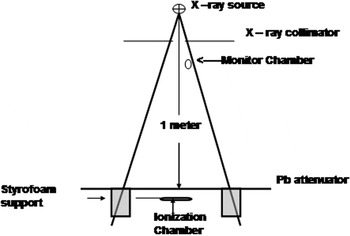
A diagnostic X-ray unit (Philip Super 80 CP) was chosen for this study due to its stable exposure history. Figure 1, shows the schematic diagram of the experimental set up used in exposure measurement. Two lead sheets of 3.2 mm thick and 30 × 30 cm2 were used to make slits of 2 mm and 5 mm width. The slit allowed a narrow region of the detector to be exposed to the X-ray beam. A Radcal 6.0 cm3 ionization chamber with an electrometer (Model 1015C), used as a monitor chamber was placed close to the collimator (Figure 1) to get relative exposure with that of the measuring ionization chamber. The readings of the monitor chamber were used to eliminate the fluctuation due to change in radiation output and measuring conditions. All measurements were performed at source to chamber distance (SCD) of 1 m. The lead sheet was supported on a specially designed Styrofoam platform. The ionization chambers of 15 cm3, 150 cm3 and 600 cm3 was used to measure partial volume response, placed under the lead sheet with 2.0 mm slit, which was moved at 2.0 mm increments over the entire length of the chamber. The X-ray unit was operated for the parameters of 81 kVp, 400 mA and 250 ms for each exposure for these chambers. For the ionization chamber of 10 cm3 volume (Computed Tomography chamber) the lead sheet with 5.0 mm slit was moved in 5.0 mm increments over the entire length of the chamber and the parameters on X-ray was selected 120 kVp, 200 mA, and 0.2 s.
Schematic diagram of the experimental set up used in this study.

RESULTS AND DISCUSSION
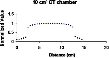
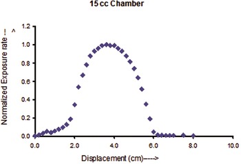
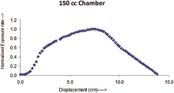
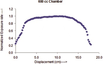
Figures 2–5 showed the normalized exposures against the relative position of the exposed area of the ionization chambers.
Spatial response of Computed radiography (CT) 10 cm3 ion chamber of exposure rate.

Spatial response of 15 cm3 ion chamber of exposure rate.

Spatial response of 150 cm3 ion chamber of exposure rate.

Spatial response of 600 cm3 ion chamber of exposure rate.

The ionization chambers of 10 cm3 (Keithley Computed Tomography Chamber, Model 500–200), 15 cm3 (Keithley, Model 96035 B), 150 cm3 (Keithley, Model 96020C) and 600 cm3 (Keithley, Model 96050) volumes exhibit ± 10% variation of plateau region from 3.5 cm to 11.7 cm (FWHM ranged 2.7–12.7 cm), 2.9 cm to 4.5 cm (FWHM ranged 2.16–5.41 cm), 5.2 cm to 8.7 cm (FWHM ranged 2.1–10.7 cm) and 3.4 cm to 13.2 cm (FWHM ranged 1.2–17.2 cm), respectively, from the edge of the chamber. Beyond this plateau region the exposure measurement decreases relative to the chamber position exhibiting a partial volume effect. A similar enhanced region (30–50 cm) was observed by Kofler et al.Reference Kofler, Gray and Daly10 for Radcal 60 cm3 ion chamber Mode 10 × 5–60.
The response of the 10 cm3 and 600 cm3 chambers, along the axis of the chamber, is relatively constant and have broad plateau compared to that of the 15 cm3 and 150 cm3 chambers. The 15 cm3 and 150 cm3 chambers exhibit narrow regions of sensitive volume that suggests that the use of these chambers without knowing the position of their sensitive volume will underestimate the radiation dose. For all the chambers the sensitive volume decreases rapidly beyond the ± 10% variation of plateau region. Similar results were shown by Kofler et al.Reference Kofler, Gray and Daly10 in Radcal CT chamber.
CONCLUSION
The chambers used in this study are commonly used in diagnostic radiology for quality control programs and also used in radiation therapy for quality control program in radiation therapy for cone beam CT and 4D CT, simulator and radiation protection survey of the facility. It is apparent that the spatial dependence of ion chambers sensitivity varies from chamber to chamber. Similar results were reported by Kofler et al.Reference Kofler, Gray and Daly10 in Radcal chamber of various sizes and shapes. It is therefore, important that ion chamber behavior and the manufacturer’s specifications such as dose and dose rate changes, operating voltage, X-ray energy and/or spectral characteristics should be strictly adhered to. Any deviation or cost cutting measures, where one chamber is used for all radiation measurements, can result in erroneous and misleading results.