Introduction
Kidneys perform several essential homeostatic functions, including waste removal, electrolyte fluid balance, acid-base metabolic balance of the blood, and the production and modification of hormones that regulate blood pressure, calcium, and potassium homeostasis, as well as stimulating the production of red blood cells. Reference Murray and Paolini1 As reviewed by Kataria et al., numerous studies have documented adverse renal effects in response to gradual exposure to a wide range of environmental chemicals. Reference Kataria, Trasande and Trachtman2 Certain chemicals, known as endocrine disruptors (EDs), can cause kidney injury, mitochondrial oxidative damage, and nephrotoxicity. Reference Xu, Nie, Ding and Hou3 Schug et al. concluded that human exposure to numerous EDs is closely related to the development of diseases in offspring and subsequent generations. Reference Schug, Janesick, Blumberg and Heindel4
In both humans and animals, endocrine disruption is a mechanism related to the functioning of the endocrine system. EDs can act in several ways, including directly damaging an endocrine organ, altering its function, interacting with hormone receptors, and altering the metabolism of a specific hormone. Reference European5 Many renal diseases (RD) such as hypertension may originate early in life. Reference Luyckx, Bertram and Brenner6
The prenatal and postnatal periods, marked by intense renal development – both structural and functional – are particularly vulnerable to various stressors, including chemical exposures, nutritional deficiencies, and chronic behavioral stress. As such, these periods are referred to as “sensitive windows of exposure.” Early-life stressors can program the individual, via multiple molecular mechanisms, to develop hypertension or kidney disease in adulthood. Reference Albertoni Borghese, Oronel, Ortiz and Majowicz7 In a recent study, Hsu et al., demonstrated that perinatal exposure to such stressors may impair renal development through hormonal receptor dysfunction and epigenetic modifications. These alterations can predispose individuals to early-onset RD, including hypertension and kidney disease, even in the absence of traditional risk factors. Reference Hsu and Tain8
A wide range of early-life environmental insults can initiate renal programming – a process associated with reduced nephron endowment and various molecular alterations. These include dysfunction of the renin-angiotensin system (RAS), changes in sodium transporter expression, increased renal sympathetic activity, glucocorticoid effects, and oxidative stress. Renal programming represents a “first hit” to the kidneys, rendering them more susceptible to subsequent postnatal insults (“second hits”), which may increase the risk of developing chronic kidney disease (CKD) later in life. Reference Tain and Hsu9
The World Health Organization (WHO) has compiled lists of chemicals suspected of causing endocrine system disruption. Reference Petrović, Eljarrat, de Alda and Barceló10 Maqbool et al. reported that approximately 800 chemicals used in everyday life can disrupt the hormonal environment, although only a small fraction has been assessed, and their mechanisms of action have been elucidated. Reference Maqbool, Mostafalou, Bahadar and Abdollahi11 Furthermore, studies focusing on the combined action of several EDs are rare, with most addressing isolated substances or small groups. Given the need to understand the actions of EDs in combination, Christiansen et al. developed a mixture of 13 chemical compounds selected to represent a range of other EDs, thereby mimicking human exposure. Reference Christiansen, Kortenkamp and Axelstad12
The association between maternal exposure to environmental risk factors and an increased likelihood of developing diseases in adulthood is known as “developmental programming” or “developmental origins of health and disease” (DOHaD). This hypothesis gained momentum after numerous observational studies, followed by epidemiological investigations showing that prenatal insults could predispose offspring to noncommunicable diseases later in life. Reference Barker13
An increasing number of studies have shown that EDs can act on the placenta, supporting evidence of the transplacental transfer of these compounds. In the study by Zhu et al., prenatal exposure to phthalates in humans was associated with increased placental thickness and a more circular placental shape. Reference Zhu, Gao and Huang14 In experiments with mice, Tait et al., administered BPA orally at doses of 0.5 mg/kg or 50 mg/kg body weight per day from GD1 to GD11, observing significant alterations in placental anatomy and structure. These findings highlight the placenta as a key target of BPA, even at environmentally relevant exposure levels. Reference Tait, Tassinari, Maranghi and Mantovani15 Furthermore, Li et al., suggest that passive diffusion may be a plausible mechanism for the placental transfer of such compounds, as the placental transfer efficiency of bisphenols is below 100%, with lower molecular weight facilitating translocation across the placental barrier. Reference Li, Zhuang and Song16
RDs are prevalent worldwide and ∼ 10% of the global population is affected by CKD. Reference Lozano, Naghavi and Foreman17 Considering kidney disease and the growing burden of various global noncommunicable diseases, the WHO has notified the population about policies for the prevention and control of noncommunicable diseases. These policies consist of actions aimed at understanding the distribution and risk factors in the population and identifying their social, economic, and environmental determinants, with the goal of supporting the prevention and control of such diseases. 18 Moreover, several studies have shown that kidney diseases are more prevalent during aging and are associated with hypertension, obesity, diabetes, and metabolic syndrome. Consequently, the DOHaD concept has become a key prevention strategy for reducing the risk of chronic diseases in future generations. Reference Baird, Jacob and Barker19
Therefore, the present study aimed to investigate whether exposure to a mixture of EDs during the perinatal period causes tissue alterations in different renal compartments during aging.
Materials and methods
Animals and experimental environment
A total of 24 adult female Sprague-Dawley (SD) rats (120-days-old, weighing ∼ 300 g) and 12 adult male SD rats (90-days-old, weighing ∼ 300 g) were obtained from the Multidisciplinary Center for Biological Research in the Area of Laboratory Animal Science (CEMIB/UNICAMP) and housed at the Small Mammal Animal Facility at UNOESTE. The animals were distributed in a ratio of 2–3 females and 1 adult male for mating per polypropylene cage (41 × 34 × 16 cm) with a stainless-steel lid in a grid lined with autoclaved white pine shavings. The cage and bedding were changed twice weekly. Throughout the experimental phase, environmental conditions of the animal facility were controlled, including temperature (average of 23°C to 25°C), relative humidity (55 ± 10%), light cycle (12 h light/12 h dark), and continuous air exhaustion. All the animals received a commercial phytoestrogen-free diet (NUVILAB CR1/Nuvital-PR) and 500 mL of filtered water in glass drinking bottles containing metal lids and spouts. Water and food were provided ad libitum. The handling procedures, drug administration, anesthesia, and euthanasia of the animals were approved by the Ethics Committee on the Use of Animals at UNOESTE (CEUA Protocol 6034).
ED mixture
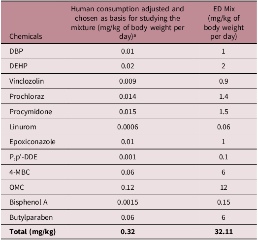
The compounds DBP (CAS No. 175606-05-0), DEHP (CAS No. 117-81-7), Vinclozin (CAS No. 50-471-44-8), Prochloraz (CAS No. 67,747-09-5), Procymidone (CAS No. 32,809-16-8), Linuron (CAS No. 330-55-2), Epoxiconazole (CAS No. 133855-98-8), p,p’-DDE (CAS No. 72-55-9), 4-MBC (CAS No. 36,861-47-9), OMC (CAS No. 5466-77-3), BPA (CAS No. 80-05-07), and Butylparaben (CAS No. 94-26-8) were purchased from Sigma-Aldrich (St. Louis, Missouri, USA). The ED mixture was diluted in corn oil (vehicle) to a concentration of 32.11 mg/kg/day and administered orally (by gavage) to the animals during the gestation and lactation periods. The composition of the mixture is detailed in Table 1 and was adapted from Christiansen et al. Reference Christiansen, Kortenkamp and Axelstad12
Mixture composition, adjusted individual human consumption of the chemical compounds, and the concentration of ED Mix administered

a Based on Christiansen et al.11 for detailed information on human consumption estimates and for the adjusted concentrations that were chosen as basis for the mixture.
Experimental design
The presence of sperm in the vaginal smear and positive vaginal cytology in the estrous phase were considered indicators of gestational day 0 (GD0). Pregnant females were housed individually in cages and randomly assigned to two experimental groups: a Control (vehicle: corn oil, 2 mL/kg/day by gavage) and an ED Mix Group (32.11 mg/kg/day of the ED mixture diluted in corn oil, 2 mL/kg/day by gavage). The animals in the ED Mix group received the ED mixture developed by Christiansen et al. Reference Christiansen, Kortenkamp and Axelstad12 and reproduced by BOBERG et al. Reference Boberg, Johansson and Hadrup20 ISLING et al. Reference Isling, Boberg and Jacobsen21 JOHANSSON et al. Reference Johansson, Jacobsen and Hass22 and MANDRUP et al. Reference Mandrup, Johansson and Boberg23 The ED mixture was adapted from the original formulation by removing the acetaminophen because of its hepatotoxic potential.
Pregnant or lactating rats received treatment from GD7 until postnatal day 21 (PND21) during the same period (8–10 AM). Pregnant or lactating rats were kept in individual cages and weighed every other day to calculate the volume of ED mixture to be administered and to monitor for signs of clinical toxicity.
After birth, the number of pups per litter was reduced to eight (with a male-to-female ratio of 1:1 whenever possible) and litters with fewer than seven pups were excluded from the study. At PND22, male offspring were weighed and assigned to the experimental groups, with 8 animals in the control group (Ctrl) and 9 animals in the ED-exposed group (ED Mix). Each animal was selected from a different litter. The animals were kept until they reached 440 days of age, received water and food ad libitum, and were euthanized by the intramuscular administration of ketamine and xylazine, followed by exsanguination. The left and right kidneys were removed, weighed, and subjected to histological analysis (Fig. 1).
Experimental design. Pregnant Sprague-Dawley rats were exposed to corn oil (Ctrl) or an endocrine disruptor mixture (ED Mix) from gestational day 7 (GD7) to postnatal day 21 (PND21). Male offspring were weaned at PND22 and euthanized at PND440 for renal histological analyses using PAS, H&E, and picrosirius staining.

Organ-somatic index
The weights of the rats were recorded immediately after intramuscular administration of the anesthetics, followed by euthanasia. The weight of each kidney was recorded and the organ coefficient was calculated as organ weight/body weight × 100%.
Renal structure analysis
Kidney fragments were fixed by immersion in metacarns (6 methanol: 3 chloroform: 1 acetic acid) and stored in 70% alcohol. Subsequently, the material was dehydrated using increasing concentrations of ethanol, cleared in xylene, and embedded in Paraplast (Oxford Labware, St Louis, MO, USA). The paraplast-embedded fragments were sectioned at a thickness of 4 μm using a rotary microtome and subjected to the following staining protocols:
-
1. Periodic Acid-Schiff (PAS): General histopathological analysis.
-
2. Hematoxylin-Eosin (HE): fractal dimension, karyometry, glomerular volume, nuclear perimeter, and elongation factors.
-
3. Picrosirius: Collagen fiber analysis.
Histopathological analysis
Histopathological analyses were performed blindly by an experienced observer using a standard optical microscope (NIKON Laboratories, Tokyo, Japan). The kidney parameters evaluated included congestion, cytoplasmic vacuolization, inflammatory infiltration, hemorrhage, nuclear vacuolization, tubular fusion, and necrosis. Each parameter for each animal was scored between 0–3 (0, no alteration; 1, mild; 2, moderate; and 3, severe). The maximum score for each sample was 21, and the minimum score was 0. Two histological sections were evaluated per animal, spaced 50 µm apart. For each kidney, the scores of different parameters were summed to determine the mean histopathological score of the organ. Reference Sümer, Sentürk, Demirel and Yeşilada24
Karyometric analysis
Karyometric analysis was performed on histological sections stained with HE from eight animals in the Ctrl and nine animals in the ED Mix. Four histological fields per section were photographed at 100X magnification, and measurements were taken from five random nuclear regions per photograph. Cross-sectional nuclear areas (μm2) and perimeters (μm) were determined for the nuclei of 200 epithelial secretory cells to obtain the shape factor parameter [ = 4π × nuclear area / (nuclear perimeter)]. Reference Kataria, Trasande and Trachtman2 The shape factor measures the roundness, with values indicating the shape of the nucleus ranging from 0 to 1, where 1 represents a perfectly circular shape.
Fractal analysis
Histological sections stained with HE from eight animals in the Ctrl and nine animals in the ED Mix were analyzed, with 10 histological fields per section photographed at 40X magnification, binarized for reading and fractal dimension analysis. For fractal analysis, the Image J software (National Institutes of Health, USA – NIH), available for free online (https://rsbweb.nih.gov/ij/), was utilized using the box-counting method. The software considered box counting in two dimensions, allowing for the quantification of pixel distribution in that space without considering the image texture. Thus, two images with the same pixel distribution, one binarized and the other grayscale, had the same fractal dimension. Consequently, the analyses were guided by the relationship between resolution and the evaluated scale, and the results were quantitatively expressed as the fractal dimension of the object, calculated as DF = (log Nr/log r - 1), where Nr is the number of identical elements needed to fill the original object, and r is the scale applied to the object. Thus, the fractal dimension was always calculated to be between 0 and 2 without distinguishing the different textures.
Analysis of glomerular volume and Bowman’s space area
A minimum of 100 glomeruli were selected per group, proportionally distributed among the 8 animals in the Ctrl group and the 9 animals in the ED Mix group. The minor and major diameters of each glomerulus were measured using the ImageJ software (National Institutes of Health, USA). The glomerular volume was calculated using the following formula: 4(d(G)/23 /3, where d(G) represents the arithmetic mean of the major and minor diameters. Reference Yildiz, Mağden, Abdulkerim, Özçicek, Hür and Candan25 Additionally, in the same glomerulus, the area of Bowman’s space was calculated by subtracting the area of the glomerulus from the area occupied by the glomerular capillaries.
Analysis of nuclear perimeter and elongation factor
This analysis was performed on histological sections stained with HE from eight animals in the Ctrl and nine animals in the ED Mix. Four histological fields per section were photographed at 100X magnification, and measurements were obtained from five nuclear regions per histological section. For each nucleus, the perimeter (μm) and the elongation factor (minor diameter/major diameter) were assessed. For the elongation factor, values equal to 1 are associated with circular nuclei, whereas values less than 1 indicate a more elongated shape.
Quantification of type I and type III collagens
Histological sections stained with picrosirius from eight animals in the Ctrl and nine animals in the ED Mix were analyzed, with 10 histological fields per section photographed at 40X magnification. The slides were analyzed using polarized light, which differentiates collagen types based on the intensity of birefringent fibers. Specifically, reddish coloration represents type I collagen and greenish coloration refers to type III collagen. Reference Junqueira, Cossermelli and Brentani26,Reference Montes, Krisztan, Shigihara, Tokoro, Mourão and Junqueira27 Collagen fibers were differentiated and quantified using the RGB measurement tool in ImageJ (Image J, 2004).
Statistical analysis
Data were analyzed using the Shapiro–Wilk normality test. The biometric data (organosomatic index and absolute kidney weight), glomerular volume analysis, elongation factor analysis (glomerulus), fractal analysis by H&E (cortex, medulla, and glomerulus), histopathological analysis, and fractal analysis by picrosirius (cortex) were parametric. Subsequently, the Student’s t-test was used to assess the significance between groups using Prism software (version 5.0; GraphPad) (significance at p < 0.05). Conversely, the nuclear perimeter, elongation factor analysis (cortex and medulla), quantification analysis of type I and type III collagen (cortex, glomerulus, and medulla), and fractal analysis by picrosirius (glomerulus and medulla) did not exhibit a normal distribution; thus, the nonparametric Mann–Whitney test was applied (significance at p < 0.05).
Results
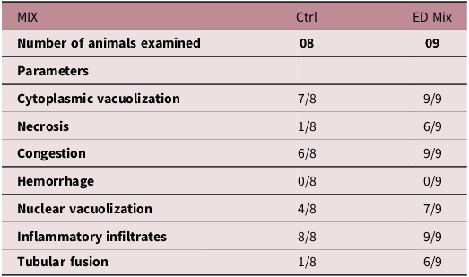
The exposure to EDs mixture did not alter the organosomatic index or influence absolute kidney weight (Table 2). Histopathological analysis addressed cytoplasmic vacuolization, necrosis, congestion, hemorrhage, nuclear vacuolization, inflammatory infiltration, and tubular fusion. After analyzing and scoring these parameters, the group exposed to the ED mixture showed a biological trend (p = 0.0687) for a higher number of histopathological alterations (Table 3 and Fig. 2). The most prevalent alterations in the ED Mix group were necrosis (Fig. 2, circle) and tubular fusion (Fig. 2, dashed circle) (66.6% in the ED Mix vs. 12.5% in the Ctrl) (Table 3).
Exposure to the ED mixture induces histopathological changes in rat kidneys. Histological sections and histopathological scores in the kidneys of rats exposed to an ED mixture during gestation and lactation. Details of renal lesions identified in the experimental groups: (a) necrosis (circle), (b) tubular fusion (dashed circle), (c) venous congestion (asterisk) and inflammatory infiltration (arrowhead), (d) inflammatory infiltration (arrowhead), (e) cytoplasmic vacuolization (square), (f) venous congestion (asterisk) and inflammatory infiltration (arrowhead). Eight animals in the Ctrl and nine animals in the ED Mix group. Magnification: 40 ×. Staining: PAS. (g) graphical representation of the mean histopathological scores. Results expressed as mean ± standard deviation (SD). Statistical analysis performed using student’s t-test (p < 0.05).

Biometric data of offspring from rats exposed during gestation and lactation to a ED mixture based on human exposure

The results are expressed as mean ± SD. p<0.05 compared to the Ctrl group.
Histopathological score of rats exposed to the ED mixture during gestation and lactation

Fig. 3 illustrates that exposure to the ED mixture modified the nuclear phenotype of renal cells and that these changes were dependent on the compartment analyzed. Karyometric analysis indicated that the ED mixture increased the form factor in the cortical (p < 0.0001) and medullary (p = 0.0107) cells, resulting in more rounded nuclei (Fig. 3, arrows; Fig. 4a and c). Nuclear perimeter analysis corroborated these results, indicating the presence of larger nuclei in the cortical (p < 0.0001) and medullary (p = 0.0032) cells of animals exposed to the ED mixture (Fig. 4d and f). Conversely, the elongation factor did not differ between groups in any of the analyses (Fig. 4j, k, and l).
Exposure to the ED mixture alters the nuclear phenotype of renal cells in a compartment-dependent manner. Histological sections of different renal compartments (cortex, glomerulus, and medulla). The cortical and medullary regions in the ED Mix group showed more rounded nuclei (H, L, arrows), while in the Ctrl group, they were more elliptical (B, F, arrows). Magnification 40X (A, E, G, K). Magnification 100X (B, C, D, F, H, I, J, L) (eight animals in the Ctrl and nine animals in the ED Mix). Staining: HE.

Exposure to the ED mixture induces significant quantitative changes in nuclear morphology. Graphical representation of karyometric analysis (a, b, c), nuclear perimeter (d, e, f), fractal analysis (g, h, i), and elongation factor (j, k, l). Data are presented as median [interquartile range] for panels a, b, c, d, e, f, j, and l, and were analyzed using the Mann–Whitney test. Data are presented as mean ± standard deviation (SD) for panels g, h, i, and k, and were analyzed using the student’s t-test. P < 0.05 compared to the Ctrl group. Eight animals in the Ctrl group and nine animals in the ED Mix group.

In addition to changes in nuclear shape, the pattern of cellular distribution was also altered. The fractal analysis indicated differences between the experimental groups, showing in the ED Mix group a reduction in the fractal dimension in glomerular cells (p < 0.0001) and an increase in medullary cells (p = 0.0018) (Fig. 4h and 1). The glomeruli also exhibited volume changes, as indicated in Figure 5, with a reduction observed in animals exposed to the mixture (p = 0.0256) (Fig. 5c). However, no changes were observed in Bowman space (Fig. 5d). In the renal stroma, exposure to the DE mixture altered the quantity and distribution of collagen fibers (Fig. 6 and Fig. 7, white arrows) in a compartment-dependent manner. An increase in the proportion of type I and type III collagen fibers was observed in the cortex and medulla (p < 0.0001) (Fig. 7a, c, d, and f) and a reduction was observed in the glomerulus (p < 0.0001) (Fig. 7b and e), which corroborates the decrease in glomerular volume observed in animals exposed to the ED mixture. Furthermore, the distribution pattern of collagen fibers was altered (p < 0.0001) (Fig. 7g, h, and i), demonstrating that the DE mixture also affected renal stromal remodeling (Graphical Abstract).
Exposure to the ED mixture decreases glomerular volume while preserving the integrity of Bowman’s space. Histological sections of glomeruli from rats exposed to a mixture of twelve EDs during gestation and lactation. Morphological characteristics of the glomerulus in the Ctrl (a) and ED Mix (b) groups. Graphs represent the quantification of glomerular volume (c) and Bowman’s capsule space area (d). Results expressed as mean ± SD, and were analyzed using the Student’s t-test (eight animals in the Ctrl and nine animals in the ED Mix). Magnification 100X. Staining: HE. *p < 0.05 compared to the Ctrl group.

Exposure to the ED mixture promotes stromal remodeling in the kidney, altering collagen distribution and content. Histological sections of rat kidneys exposed during gestation and lactation to a mixture of twelve EDs. Morphological characteristics of the renal stroma in Ctrl (A and B – cortex, E and F – glomerulus, I and J – medulla) and ED Mix (C and D – cortex, G and H – glomerulus, K and L – medulla).

The ED mixture changes collagen quantity in kidney stroma. Graphs represent the quantification of type I collagen (m, n, o), type III collagen (p, q, r), and fractal dimension (S, T, U). In m, n, o, p, q, r, t, and u, results are expressed as median [interquartile range], Mann–Whitney test. In S, results are expressed as mean ± SD, Student’s t-test. (eight animals in the Ctrl and nine animals in the ED Mix). Magnification 100X. Staining: picrosirius. *p < 0.05 compared to the Ctrl group.

Discussion
In the last three decades, the importance of the environment during early life in the development of chronic diseases in aging and future generations has been reported. Reference Baird, Jacob and Barker19 The developmental origins of the disease pathogenesis model are supported by biological evidence obtained from animal experiments. These studies have shown that maternal dietary alterations during pregnancy can modify the physiological processes of the offspring, and that these modifications are long-lasting. Reference Bertram and Hanson28
Experiments in animal models have clarified that the effects of nutrition in early-life act through changes in the development of organs and tissues, such as the pancreas, liver, kidneys, skeletal muscle, and adipose tissue. Similar to the pioneering studies on this topic conducted in Hertfordshire, UK, which explored the association between early life and chronic diseases in adulthood, Reference Kett and Denton29 animal model studies reviewed by Bertram and Hanson showed significant alterations in the renal structure of aged rats solely due to maternal exposure during gestation and lactation. Reference Bertram and Hanson28
The impact of environmental chemicals on public health and clinical well-being has long been recognized. The liver is considered the primary target organ of most chemicals implicated in the induction of toxic effects after environmental exposure. However, emerging data and the present study suggest that the kidney is an important site of injury following exposure to chemicals, although substantial gaps remain regarding the specific effects on renal function. Reference Kataria, Trasande and Trachtman2
In humans, renal development occurs in utero between weeks 5th and 36th weeks. By the end of gestation, nephrogenesis is complete, and the basic renal architecture, including vascular, neural, and hormonal networks, is established. Like humans, however, kidney maturation in animals continues during the weaning and early postweaning periods. Reference Kett and Denton29 Given the long period of renal development, it is possible that the kidney is sensitive to adverse intrauterine and neonatal effects, as demonstrated in the present study.
Kett et al. used the term renal programming to describe a series of insults that the kidney may suffer during its development. The consequences of this programming may manifest grossly as a reduced kidney size or, more subtly, as changes in the activity of the renal renin-angiotensin-aldosterone system. Reference Kett and Denton29 In this study, we did not deduct any change in renal weight between groups; despite this discrepant result, other authors have demonstrated that kidney programming can occur in the absence of any effects on birth weight. Reference Denton, Kett and Dodic30,Reference Moritz, Singh, Probyn and Denton31
Sangai et al. evaluated the kidneys of rats exposed to BPA for 30 days and identified degenerative changes, increased vacuolization, glomerular disorganization, and increased space between the glomerulus and capsule wall. Reference Sangai and Verma32 Our study used a scoring system to evaluate histological changes collectively, and a biological trend was observed, with a higher predisposition to lesions, such as necrosis and tubular fusion, in the group exposed to the DE mixture.
Cytoplasmic vacuolization, although mostly reversible, can be caused by exposure to various chemicals and precedes elevated creatinine levels, Reference Carraro-Eduardo, Santino, Macedo and da33 an important marker of renal function. Other significant findings in the ED Mix group included venous congestion, necrosis, and tubular fusion alterations, indicating possible tubular damage and attempts at regeneration in the group exposed to EDs. Kata et al. evaluated renal histology in rats exposed to the pesticide Malathion for six days and observed inflammation in various tissues, including the kidney; Reference Kata34 similar to our study, congestion of blood vessels was noted, resulting in inadequate oxygen supply and necrosis in adjacent regions.
Karyometric analysis, or the quantification of the nuclear characteristics of cells under optical microscopy, has been successfully applied to classify various types of cancer. Reference Van der Poel, Mulders and Oosterhof35 Tosi et al. reported a positive correlation between morphometric results and survival in patients with early-stage renal neoplasia. Patients with more circular nuclei in renal tumor cells have a higher five-year survival rate. Reference Tosi, Luzi and Baak36 Our study revealed an increase in the nuclear form factor in the cortex and medulla, indicating that the nuclei of epithelial cells in these regions were rounder in rats exposed to the EDs mixture. Conversely, the nuclear perimeter was larger in the cortex and medulla, which are negative prognostic factors of renal tumor cells. This predictor assumes that a larger nucleus is a morphological manifestation of abnormal DNA content. Reference Barlogie, Raber and Schumann37,Reference Ruiz, Hernández, Martinez, Vera and Jimenez-Cruz38 These discordant results may be explained by the absence of established tumor processes in the animals used in our study. Moreover, the value of this analysis for benign cells and its clinical correlations require further investigation.
Fractal analysis is a powerful tool for understanding complex structures such as those found in biological systems, including cells. It can be used to quantify the spatial organization and complexity of tissue structures. Reference Barnsley and Demko39 In renal cells, this analysis can be applied in various ways, such as cellular morphology, tubular structure, and renal microcirculation. Li et al. performed fractal analysis in patients with hypertension and suggested that decreased fractal complexity was associated with the progression of CKD. Reference Li, Ma, Zhang and Wu40 Wang et al. used fractal geometry as an early evaluation method for diabetes-related renal damage. Reference Wang, Liu and He41 In our study, fractal analysis showed alterations in the glomerulus and medulla of the ED Mix group, indicating that perinatal exposure to EDs can alter renal structure and cellular homogeneity.
The group exposed to EDs during gestation and lactation exhibited a significant reduction in glomerular volume. Supporting this result, Moraes et al. observed that rats exposed to smoke from approximately 40 cigarettes/day for 28 days had a smaller glomerular volume, which was clinically reflected in a lower urinary volume and increased serum glucose and creatinine levels, impacting renal homeostasis. Reference Moraes, Breda-Stella and Carvalho42
Some authors suggest that exposure to EDs affects the glomeruli in ways that have downstream consequences on the RAS. In a study by Wei et al., pregnant rats were exposed to Di-(2-ethylhexyl)phthalate (DEHP) at doses of 0.25 and 6.25 mg/kg body weight/day GD0 PND21. The findings revealed that maternal DEHP exposure impairs renal development in the offspring, delaying glomerular maturation and leading to a reduced nephron count. This structural alteration contributes to elevated blood pressure in adulthood, possibly through the suppression of the RAS. Reference Wei, Song and Wei43
Similarly, La Merrill et al. demonstrated that perinatal exposure to dichlorodiphenyltrichloroethane (DDT) – a pesticide still used in some developing countries for vector-borne disease control – can result in long-term adverse outcomes, including hypertension and cardiac hypertrophy in adult offspring. One of the key mechanisms driving this hypertension was the hyperactivation of the RAS, accompanied by increased renal expression of sodium channels. Additionally, perinatal DDT exposure induced cardiac hypertrophy, likely as a compensatory response to blood pressure overload. These findings reinforce the hypothesis that exposure to endocrine-disrupting chemicals during the perinatal period can compromise renal and cardiovascular development, thereby increasing the risk of hypertension and cardiac dysfunction later in life. Reference La Merrill, Sethi, Benard, Moshier, Haraldsson and Buettner44
Renal fibrosis is characterized by the pathological deposition of collagen, with collagen types I and III being the main components of this accumulation. Baues et al. and Stanchev et al. demonstrated that type I collagen is a typical interstitial protein; thus, renal fibrosis is characterized by a significant increase in this protein. Reference Baues, Klinkhammer and Ehling45,Reference Stanchev, Stamenov, Kirkov, Dzhambazova, Nikolov and Paloff46 Our study found changes in the distribution of collagen fibers in all three evaluated renal compartments, along with an increase in the deposition of type I and III collagen in the cortex and medulla, and a reduction in the glomerulus in the ED Mix group. These data hold significant clinical relevance, as fibrosis is a common finding in CKDs, regardless of the etiology. Reference Baues, Klinkhammer and Ehling45,Reference Panizo, Martínez-Arias and Alonso-Montes47
The results of the collagen analyses are consistent with those of other studies, such as those by Zhu et al., who exposed pregnant rats to DBP during gestation and observed an increase in renal dysplasia and fibrosis in adult offspring, Reference Zhu, Chen and Wang48 as well as with the study by Priego et al., who analyzed the effects of BPA exposure for five weeks in nephrectomized rats, leading to inflammatory infiltration, tubular injury, and renal fibrosis. Reference Priego, Parra, Mas, Morgado-Pascual, Ruiz-Ortega and Rayego-Mateos49 Both studies explain the effects of increased pro-inflammatory genes and significant oxidative stress, reinforcing the deleterious effects of environmental toxins and their profibrotic effects.
Conclusion
In conclusion, the ED mixture can alter the nuclear phenotype in different renal compartments, in addition to promoting an increase in collagen fibers and stimulating renal fibrosis. These changes were observed in aged animals, demonstrating the potential of perinatal ED exposure to alter the renal tissue microenvironment. These findings underscore the ongoing need for research to elucidate the underlying mechanisms and long-term consequences of such exposure as well as the importance of preventive measures to protect renal and overall health during critical stages of development.
Acknowledgements
The authors are grateful to CAPES (Coordination of Superior Level Staff Improvement) for providing a scholarship to K. D. Hinokuma.
Financial support
Grant 2018/24044, São Paulo Research Foundation (FAPESP).
Competing interests
The authors declare no competing interests.
Ethical standards
All experiments were conducted according to the guidelines established by the Brazilian Association for Animal Experimentation (COBEA) and were approved by the Ethics Committee in Animal Use of the University of Western São Paulo (Protocol number 6034).